




Abstract
Recently, the synthesis of radiolabeled plant origin compounds has been increased due to their high uptake on some cancer cell lines. Eugenol (EUG), a phenolic natural compound in the essential oils of different spices such as Syzygium aromaticum (clove), Pimenta racemosa (bay leaves), and Cinnamomum verum (cinnamon leaf), has been exploited for various medicinal applications. EUG has antiviral, antioxidant, and anti-inflammatory functions and several anticancer properties. The objective of this article is to synthesize radioiodinated (131I) EUG and investigate its effect on Caco2, MCF7, and PC3 adenocarcinoma cell lines. It is observed that radioiodinated EUG would have potential on therapy and imaging due to its notable uptakes in studied cells.
Introduction
Cancer is one of the major mortal diseases in the last century. Synthetic and natural drugs are used to cure this disease. However, use of synthetic drugs are expensive and they may have some side effects, such as decreased pathogen sensitivity causes unnecessary application. 1,2 For this reason, the natural drugs have gained significant importance. In the last two decades, usage of plants and their extracts was increased because of their anticancer properties in cancer researches. 3 –9 Psoralea corylifolia, 10 red clover, 11 henna, 12,13 yarrow, 14 Olea europaea L. products, 7,15 –17 and Papaver somniferum 18,19 are some examples of natural products whose extracts are examined for various purposes. For example, radioiodinated P. corylifolia extract (125I-bakuchiol) was investigated in lymphosarcoma (LSA) and radiation-induced solid thymic lymphoma (barcl-95) cells and it was reported as a favorable cytotoxic agent, promising therapy. 10
One of these plants is clove (Syzygium aromaticum). It has different names in different languages such as “Dutch (nagel), Spanish (clavo), and Portugese (cravo).” 20 It is commonly used as a spice in the Indian kitchen. 4 The clove includes some essential oils such as eugenol, caryophyllene, alpha-humulene, alpha-terpinylacetate, eugenyl, methyleugenol, actyleugenol, naphthalene, chavicol, heptanone, sesquiterpenes, methyl salicylate, pinene, and vanillin. Among them, eugenol, which has a great percent (81.1%), is the main compound of clove. 4 Eugenol (4-allyl-2-methoxyphenol) (EUG) with a molecular formula C10H12O2 (Fig. 1) and a molecular weight 164.21 g/mol is weakly acidic, slightly soluble in water and soluble in organic solvents. 6,20 It appears as a clear to pale yellow oily liquid. 5
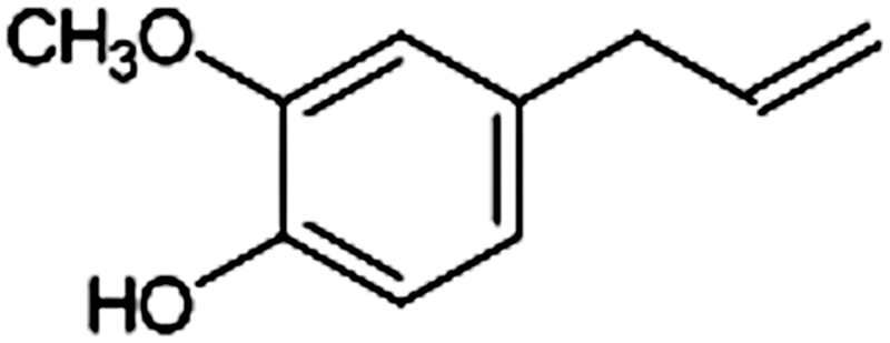
Molecular structure of EUG. EUG, eugenol.
Eugenol is used in pharmaceutical production due to its antibacterial, antifungal, antiplasmodial, antiviral, anthelmintic, anti-inflammatory, analgesic, antioxidant, and anticancer activities. 2,4,20 In the literature, it is reviewed from a vary of researches that EUG has an anticancer activity against various cancers types. 5 It is seen that most of them were studied on cancer cells in breast, colon, HeLa, and prostate cancer. 2,21 –24 One of the methods to investigate the effect of the plant on cancer is radiolabeling them with radionuclides. In the literature, there are very limited studies on the evaluation of radiolabeled plant extracts on cell lines. 7,8,12,25 One of the radionuclides that is used in radiolabeling studies is Iodine-131 (131I). It is a beta particle and gamma-emitting radioisotope (t1/2 = 8.04 days) used in preclinical studies to determine the pharmacokinetic properties of new drugs. 14,26,27
In this study, the eugenol that was obtained after extraction and purification of clove was radiolabeled with 131I, and the effect of radiolabeled eugenol on the cells was examined using MCF7, Caco2, and PC3 cell lines. The structure determination of eugenol was made and the quality control studies of radiolabeled eugenol (131I-EUG) were performed by thin layer radio-chromatography (TLRC) and high-performance liquid radio chromatography.
Experimental
General methods and materials
All reagents were of high-performance liquid chromatography (HPLC) grade and were supplied from Merck Chemical Co. and Sigma-Aldrich Chemical Co. The clove buds were obtained commercially from a gross market in which special herbs and spices are sold (Bornova-Izmir/Turkey) and identified by authors. In addition, a sample was stored for further reference. A low pressure gradient HPLC system [quaternary pump (LC-10ATvp), diode array detector (DAD; SPD-M20A), NaI(Tl) radioactivity detector (Gabi Star, Raytest), an autosampler (SIL-20A HT), fraction collector (FRC-10A) and column (RP-C18; 5 μm, 250 × 4.6 mm I.D., ODS Gl sciences Inc.)] was used. ITLC-cellulose (Merck 5565) plates (1.5 × 10 cm-sized) were used for TLRC. TLRC measurements were accomplished using a TLC scanner (Bioscan AR-2000 Scanner). Statistical significance was assessed with one-way analysis of variance and nonlinear regression by the GraphPad program for cell culture studies. Na131I was obtained from Sifa University Bornova Training and Research Hospital, Izmir, Turkey. The human breast adenocarcinoma cell line (MCF7), human prostate cancer cell line (PC3), and human colonic carcinoma cell line (Caco2) were obtained from the American Type Culture Collection. Packard TriCARB-1200 liquid scintillation counter (Ege University, Faculty of Medicine, Department of Physiology) was used to measure radioactivity of cells after incorporation studies.
Extraction procedure of Clove buds
A method similar to that previously reported was used for extraction procedure. 28 The commercially obtained clove buds (100 g) were pulverized by using a mortar and pestle. The powder sizes were homogenized using sieve with 250 μm pores. Then, 30 g clove powder was dissolved with ethanol (100 mL) and mixed for 24 hours (50°C, 1100 rpm/min). The clove buds extract (CBE) was obtained after removing ethanol by a rotary evaporator. The extract was kept at 4°C until use.
Verification of clove extract and isolation of eugenol
A procedure similar to that previously reported was used for verification analysis of CBE. 29 To validate the CBE, HPLC analysis was carried out as mentioned below. After HPLC analysis of CBE, the obtained chromatogram was compared with a study that was previously carried out. 29 The peak that belongs to eugenol was estimated within the obtained peaks. Then, the selected peak was isolated by purification using the fraction collector of HPLC system. Following isolation, structural analysis was performed using liquid chromatography mass spectrometry (LC-MS).
HPLC analysis
The aforementioned reversed-phase HPLC system with C18 column was utilized. HPLC conditions were optimized according to the previously reported method. 29 Before HPLC analysis, CBE was filtrated using 0.45 μm membrane filter. The samples were eluted at a flow rate of 1 mL/min. Ultraviolet detections were achieved at 215 nm. The mobile phase system to analyze CBE and purify EUG was a mixture of bidistilled water (A) and methanol (B) (40% A and 60% B v/v).
Structural analysis of EUG
LC-MS analysis was carried out to determine the molecular structure of the isolated peak as EUG from CBE. Bruker HCT Ultra Ion Trap Mass Spectrometry (ESI Ionization Interface) and Merck ZIC-HILIC (0.3 × 150 mm 5 μm) column was used as LC-MS system and mobile phase was a mixture of acetonitrile and ultra-pure water (1:1) with flow rate at 20 μL/min. Fragmentation of the sample was analyzed as MS/MS concerning the base molecular ion (M+H)+.
Radioiodination of eugenol with 131I
Radioiodination reaction of EUG with 131I was acquired by oxidation of iodide (I−) with the iodogen method, which is the most widely used method for radioiodination. 30,31 For this purpose, initially iodogen-coated tubes were prepared and then the radioiodination reaction was assessed.
Preparing the iodogen-coated tubes
Iodogen (1, 3, 4, 6-tetrachloro-3α,6α-diphenylglycoluril) (0.25 mg) was dissolved in dichloromethane (CH2Cl2) (0.25 mL) in a tube and the solution was shaken and evaporated. In this way, iodogen was left as a thin layer on the walls of the tube.
Radiolabeling of eugenol
Eugenol (0.05 mg/0.10 mL ethanol) and Na131I (0.10 mCi) were added into iodogen-coated tubes. Radioiodination reaction was conducted by incubating this mixture for 30 minutes at room temperature. After incubation, the mixture was removed to a new tube and quality control studies were conducted to determine the radioiodination yield.
Quality control procedure of radioiodinated eugenol (131I-EUG)
Thin TLRC method was used for the quality control studies. For this purpose, radioactive samples (131I-EUG, Na131I, and oxidized 131I) were dropped at 0.5 cm from the lower end of cellulose plates [(ITLC-cellulose (Merck 5565) (1.5 × 10 cm sized)]. The plates were developed in the mobile phase solution. Different mobile phase systems were tested. However, the best results were obtained with the mobile phase system, which was encoded as TLRC1 [2-propanol/n-butanol/0.2 N NH4OH (4:2:1)]. The cellulose plates were counted by the TLC scanner (Bioscan AR-2000 Scanner). Relative front (Rf) values of 131I-EUG, Na131I, and oxidized 131I were acquired and radioiodination yield of 131I-EUG was determined.
Structural analysis of cold iodinated EUG (127I-EUG)
Cold iodinated EUG (127I-EUG) was synthesized to gain opportunity for structural analysis of radioiodinated EUG. EUG, potassium iodide, and oxidizing agent iodogen were prepared in stoichiometrically equivalent amounts to iodinate EUG with inactive iodine (127I) to confirm the structural analysis of the compound.
Then NMR analysis of 127I-EUG was carried out for structural analysis. 1H/13C-NMR (in D2O) spectrum was taken to identify the chemical structure of cold iodinated EUG (127I-EUG). NMR spectra of 127I-EUG were acquired at Erzurum Ataturk University, Faculty of Science and Department of Chemistry. Theoretical (ACD/Labs Software) spectra were used for comparison with experimental NMR spectra.
Examining the in vitro stability of radiolabeling yield of 131I-EUG
To investigate the stability performance of 131I-EUG, radiochemical purity of 131I-EUG at room temperature until 24 hours (0, 30, 60, 120, 240, and 1440 minutes) after radiolabeling was determined by TLRC method as described above. 12
Determination of partition coefficient (logP) for 131I-EUG
Equal volume (0.30 mL) of n-octanol and phosphate buffered solution (pH7) was mixed in a centrifuge tube. Then, 0.10 mL of sample (131I-EUG) was added to this mixture. The reaction mixture was vortexed at room temperature for 1 minute and then centrifuged at 2500 rpm for 30 minutes. Samples (0.10 mL) from the n-octanol and aqueous phases were transferred into other tubes and then counted in a Cd(Te) detector equipped with a RAD 501 single-channel analyzer. The partition coefficients were calculated using these measurements according to the following equation: logP = log(CPSn-octanol layer/CPSbuffer layer) (CPS: counts per second). Each assay and measurements were conducted thrice and averaged.
Cell culture
MCF7, PC3, and Caco2 cell lines were used for cell culture studies. MCF7 cells were grown in Eagle's minimum essential medium (EMEM) supplemented with 10% fetal bovine serum (FBS), 2 mM glutamine, 1.5 g/L sodium bicarbonate, 0.1 mM nonessential amino acid, and 1 mM sodium pyruvate. PC3 cells were grown in Dulbecco's modified Eagle's medium supplemented with the reagents used for MCF7 cells. Caco2 cells were cultured in EMEM supplemented with 20% FBS and the same other reagents as used for MCF7. In all experiments, cells were grown at 37°C in a humidified incubator equilibrated with 5% carbon dioxide (CO2). The cells were maintained in exponential growth by subculturing with trypsin-ethylenediamine-tetraacetic acid (0.25% by w/v in Hanks' balanced salt solution). Cells were then pelleted and resuspended in the cell medium.
In vitro cytotoxicity assays
Cytotoxicity assays of EUG were conducted on MCF7, PC3, and Caco2 cell lines. The half maximal inhibitory concentration (IC50) values were determined colorimetrically with the WST-8 {2-(2-methoxy-4-nitrophenyl)-3-(4-nitrophenyl)-5-(2,4-disulfophenyl)-2H-tetrazolium, monosodium salt} test. Cell suspensions (1 × 10⁵ cells/mL per well) were grown in 96-well plates. EUG concentrations (6.25, 12.50, 25.00, 50.00, and 100.00 μg/mL) were prepared in the medium and added to the wells. The medium without cell or any reagent was used as a negative control. Each parameter of the assay was repeated thrice. Cells were incubated until 48 hours after the beginning of incubation at 37°C in an incubator with relative humidity and 5% CO2 atmosphere. After incubation for 24 and 48 hours, 10 μL of WST solution was added to each well and incubated for 4 hours more. Then, absorbance values (optical density) of each well at 450/690 nm were measured by spectrophotometer (Thermo Scientific Varioskan Flash multimode reader, Vantaa, Finland). The absorbance value of the negative control was approved as zero (0). Cell viabilities (%) were calculated by using the following formula: measured absorbance value/absorbance value of negative control × 100.
In vitro incorporation assays
MCF7, PC3, and Caco2 cells were sown in 24-well plates and cultured to confluence. The trypan blue exclusion method was used to assess cell viability. Monolayers were washed thrice with phosphate buffered saline (PBS), and 131I-EUG in the medium (1 μg-1 μCi 131I-EUG per well) was applied to the cells. In addition, 131I was also applied to the cells to check its incorporation as the control group. The cells were incubated for 60, 120, and 240 minutes to investigate time-dependent incorporations of 131I-labeled EUG and 131I. After incubation, the cells were washed thrice with PBS and suspended by treating with 200 μL of RIPA lyse buffer solution. Part of the lysed cell suspension (50 μL) was used for determination of protein quantity by the bicinchoninic acid method and 100 μL was collected for radioactivity measurement by counting cells in a Packard TriCARB-1200 liquid scintillation counter. Also, 131I-EUG and 131I in the culture medium were used to measure initial radioactivity counts. Each assay was repeated 6 times both for 131I-EUG and 131I.
Statistical analysis
Statistical significance was assessed by one-way analysis of variance and linear regression using the GraphPad program. p-Value <0.05 was considered statistically significant.
Results and Discussion
The objective of this study is to investigate the effect of a plant originated and radioiodinated compound on various adenocarcinoma types (Caco2, MCF7, and PC3).
The plant-originated compound, EUG, was isolated from CBE. It is known that EUG is one of the main components of clove, which is known with several activities (anticancer, antibacterial, antifungal, antiplasmodial, antiviral, anthelmintic, anti-inflammatory, analgesic, and antioxidant). 2,4,20
The previously reported method was utilized with some modifications to extract clove buds. 28 The CBE was analyzed by HPLC. HPLC conditions were set out according to literature search. 29 Figure 2 shows the HPLC chromatogram of CBE. It's estimated that the biggest peak is EUG. It is known that EUG is the one of the main components of clove with the highest percent (% 81.1). 4 The second peak with retention time 12.456 minutes was isolated as EUG utilizing fraction collector of HPLC system.

HPLC chromatogram of clove buds extract. HPLC, high-performance liquid chromatography.
The purified EUG was analyzed with HPLC as shown in Figure 3. In addition, the LC-MS method was used for structural analysis of EUG. According to results of LC-MS analysis (Fig. 4), molecular ion at m/z 164.80 at positive mode was determined as the molecular weight of EUG. After the determination of the EUG, whole CBE solution was purified utilizing fraction collector of HPLC system.
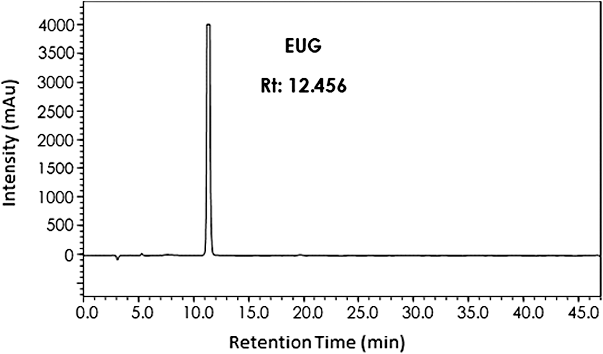
HPLC chromatogram of purified EUG.

Liquid chromatography–mass spectrometry spectrum of purified EUG.
EUG was radioiodinated with iodine-131 (131I), which is a convenient radioisotope for therapy, including imaging potential. 25 Iodogen method, which is commonly based on the oxidation of iodide (I−), was used for radioiodination reaction. Radiolabeling yield of 131I-EUG was determined by the TLRC method. Various mobile phase systems were tested during TLRC experiments; however, the best results were obtained with the mobile phase solution of TLRC1 [2-propanol/n-butanol/0.2 N NH4OH (4:2:1)]; 131I and radioiodinated EUG were migrated toward the end of the solvent, respectively, with Rf values of 0.714 and 0.845, while oxidized 131I was migrated toward the middle of the solvent with an Rf value of 0.234 (Table 1). Radiolabeling yield of 131I-EUG was determined as 98.59 ± 1.02% (n = 6).
EUG, eugenol; TLRC, layer radio chromatography.
Cold iodination of EUG (127I-EUG) was carried out to identify the molecular structure of the radioiodinated EUG. The proposed molecular structure of 127I-EUG, which was plotted by the ACD/LogPAlgorithm (Version 12.01) program, is presented in Figure 5. In addition, theoretical δ (ppm) values of 1H-NMR (D2O) and 13C-NMR (D2O) for cold iodinated EUG (127I-EUG) were obtained by using the ACD/LogP Algorithm (Version 12.01) program. Then, theoretical and experimental δ (ppm) values were compared, which are presented in Table 2. In this study, it is proposed that the attachment of iodide was from the ortho position of the aromatic ring as shown in Figure 5. It was observed that the theoretical and experimental data were compatible with each other. It is known that the iodination mechanism with iodogen method is based on oxidation of iodide (I−) to I+, and I+ reacts with aromatic rings. 26
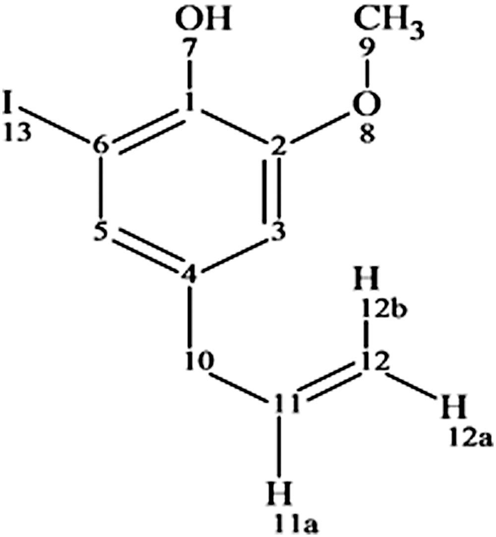
Proposed molecular structure of the 127I-EUG [plotted by ACD/LogPAlgorithm (Version 12.01) program].
To evaluate the in vitro stability of radioiodinated EUG, radiolabeling yield was determined by TLRC method at 30, 60, 120, 240, and 1440 minutes after the labeling procedure. It is observed that the labeling yield of 131I-EUG was stable until 24 hours.
The logarithm of the octanol/water coefficient is generally expressed as lipophilicity of the complex. 32 In this study, experimental LogP value of 131I-EUG, which gives information about the lipophilicity of 131I-EUG, was determined as −1.50 ± 0.15. As a result of lipophilicity study, it is observed that radioiodinated EUG has a hydrophilic structure.
Cell viabilities (%) of EUG on Caco2, MCF7, and PC3 cells were evaluated by the WST cytotoxicity assay and the results are shown in Figure 6. The viabilities were examined according to different concentrations (6.25, 12.50, 25.00, 50.00, and 100.00 μg/mL) of EUG. Viability percentages for all cell types were decreased when the concentration was increased. After 24 hours, the cell viabilities according to cell line type were similar. However, after 48 hours, the viabilities were varied according to the cell line. At 48 hours, maximum cell viabilities were observed for Caco2 cell line and minimum cell viabilities were seen for MCF7 cell line when compared with all cell lines. The efficacy of a compound on inhibition of biological function, which is expressed with the half maximal inhibitory concentration (IC50), was determined for EUG on Caco2, MCF7, and PC3. IC50 values of EUG at 48 hours are given in Table 3. It is seen that EUG has a higher cytotoxicity on MCF7 cell line than PC3 and Caco2. In the literature, cytotoxicity assays with MTT method for different extracts of clove were carried out on HeLa (cervical cancer), MCF7 (ER+) and MDA-MB-231 (ER−) breast cancer, DU-145 prostate cancer, and TE-13 esophageal cancer cell lines. It was reported that in the examined five cancer cell lines, the extracts showed different patterns of cell growth inhibition activity, with the oil extract having maximal cytotoxic activity. 4 In another study, cell proliferation assays of EUG were conducted on MCF7, T47-D, and MDA-MB-231 cell lines and it was reported that EUG has a cytotoxic effect on estrogen-positive and estrogen-negative breast cancer cells. 33

Cell viabilities (%) of EUG on Caco2, MCF7, and PC3 cells.
Recently, there are a lot of researches in which medicinal plants are used for early diagnosis, staging, or therapy of cancer. 2 –5,8,34 –36 In this study, similar in recent years, efficacy of radioiodinated EUG was examined on different cancer cell lines. Time-dependent (60, 120, and 240 minutes) incorporation of 131I-EUG and 131I on Caco2, MCF7, and PC3 cell lines was investigated. Results of the incorporation studies are presented in Table 4. Incorporation percentage of 131I on Caco2 cell line is 0.20 ± 0.18 in 60 minutes, 0.63 ± 0.39 in 120 minutes, and 0.31 ± 0.20 in 240 minutes. Incorporation percentage of 131I on MCF7 cell line is 0.66 ± 0.20 in 60 minutes, 1.03 ± 0.02 in 120 minutes, and 0.98 ± 0.06 in 240 minutes and incorporation percentage of 131I on PC3 cell line is 0.48 ± 0.12 in 60 minutes, 1.16 ± 0.66 in 120 minutes, and0.52 ± 0.12 in 240 minutes. It is shown from these results that, the incorporation percentage of 131I on whole cell lines was ∼0.6%, which means 131I alone has no effect on the cell lines. The low incorporation of 131I was similar with the studies in the literature. 7,35
Minimum and maximum incorporation of 131I-EUG were observed on PC3 cells in 60 and 240 minutes, respectively. Incorporation percents of 131I-EUG in 60 minutes are 25.60 ± 3.09, 27.11 ± 5.12, and 21.79 ± 2.40, respectively, on Caco, MCF7, and PC3 cells. It is observed that there is no important variation of incorporation values of 131I-EUG in 60 minutes for whole cell lines. However, there are dramatically increases in 120 minutes as 1.48-fold and 2.49-fold for Caco2 and PC3. Uptake values of 131I-EUG were increased at 120 minutes for Caco2 and PC3, while it was stable for MCF7 cells. The incorporation values were stable from 120 to 240 minutes on Caco2 and PC3. On the other hand, the incorporation of 131I-EUG on MCF7 was significantly increased (1.69-fold) at 240 minutes.
Briefly, the incorporation behavior of 131I-EUG on Caco2 and PC3 was similar and time dependent. In addition, maximum incorporation values were observed on PC3 cells. Consequently, 131I-EUG has noteworthy incorporation values on whole cell lines.
Conclusions
In this study, clove buds were extracted and EUG was isolated using HPLC system from CBE. Molecular structure of EUG was carried out using the LC-MS method. Then, EUG was radioiodinated with 131I, which is an ideal radioisotope for therapy, enabling imaging. Also, cold iodinated EUG was synthesized and 1H/13C-NMR analysis was performed for structural analysis of radioiodinated EUG. It seen that EUG was radioiodinated in high yields (% 98.59 ± 1.02) and stable until 24 hours. IC50 values were determined for 48 hours on Caco2, MCF7, and PC3 cells. Incorporation studies demonstrated that EUG has considerably high uptakes in Caco2, MCF7, and PC3 cells.
Consequently, plant-originated EUG has a favorable potential on Caco2, MCF7, and PC3 cells. Since this study has been based on in vitro cell culture experiments, further investigations using animal models may be needed to examine the promise of EUG on cancer therapy.
Footnotes
Disclosure Statement
No competing financial interests exist.