
Abstract
Skeletal metastasis is common in advanced stages of various cancers, particularly of the prostate and breast carcinoma. 188Re-HEDP (1-hydroxyethane 1, 1-diphosphonic acid) is a clinically established radiopharmaceutical for bone pain palliation of osseous metastasis, and it takes advantage of high bone affinity. The present work aims at elucidating the possible mechanisms of cell killing by 188Re-HEDP in osteosarcoma cells and biodistribution studies in mice.188Re-HEDP complex was prepared by using lyophilized HEDP kits prepared in-house. In vitro cellular uptake in mineralized bone matrix was found to be 13.41% ± 0.46% (at 2 hours), which was reduced to 2.44% ± 0.12% in the presence of excess amounts of unlabeled HEDP ligand. Uptake of 188Re-HEDP in bones of normal Swiss mice in vivo and mineralized bone in vitro indicated its affinity toward the bone matrix. The study also revealed that cellular toxicity and G2/M cell cycle arrest were dose dependent. At higher doses, G2/M cell cycle arrest was observed, which might be the major cause of cell death and a possible mechanism of bone pain relief.
Introduction
Malignant tumors, particularly prostate and breast carcinoma, often undergo metastasis to bone. 1 Approximately 65% of patients with prostate or breast cancer and 35% of those with advanced lung, thyroid, and kidney cancers will have symptomatic skeletal metastases. 2 Severe bone pain is a clinical symptom caused by such metastasis, which has a significant impact on the quality life of patients. 3 In advanced stages of such malignancies, where chemotherapy and/or hormonal therapy fail to respond, radiotherapy is recommended for bone pain palliation. Although radiotherapy is not curative, it can significantly improve the quality of life of the patient with minimal side-effects compared with other therapeutic modalities. Spread of metastatic lesions in patients' bodies is the deciding factor for the selection of radiotherapy. For localized disease, local external beam radiation therapy is desirable; whereas for widespread metastasis, radionuclide therapy using appropriate radiopharmaceutical is preferred. 4 –6 Recently, Alpharadin (223RaCl3), an α-emitting radiopharmaceutical, is gaining popularity for bone pain palliation. 5
Rhenium-188 is an attractive therapeutic radioisotope. Its high-energy β particle (Eβmax—2.1 MeV) has been effectively utilized for a variety of therapeutic applications such as bone pain palliation due to osseous metastasis, nonresectable liver cancer, nonmelanoma skin cancers, radiosynovectomy of big joints, etc. 7 –9 High-energy β radiation (maximum soft tissue range, 11 mm; average soft tissue range, 3.8 mm) from 188Re is especially useful for therapy of large-volume tumors and synovectomy of large joints; concomitant γ emission (Eγ—155 keV, 15% abundance) is useful for the postinjection monitoring of the radiopharmaceuticals in vivo. In addition, versatile chemistry of 188Re, such as 99mTc, allows radiolabeling with a variety of biological vectors for various therapeutic applications. Another advantage of 188Re over other reactor-produced therapeutic radioisotopes is its availability from 188W/188Re generator. The concept and types of radionuclide generators developed for medical applications have been thoroughly reviewed elsewhere. 10 –13
Bisphosphonates have a high affinity for calcium phosphates, the main mineral of bone. 14,15 This property is taken advantage of to develop bone-targeting radiopharmaceuticals for diagnosis as well as therapy. 16 –18 Among 188Re-radiopharmaceuticals for therapy, 188Re-HEDP is probably the most widely used bisphosphonate radiopharmaceutical in clinical nuclear medicine. 19,20 188Re-HEDP is a clinically proven radiopharmaceutical for bone pain palliation in patients with osseous metastasis due to breast, prostate, or other cancers. 21 Comparative clinical investigations have revealed that efficacy of 188Re-HEDP is comparable to other bisphosphonate agents such as 186Re-HEDP, 153Sm-EDTMP, and the bone seeker 89Sr. Therapeutic efficacy being comparable, 188Re-HEDP enjoys an advantage over other bone pain palliation agents based on reactor-produced therapeutic radioisotopes in terms of availability, since it is a generator-produced radioisotope. Thus, hospital radiopharmacies housing a 188W/188Re generator can prepare and use 188Re-HEDP as per demand. The preparation and clinical evaluation of a freeze-dried kit for the preparation of 188Re-HEDP has been recently reported by our group. 22
Though reports on clinical investigations using 188Re-HEDP for bone pain palliation are available, yet in vitro studies of 188Re-HEDP in cancer cells lines are rather limited. Kumar et al. had reported in vitro studies of 177Lu-EDTMP in mineralized MG63 bone carcinoma cells, which demonstrated uptake as well as cellular toxicity, along with other molecular evidences for enhanced apoptosis. 23,24 In the present work, the interaction of 188Re-HEDP with mineralized bone of human osteosarcoma cells (MG63) was studied. In addition, the underlying mechanism of cell toxicity is documented and discussed.
Materials and Methods
Chemicals for assays were purchased from M/s. Sigma Aldrich, unless stated. Flow cytometer kits were purchased from Merck KGaA (Darmstadt, Germany). Etidronic acid (HEDP) was purchased from M/s. Fluka (Germany). HPLC grade water was procured from Merck (India). Rhenium-188 as Na188ReO4 was obtained from 188W/188Re generator purchased from ITG (Germany). The Na188ReO4 was eluted from the generator with 10 mL of saline with an elution efficiency of 80%. The half-life of 188W is 69.4 days, and useful life of a 500 mCi generator for clinical purpose is ∼3 months. Ammonium perrhenate used as a carrier perrhenate source was purchased from M/s. Sigma-Aldrich. Whatman No. 3 chromatography paper was used for paper chromatography (PC) analysis. Radioactivity distributions on PC strips were determined on MiniGITA γ-radioactivity TLC scanner, Raytest (Germany). Optical density (OD) measurements of assay samples were carried out by using BioTek Universal Microplate Reader (Winooski, VT). The cell cycle study was carried out in Guava® easyCyte flow cytometer (Merck KGaA), and it was analyzed by ModFit LT version 4.1.7.
Cell culture
MG63 cell line was obtained from the National Center for Cell Sciences (NCCS, Pune, India) and cultured in MEM supplemented with 10% serum (Invitrogen, CA) and antibiotic solution. Cells were grown in a humidified 5% CO2 atmosphere incubator at 37°C and were passaged on alternate days.
Induction of bone mineralization of MG63 osteocarcinoma cells
Bone osteosarcoma (MG63) cells were harvested after trypsinization, and 1 × 10⁴ cells were plated per well in six-well plates and cultured overnight. The cell culture medium was replaced with osteogenic medium containing 10 nM dexamethasone, 10 mM β-glycerophosphate, and 50 μg/mL ascorbic acid, in addition to complete MEM media. The cells were kept in culture up to 21 days, with regular replacement with fresh osteogenic media. 25,26
Preparation of 188Re-HEDP
188Re-HEDP was prepared by using lyophilized HEDP kits following a procedure reported earlier. 22 Briefly, freshly eluted perrhenate solution (37 MBq/mL) containing ammonium perrhenate (1 μmol) as a carrier was added to the HEDP kit vial. Subsequently, the kit vial was incubated at 100°C for 20 minutes. After cooling the vial to room temperature, pH of the solution was adjusted to 7 by addition of sodium acetate solution (500 μL, 40 mg).
Quality control of 188Re-HEDP
Radiochemical purity (RCP) of 188Re-HEDP complex was determined by PC in acetone and physiological saline. About 4 μL spots of reaction mixture were made on two separate PC strips. One strip was developed in acetone, whereas the other was developed in physiological saline. In saline, 188Re-HEDP as well as perrhenate moved to the solvent front whereas reduced rhenium remained at the point of spotting. In acetone, perrhenate moved to the solvent front whereas 188Re-HEDP complex and reduced rhenium (ReO2) remained at the point of spotting. The PC strips were subsequently analyzed on a TLC scanner to determine the radioactivity distribution. From the peak area measurements, RCP of 188Re-HEDP complex was calculated.
Binding assay of 188Re-HEDP with the mineralized bone matrix
Mineralized bone cells were incubated with different activity concentrations of 188Re-HEDP (14.2, 28.5, and 42.55 KBq) for 2 hours in complete MEM media at room temperature. To study inhibition, mineralized bone was incubated with 188Re-HEDP in the presence of excess amounts of HEDP ligands under identical conditions. Subsequently, the mineralized bones were washed thrice with cold phosphate-buffered saline (PBS) and associated radioactivity was measured in an NaI(Tl) γ-counter with the energy window set to measure 155 keV γ. Percentage binding was calculated as (Radioactivity associated with mineralized bone/Total Radioactivity added) × 100.
Treatment of MG63 cells with 188Re-HEDP and HEDP
MG63 tumor cells (2 × 10⁵) were plated overnight in complete MEM medium followed by incubation with 0.037, 0.37, and 3.7 MBq of 188Re-HEDP and equivalent amounts of HEDP, separately, for 48 hours in a humidified 5% CO2 atmosphere at 37°C. After completion of the incubation period, cells were harvested and used for various studies.
Estimation of cell toxicity by lactate dehydrogenase assay in cell supernatant
Lactate dehydrogenase (LDH) assay was carried out following the instructions available with the kit (Sigma). Briefly, after completion of treatment with 188Re-HEDP/HEDP, cell supernatant was separated by centrifugation. LDH assay mixture was prepared by mixing equal volumes of freshly prepared LDH assay substrate, co-factor, and dye, before use. The reaction mixture was added along with the supernatant culture media (2:1, v/v) in 96-well plates, mixed well, and incubated for 30 minutes at ambient temperature in the dark. The reaction was terminated by adding 1/10 volume of 1 N HCl and subsequently, the OD was measured at 490 nm. The percentage release of LDH was calculated as (OD of treated sample/OD of control sample) × 100.
Cell cycle analysis
Synchronized MG63 cells were separately treated with different concentrations of 188Re-HEDP and an equivalent amount of HEDP as a vehicle control. Cells were harvested after completion of the treatment, washed with PBS, and fixed in 70% ethanol for 2 hours at 4°C. Subsequently, the cells were washed again with PBS and the supernatant was discarded. The cell pellets were re-suspended in the residual PBS in which Guava cell cycle reagent (200 μL) was added, mixed, and incubated for 20 minutes at ambient temperature. Thereafter, a cell cycle study was carried out.
Biodistribution studies
Biodistribution of 188Re-HEDP was carried out in normal Swiss mice in strict compliance with the national laws governing the conduct of animal experiments and after obtaining the requisite approval from the Institutional Animal Ethics Committee. Normal Swiss mice were used for biodistribution studies. Two groups of animals (n = 3) were administered with 188Re-HEDP (∼3.7 MBq per animal in 100 μL volume) through the lateral tail vein. Biodistribution studies were carried out at 2 and 24 hours postinjection during which the animals were sacrificed, relevant organs were excised, weighed and the radioactivity associated with them was measured in a flat-bed type NaI(Tl) counter with an energy window adjusted to measure 155 keV γ of 188Re. The activity associated with each organ/tissue was expressed as percentage injected dose per organ (%ID/organ). To calculate the activity associated with the skeleton, activity associated with both femurs was determined and the value was extrapolated, considering the total weight of the skeleton to be 10% of the body weight of the animal. Similarly, activity (%ID/organ) associated with blood and muscle was determined by extrapolation, considering their total weight to be 7% and 40% of the body weight of the animal, respectively.
Statistical analysis
Statistical analysis was carried out by using the Origin pro 2015 Software. The results are expressed as mean ± SD for the three independent experiments. The t-test was used to compare the treated and control samples, and p ≤ 0.05 was considered statistically significant.
Results and Discussion
Preparation of 188Re-HEDP and quality control
The 188Re-HEDP complex was prepared by using lyophilized HEDP kit and freshly eluted Na188ReO from a 188W/188Re generator, following the procedure reported earlier. 22 The 188Re-HEDP complex thus prepared was characterized by PC in acetone and saline. Typical radioactivity distribution in PC is shown in Figure 1. By this method, 188Re-HEDP complex could be consistently prepared in >95% RCP.

Paper chromatography pattern of 188Re-HEDP in saline
188Re-HEDP binding with mineralized bone
Bone metastases from breast and prostate cancer mostly affect the skeleton, but, in general, they can affect any other osseous site. Once metastasized, the bones undergo activation of osteoblasts (mostly in prostate cancer metastasis) and osteoclasts (osteolytic lesion due to breast/kidney cancer metastasis), which ultimately cause formation of weak sclerotic bone. Such complications of bone often make them susceptible to fracture, a high level of blood calcium, and severe pain. 27,28
In general, bone pain-palliating agents bind to mineralized bone (bone matrix) and not to the tumor cells. Thus, MG63 cells have the potential to opt for bone mineralization on the action of osteogenic media. Hence, MG63 cells were used in these studies, because they can serve the purpose of both in vitro bone binding (in mineralized bone) and toxicity studies (MG63 cells). Bone mineralization of MG63 cells was carried out following a reported procedure. 23,25,26 Subsequently, 188Re-HEDP complex was incubated with mineralized MG63 bone matrix to study binding. About 13.41% ± 0.46% binding of 188Re-HEDP with mineralized MG63 was observed on incubation for 2 hours. However, in the presence of 100-fold excess of free HEDP ligand, the binding decreased to 2.44% ± 0.12% (Fig. 2).
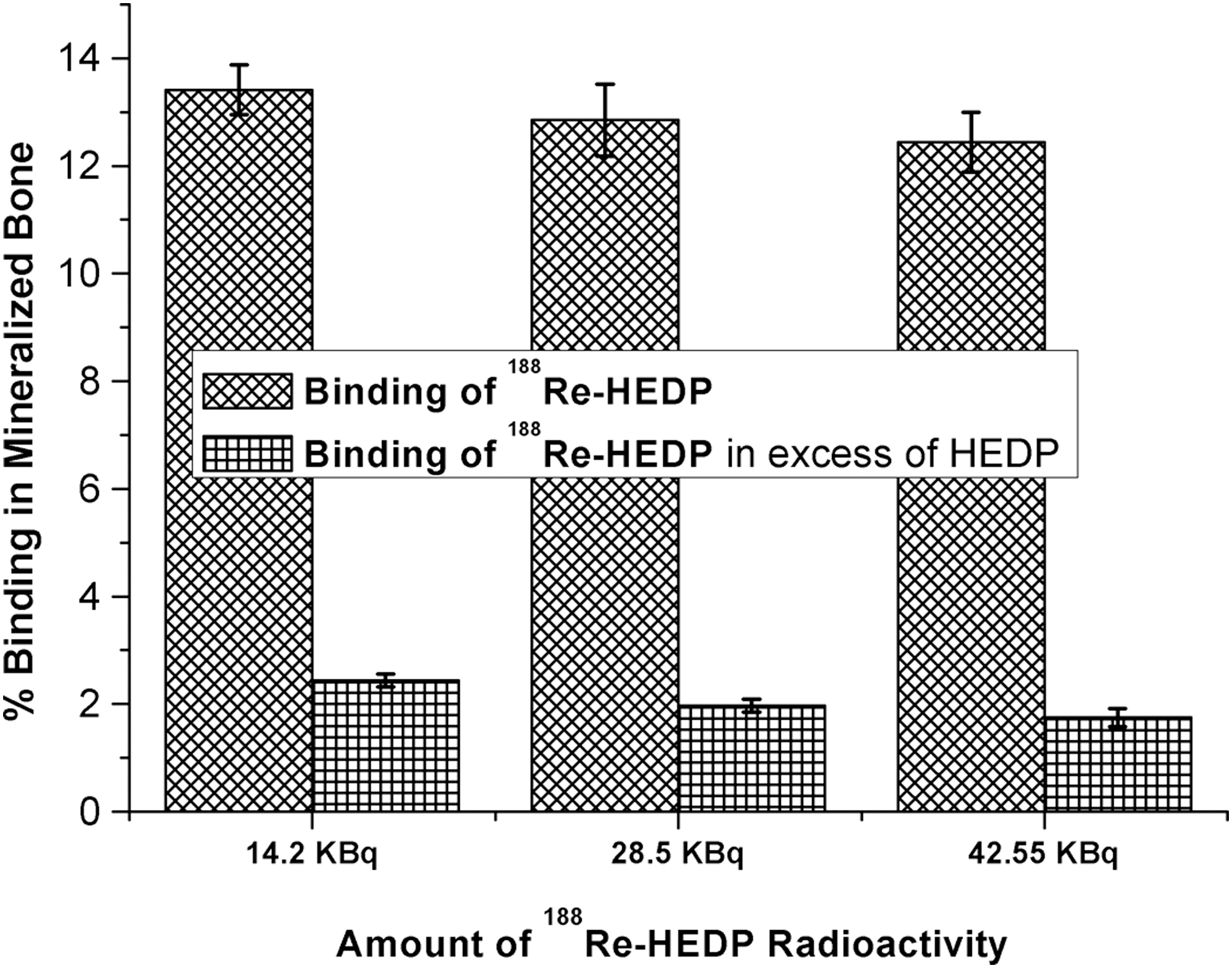
Study of 188Re-HEDP binding toward mineralized bone. Equal amounts of mineralized bones were added with various amounts of 188Re-HEDP radioactivity with and without HEDP for 2 hours.
Very few reports are available on the mechanism of uptake of bone-seeking radiopharmaceuticals with bone cell lines and minerals. 23,29 –31 Some studies have reported the in vitro filtration method to evaluate the binding with minerals such as amorphous calcium phosphate (ACP), hydroxyapetite (HA), tricalcium phosphate (TPO), etc; whereas others have reported bone model set-ups with collagen fibers. 32,33 The MG63 cell binding studies with 188Re-HEDP were carried out based on the previous report on the binding study of 177Lu-EDTMP with mineralized MG63 bone cell lines. 23 Though no significant binding of 188Re-HEDP to MG63 cells was observed,188Re-HEDP showed a binding of 13.41% ± 0.46% with mineralized MG63 cells. This indicated the effect of the mineralization process and the osteoblastic effect on the uptake of 188Re-HEDP in osteosarcoma cell lines.
Effect of 188Re-HEDP on cell toxicity of MG63 cells
Cell toxicity due to high-energy β radiation from 188Re-HEDP is obvious and even in the clinical scenario, bone marrow toxicity has been observed. 2,3 However, a therapeutic dose of 188Re-HEDP is such that pain palliation is achieved without significant bone marrow toxicity. As mentioned earlier, cellular uptake studies of bone-seeking radiopharmaceuticals are very limited in literature. Likewise, in vitro cellular toxicity data for bone-seeking radiopharmaceuticals are also not abundant.
Cellular toxicity of 188Re-HEDP was estimated by LDH assay, which showed no significant cell death at lower doses (0.037 and 0.37 MBq) of 188Re-HEDP compared with the control. However, a significant (p ≤ 0.05, n = 3) increase of toxicity was observed at higher doses (3.7 MBq) of 188Re-HEDP (37.72% ± 6.5%) compared with the control (Fig. 3). It is interesting to note that in a previous study reported by our group, 23 the average cell death observed with a dose of 37 MBq of 177Lu-EDTMP was only about 12%, which is significantly lower than the 37.72% ± 6.5% cell killing achieved with one-tenth dose of 188Re-HEDP. This could be attributed to the low energy β of 177Lu (Eβmax—0.497 Mev) compared with 188Re (Eβmax—2.12 Mev) and the dose rate. A significant level of cell death at relatively lower doses of 188Re-HEDP encouraged us to study the cell cycle and the possible mechanism of cell death.
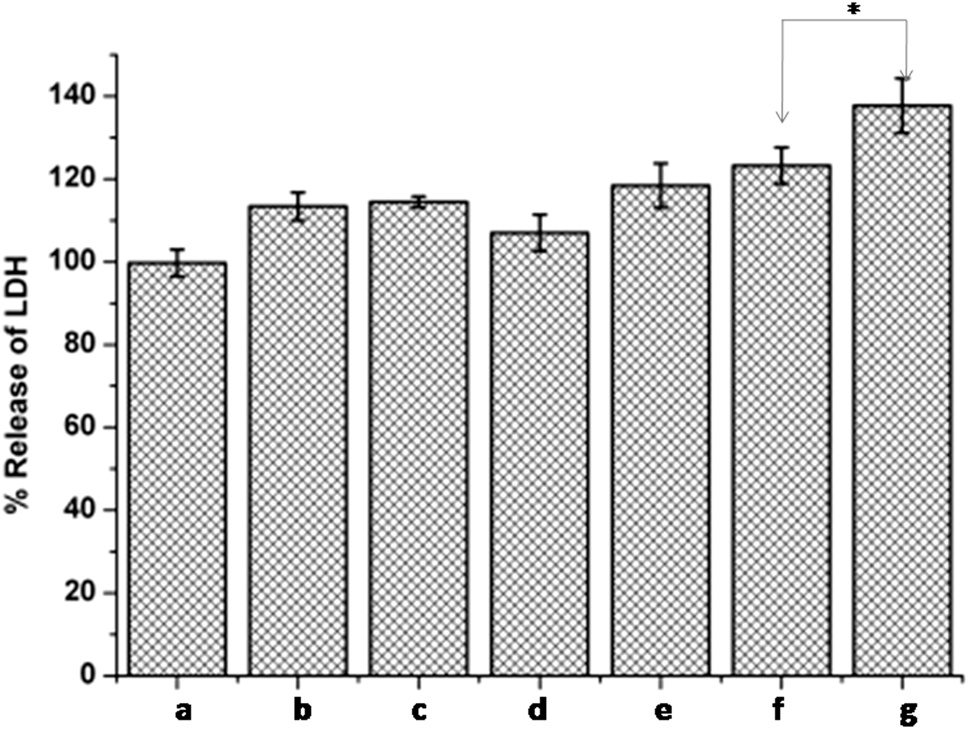
Estimation of cell toxicity by lactate dehydrogenase assay. MG63 cells treated with various concentrations of 188Re-HEDP (*significant difference at p ≤ 0.05, n = 3 t-test), where (a) Control, (b) Cells treated with an equivalent amount of HEDP in 0.037 MBq of 188Re-HEDP, (c) Cells treated with an equivalent amount of HEDP in 0.37 MBq of 188Re-HEDP, (d) Cells treated with an equivalent amount of HEDP in 3.7 MBq of 188Re-HEDP, (e) Cells treated with 0.037 MBq of 188Re-HEDP, (f) Cells treated with 0.37 MBq of 188Re-HEDP, and (g) Cells treated with 3.7 MBq of 188Re-HEDP.
Effect of 188Re-HEDP on MG63 cell cycle
Cell cycle analysis was performed to understand the underlying mechanism of cell death, and it was observed that the 188Re-HEDP induces G2/M arrest in MG63 cell lines at 3.7 MBq of 188Re-HEDP (Fig. 4). In response to DNA damage, G1/S and G2/M cell cycle checkpoints are essential for the maintenance of genomic stability and integrity. 34,35 An evident response was observed on cell cycle pattern in the MG63 cell line due to the 188Re-HEDP treatment, exhibiting a positive correlation with the concentration and dose of 188Re-HEDP treatment (Fig. 4. Table 1, p ≤ 0.05, n = 3). It has already been reported in a number of previous studies that the tumor cell cycle G2/M retardation is induced by γ-radiation. 36,37 The cell cycle arrest was also observed in the human breast carcinoma cell line, MCF-7, when incubated with a β-emitting radioisotope such as 89Sr, which is used for bone pain palliation. 38 Likewise, Eriksson et al. also successfully demonstrated that HeLa Hep2 cells exhibited a transient G2/M-phase arrest on incubation with a β-emitting radioisotope such as 131I. 39 These results indicate that G2/M-phase arrest may be one of the possible mechanisms of the growth inhibitory effect of β-emitting radioisotopes. Such reports are akin to our observation that 188Re-HEDP being a β-emitting radiopharmaceutical induces G2/M arrest in MG63 cells. However, G2/M cell cycle arrest is generally followed by apoptosis. 34 Hence, G2/M arrest might be the cause of cell death in MG63 cells. However, additional molecular evidence is required to confirm this observation.

Analysis of MG63 cells cycle by a flow cytometer, where
Biodistribution studies
Biodistribution studies were carried out in normal Swiss mice of 188Re-HEDP to demonstrate the uptake of 188Re-HEDP in bone. Distribution of 188Re-HEDP in various organs and tissues is shown in Figure 5. The results showed that 188Re-HEDP has a high affinity for bone. The bone uptake was 65.5% ± 7.6% ID/organ at 2 hours, which increased to 79.8% ± 3.1% ID/organ at 24 hours postinjection.
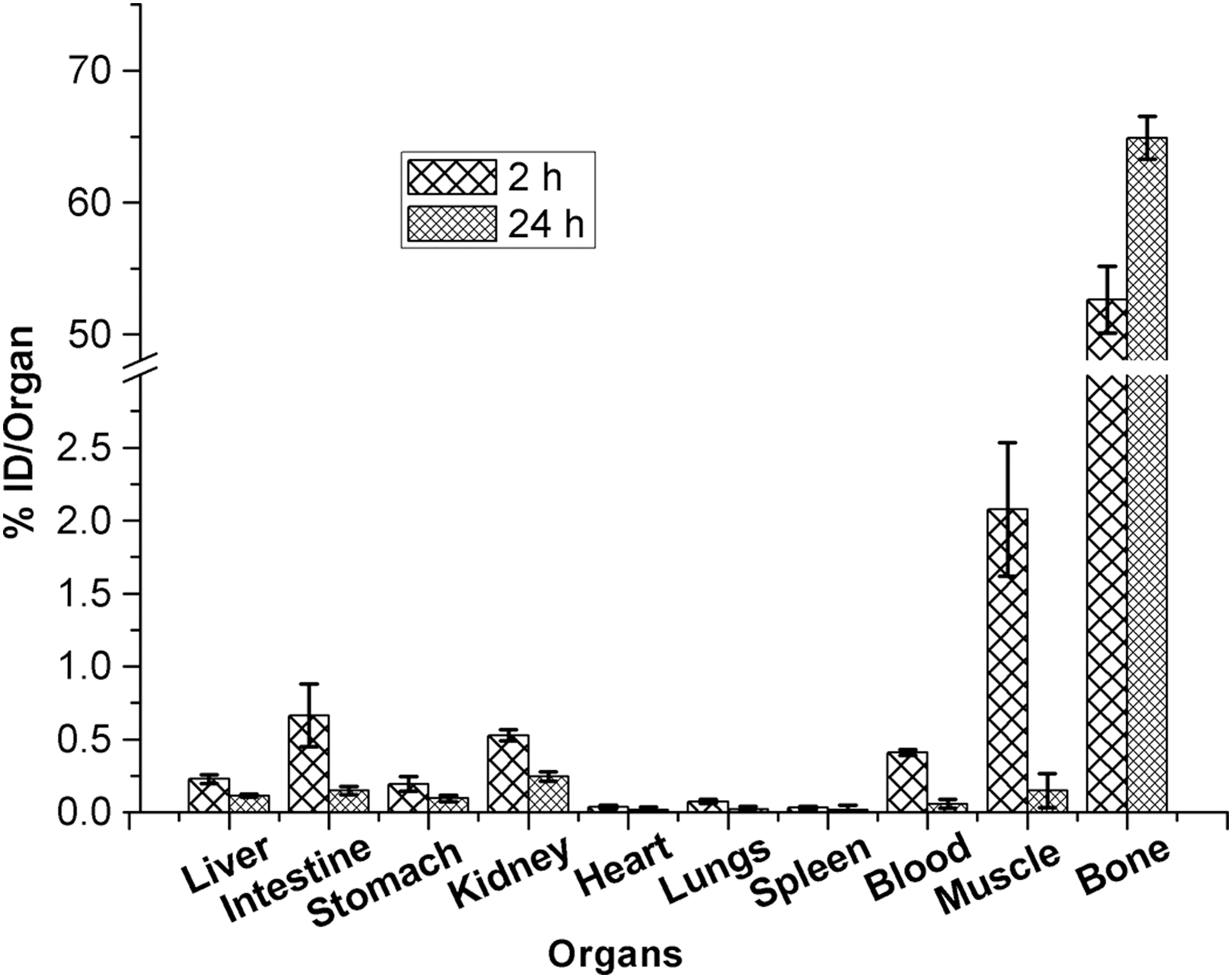
Biodistribution of 188Re-HEDP in different organs (% Injected Dose/Organ) of Swiss mice, sacrificed after 2 and 24 hours postinjection.
Conclusion
The present study focused on the cellular uptake, cellular toxicity, and probable mechanism of cell death induced by 188Re-HEDP. 188Re-HEDP was prepared under optimized conditions with excellent RCP and showed significant uptake in mineralized bone, in vitro, as well as in normal bone of mice in vivo. Further, the same complex induced significant G2/M cell cycle arrest in MG63 cells, eventually resulting in considerable cellular toxicity and tumor cell death, which might be responsible for the relief from bone pain. Since 188Re-HEDP has preferential uptake to the metastases lesions on bone, it may work for both osteoblastic and osteoclastic bone metastasis. This study provided some insights into the mechanistic aspects operating at the molecular level, which may be responsible for the pain-palliating effect of radiolabeled phosphonates: radiopharmaceuticals, in general, and 188Re-HEDP, in particular.
Footnotes
Acknowledgment
The authors are grateful to the group director, Radiochemistry and Isotope group, for the support to carry out this work.
Disclosure Statement
No competing financial interests exist.