
Abstract
Aberrant signaling of the epidermal growth factor receptor (EGFR) plays a crucial role in the tumorigenesis of many cancer types, including head and neck squamous cell carcinoma (HNSCC), making it a compelling drug target. After initial promising results of EGFR-targeted therapies such as cetuximab, the problem of therapeutic resistance is emerging and new treatment options are necessary. In contrast to first-generation EGFR inhibitors, MEHD7945A (duligotuzumab) is a monoclonal antibody with dual EGFR/HER3 specificity. Consequently, treatment with MEHD7945A may result in a more pronounced therapeutic benefit. In this study, sensitivity to MEHD7945A as a single agent and in combination with cisplatin was investigated in cetuximab-sensitive and -resistant HNSCC cell lines under normal and reduced oxygen conditions. The results demonstrated that sensitivity to MEHD7945A was cell line dependent and influenced by oxygen concentration. An additive, but not synergistic, interaction between MEHD7945A and cisplatin was observed. In conclusion, MEHD7945A has the potential to partially overcome cetuximab resistance. Nevertheless, further research is warranted to determine additional resistance mechanisms to cetuximab treatment besides HER3 signaling. Unraveling these mechanisms will ultimately lead to the development of new therapeutic strategies to improve the response to EGFR blockage.
Introduction
Head and neck squamous cell carcinoma (HNSCC) is the sixth most common cancer in the world, with more than 650,000 new cases diagnosed each year, and accounting for 90% of malignant neoplasia of the upper respiratory system. Despite recent advances in the management of locally advanced HNSCC, the overall survival has improved only marginally over the last three decades. HNSCC progression often involves the accumulation of a number of genetic and epigenetic alterations in tumor suppressor proteins, such as p53, p16, and RB, concomitant with the aberrant activity of signaling molecules such as epidermal growth factor receptor (EGFR) that drive the unrestricted growth of HNSCC cells. 1,2
About 90%–95% of HNSCCs exhibit an enhanced expression of EGFR. 1 As a result, EGFR is a clinically validated therapeutic target for several cancer types, 3 –5 which has led to the recent development of novel molecular targeted therapies. 2,6 In the past decades, two strategies targeting EGFR function have been extensively investigated in laboratory and clinical settings—the use of either monoclonal antibodies (such as cetuximab or panitumumab) or small-molecule tyrosine kinase inhibitors (TKIs such as erlotinib or gefitinib). 7 The clinical activity of these first-generation EGFR inhibitors has been well established in HNSCC and available data suggest that the combination with conventional anticancer therapies such as radiotherapy or chemotherapy improves their efficacy. 8,9 However, both intrinsic resistance and acquired resistance to EGFR inhibitors represent a major therapeutic challenge. 10,11
To overcome these intrinsic and acquired resistance mechanisms, next-generation EGFR inhibitors, such as MEHD7945A (duligotuzumab), are being developed, which block multiple EGFR family members. MEHD7945A is a monoclonal antibody with dual EGFR/HER3 specificity and it has been reported that this novel compound shows efficacy in multiple tumor models. 12 Interestingly, an analysis of various solid tumor samples demonstrated that median mRNA expression of heregulin (HRG, the ligand binding to HER3) is significantly higher in HNSCC tumors than in the other tumor types. 13 Furthermore, several studies have already suggested that compensatory HER3 signaling might be a mechanism underlying resistance to EGFR inhibition. 14 –17 As a result, dual inhibition of both EGFR and HER3 might be a novel therapeutic strategy to overcome resistance and increase the effect of EGFR-targeted therapy in HNSCC. Indeed, it has already been observed in preclinical settings that combining an anti-HER3 monoclonal antibody with cetuximab resulted in a more potent antitumor activity through simultaneous inhibition of HER3 and EGFR. 17,18 Furthermore, MEHD7945A showed superior activity compared with mono-specific EGFR- or HER3-targeting antibodies in the FaDu HNSCC cell line, 12 as well as in human xenograft models derived from HNSCC and NSCLC tumors with acquired resistance to EGFR inhibitors. 19
Therefore, the present study aims to provide key preclinical data concerning the efficacy of MEHD7945A in monotherapy as well as in combination with cisplatin in HNSCC cell lines with different sensitivity to cetuximab. As the possible association of human papillomavirus (HPV) status and efficacy of EGFR inhibition is still unclear, both HPV-negative and HPV-positive cell lines were included. In addition, oxygen deficiency (hypoxia) is a common characteristic of human solid tumors, and hypoxic tumor regions often contain viable cells that are more resistant to chemotherapy and/or radiotherapy. 20 Since previous studies demonstrated a link between hypoxia and EGFR signaling, 21 the cytotoxicity of MEHD7945A was investigated under both normoxic and hypoxic conditions.
Materials and methods
Cell lines and culture conditions
The human HNSCC tumor cell lines included in this study are CAL-27, SC263, LICR-HN1, SQD9, SCC22b, and 93-VU147-T. CAL-27 was obtained from American Type Culture Collection (ATCC, Rockville MD). SC263 and SQD9 were kindly provided by Prof. Dr. Sandra Nuyts (University Hospital Leuven, Leuven, Belgium); LICR-HN1 and SCC22b were kindly provided by Prof. Dr. Olivier De Wever (University of Ghent, Ghent, Belgium); and 93-VU-147T was kindly provided by Dr. Josephine Dorsman (VU University Medical Center Amsterdam, Amsterdam, The Netherlands). All cell lines were HPV negative, with the exception of 93-VU-147T. All HNSCC cell lines were grown as monolayers in Dulbecco's modified Eagle's medium (DMEM), supplemented with 10% fetal calf serum, 2 mM glutamine, and 1% penicillin/streptomycin. All media and supplements were obtained from Life Technologies (Merelbeke, Belgium). Cultures were maintained in exponential growth in a humidified 5% CO2/95% air atmosphere at 37°C. Cells were confirmed free of mycoplasma infection through regular testing (MycoAlert™, Plus Mycoplasma detection kit; Lonza, Verviers, Belgium). Hypoxic conditions (1% O2, 5% CO2, 95% N2) were achieved in a Bactron IV anaerobic chamber (Shel Lab, Cornelius, OR) as described previously. 22 Measurements with the ToxiRae II air oximeter (Rae BeNelux, Hoogstraten, Belgium) confirmed that the oxygen conditions in the gas phase were stable at 1% O2. Hypoxic incubation was initiated after cells had been cultured under normoxic conditions overnight, allowing attachment to culture plates.
Generation of cetuximab-resistant clones
Dose–response studies of cetuximab (Merck, Darmstadt, Germany) were previously performed to select cetuximab-sensitive cell lines. As such, CAL-27, LICR-HN1, SQD9, and 93-VU147-T were identified as intrinsically resistant to cetuximab, while SC263 and SCC22b were cetuximab sensitive. 23 Cetuximab-resistant variants of the SC263-sensitive cell line were derived by continuous exposure of the original parental cell line to cetuximab, starting with the IC50 concentration of cetuximab for 10–14 days. In parallel, parental cells were exposed to the vehicle for cetuximab, that is, phosphate-buffered saline (PBS). After 10 dose doublings, cetuximab-resistant daughter cells (SC263-R) were generated, showing resistance toward high concentrations of cetuximab. Next, the stability of cetuximab resistance was confirmed in a drug-free culture system. SC263-R cells still exhibited a resistant phenotype even after culture in drug-free medium for at least 6 weeks. 23
EGFR and HER3 expression analysis
The baseline extracellular protein expression level of EGFR and HER3 under both normoxic and hypoxic conditions was assessed using flow cytometry. Cells were fixed in 4% formaldehyde (10 minutes) under normoxic or hypoxic conditions after EGFR and HER3 PE-conjugated antibody incubation (R&D Systems). Corresponding isotype controls (rat IgG2A and mouse IgG1) were included for all samples and served as negative controls. Dead cells were excluded from the analysis by staining with the Live/Dead Fixable Far Red Dead Cell Stain kit (Thermo Fisher). All samples were measured on the FACScan flow cytometer (BD Biosciences).
Cytotoxicity experiments
Cytotoxicity studies were performed in 96-well plates and optimal seeding densities for each cell line were determined to ensure exponential growth for the entire duration of the assay. Cells were counted automatically with a Scepter 2.0 device (Merck Millipore SA/NV, Overijse, Belgium). After an incubation period overnight, cells were treated with MEHD7945A alone (0–5000 nM for 96 or 144 hours), cisplatin alone (0–10 μM for 24 hours), or with a combination of both. For the combinatorial treatment schedule, cells were treated with MEHD7945A for 96 hours immediately followed by cisplatin for 24 hours. Cell survival was determined by the sulforhodamine B assay, as previously described. 24,25
Statistical analysis
All experiments were performed in triplicate and each concentration was tested three times within the same experiment, except for the experiments assessing the cytotoxic effect of MEHD7945A in the SC263-PBS cell line after treatment for 144 hours, in the cell lines SCC22b and SC263 after treatment under hypoxic conditions and in combination with cisplatin, which were performed twice.
Flow cytometric data were analyzed using FlowJo, v10.1. The percentage of EGFR and HER3-positive cells (overton) was determined in comparison with the corresponding isotype control. Furthermore, the signal for aspecific binding was subtracted from the measured mean fluorescence intensities (denoted as ΔMFI). This parameter indicates the amount of extracellular expression of EGFR and HER3 on individual cells. Statistical analysis was performed using RStudio. A linear mixed model was fitted with either overton or ΔMFI as the dependent variable and with oxygen status, resistance status, and their interaction as fixed effects. A random intercept for cell line was added to account for the dependence between observations within the same cell line.
Survival rates were calculated as follows: [mean optical density (OD) of treated cells/mean OD of control cells] × 100%. IC50 values (i.e., drug concentration causing 50% growth inhibition) were calculated using WinNonlin software (Pharsight, Mountain View). Possible synergism between cisplatin and MEHD7945A was determined by calculation of the combination index (CI) using the additive model as described by others. 26 –28 CI <0.8, CI = 1.0 ± 0.2, and CI >1.2 indicated synergism, additivity, or antagonism, respectively. Possible significant differences in IC50 values (p < 0.05) were evaluated with the Mann–Whitney U test, using SPSS v23.0 software.
Results
EGFR and HER3 expression under normoxic and hypoxic conditions in a panel of HNSCC cell lines with different sensitivity to cetuximab
Since MEHD7945A is a dual specific antibody against EGFR and HER3, the authors examined the basal extracellular protein expression level of EGFR and HER3 under both normoxic and hypoxic conditions in a panel of HNSCC cell lines with different sensitivity to cetuximab. First, the percentage of EGFR and HER3-positive cells (overton) was determined (Fig. 1A, B). All HNSCC cell lines showed high levels of EGFR and HER3-positive cells, with one exception; the intrinsically cetuximab-resistant LICR-HN1 cell line demonstrated a lower percentage of EGFR-positive cells, that is, 19.98% ± 7.87%, compared with the other HNSCC cell lines (percentages ranging between 52.72% ± 3.38% and 80.25% ± 8.60%). However, no significant difference was found in the percentage of EGFR and HER3-positive cells between cetuximab-sensitive and (intrinsically and acquired)-resistant HNSCC cell lines (p = 0.587 and p = 0.922 for EGFR and HER3, respectively).
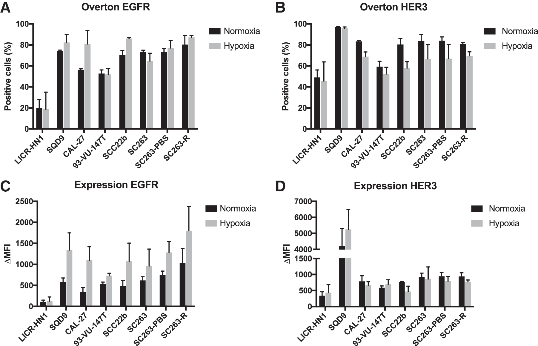
EGFR and HER3 expression under normoxic and hypoxic conditions in a panel of HNSCC cell lines with different sensitivity to cetuximab. Expression analysis was performed using flow cytometry. All HNSCC cell lines, except for LICR-HN1, showed a high percentage of EGFR
Second, the expression level of EGFR and HER3 on these receptor-positive cells was determined by the difference in ΔMFI between the receptor and corresponding isotype (Fig. 1C, D). LICR-HN1 demonstrated lower levels of EGFR expression in comparison with other HNSCC cell lines. Nevertheless, both EGFR and HER3 were highly expressed in all other HNSCC cell lines with ΔMFI values above 346.7 ± 101.0 and 586.7 ± 54.1, respectively. Remarkably, the intrinsically cetuximab-resistant cell line SQD9 demonstrated enormous levels of HER3 expression (ΔMFI = 4229.7 ± 1068.2) compared with the other cell lines (ΔMFI ranging from 339.3 ± 121.9 to 791.0 ± 140.5). However, no significant difference in ΔMFI of receptor-positive cells was observed between cetuximab-sensitive and (intrinsically and acquired)-resistant HNSCC cell lines (p = 0.645 and p = 0.911). In addition, no significant effect of HPV status was detected for the percentage of EGFR and HER3-positive cells (p = 0.802 and p = 0.319) as well as ΔMFI of receptor-positive cells (p = 0.764 and p = 0.628). Hence, the percentage of EGFR and HER3-positive cells and ΔMFI of receptor-positive cells seem to be cell line specific.
Next, the effect of reduced oxygen levels on the expression of EGFR and HER3 was investigated. The authors also tested if the interaction between oxygen status and cetuximab resistance status was significant. Hereby, cetuximab resistance status did not significantly influence the effect of oxygen status on EGFR and HER3 expression. When looking at the effect of oxygen availability across all cetuximab resistance statuses, there was a significant increase in the percentage of EGFR-positive cells (p = 0.025) and ΔMFI of EGFR-positive cells (p < 0.001) under hypoxic conditions in all HNSCC cell lines. In contrast, for HER3, the percentage of positive cells was significantly decreased under hypoxic conditions in all HNSCC cell lines (p < 0.001). However, no significant change in ΔMFI of HER3-positive cells was observed under reduced oxygen levels (p = 0.698).
Overall, these results demonstrate that HNSCC cell lines with different sensitivity to cetuximab contain a high percentage of EGFR and HER3-positive cells and that both receptors are generally highly expressed on these positive cells under both normal and reduced oxygen conditions. This means that the HNSCC cell lines used in this study are valid candidates for treatment with MEHD7945A, according to their target expression.
MEHD7945A is only partially able to overcome intrinsic resistance to cetuximab in a panel of HNSCC cell lines exposed to normoxic or hypoxic conditions
The dose–response curves of the HNSCC cell lines for MEHD7945A after 96 hours of treatment are shown in Figure 2. A clear concentration-dependent cytotoxic effect of MEHD7945A after 96 hours of treatment under both normal and reduced oxygen conditions was observed in both cetuximab-sensitive HNSCC cell lines, with IC50 values under normoxia of 4 ± 2 nM and 61 ± 33 nM for the SCC22b and SC263 cell lines, respectively (Table 1). This cytotoxic effect was also observed in one of four intrinsically cetuximab-resistant HNSCC cell lines (CAL-27), with an IC50 value of 84 ± 44 nM under normoxia (Table 1). In contrast, MEHD7945A did not demonstrate a cytotoxic effect in two of four intrinsically cetuximab-resistant cell lines (LICR-HN1 and SQD9). Furthermore, only a limited cytotoxic effect was observed in the HPV-positive, intrinsically cetuximab-resistant cell line 93-VU-147T.
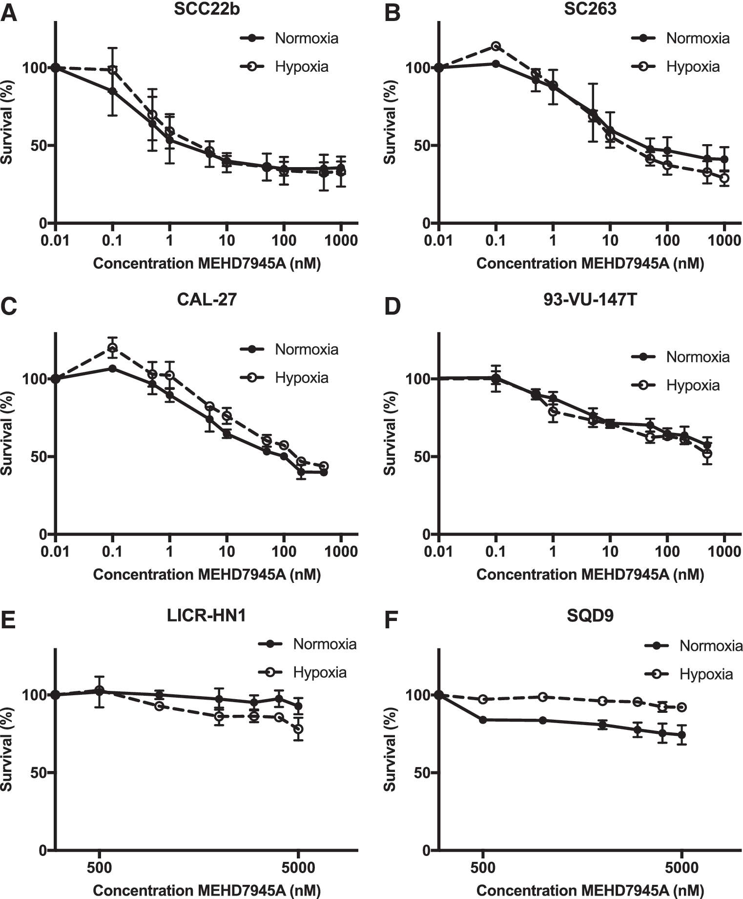
Dose–response curves for cetuximab-sensitive and intrinsically cetuximab-resistant HNSCC cell lines after MEHD7945A treatment for 96 hours under normoxic and hypoxic conditions. Dose–response curves for the cetuximab-sensitive cell lines SCC22b
p < 0.05: significant difference in IC50 value between MEHD7945A treatments under normoxic and hypoxic conditions.
/, IC50 value or p-value cannot be calculated; ND, not determined.
The effect of MEHD7945A on cell survival was not affected by the presence or absence of oxygen in cetuximab-sensitive cells. However, the intrinsically cetuximab-resistant CAL-27 cells were less sensitive to MEHD7945A when cells were incubated under hypoxia, resulting in a significantly increased IC50 value (p = 0.034).
As extending the drug incubation period may enhance the cytotoxic effect of compounds, treatment of cell lines with MEHD7945A was prolonged from 96 to 144 hours. Furthermore, bispecific antibodies in IgG-like formats usually have longer serum half-lives due to their larger size and FcRn-mediated recycling. 29 This will lead to longer drug exposure in patients. Therefore, the authors extended the incubation period with MEHD7945A in vitro in an attempt to mimic the in vivo situation.
After 144 hours of treatment, a clear concentration-dependent cytotoxic effect of MEHD7945A was still observed in the cetuximab-sensitive cell lines SCC22b and SC263 and the intrinsically cetuximab-resistant cell line CAL-27, with IC50 values ranging from 0.7 ± 0.4 nM to 39 ± 27 nM (Table 1 and Fig. 3). Furthermore, CAL-27 cells seem to be less sensitive to MEHD7945A when cells were incubated under hypoxic conditions, resulting in an increased IC50 value (p = 0.05). In comparison with 96 hours of treatment, the cytotoxic effect of MEHD7945A after treatment for 144 hours was enhanced in the HPV-positive, intrinsically cetuximab-resistant cell line 93-VU-147T, with IC50 value of 132 ± 80 nM after 144 hours of MEHD7945A (Table 1). Furthermore, the cytotoxic effect of MEHD7945A seems also to be reduced under hypoxic conditions, with an increased IC50 value under hypoxia (p = 0.05). Despite prolonged exposure, MEHD7945A was not able to establish a cytotoxic effect in the intrinsically cetuximab-resistant cell lines LICR-HN1 and SQD9.

Dose–response curves for cetuximab-sensitive and intrinsically cetuximab-resistant HNSCC cell lines after MEHD7945A treatment for 144 hours under normoxic and hypoxic conditions. Dose–response curves for the cetuximab-sensitive cell lines SCC22b
Overall, the results showed a difference in sensitivity toward MEHD7945A between cetuximab-sensitive and intrinsically cetuximab-resistant cell lines. These data indicated that MEHD7945A was not able to establish cytotoxicity in all cell lines intrinsically resistant to cetuximab. In general, MEHD7945A maintained its efficacy under hypoxic conditions in cetuximab-sensitive HNSCC cell lines. However, in intrinsically cetuximab-resistant HNSCC cell lines, the cytotoxic effect of MEHD7945A was attenuated under reduced oxygen conditions.
MEHD7945A is not able to overcome acquired resistance to cetuximab in HNSCC cell lines
The dose–response curves of the cetuximab-sensitive and isogenic, acquired cetuximab-resistant HNSCC cell lines for 144 hours of treatment with MEHD7945A are shown in Figure 4. MEHD7945A demonstrated only a limited cytotoxic effect in the acquired cetuximab-resistant SC263-R cell line after 144 hours of treatment. These data suggest that MEHD7945A does not have the potential to overcome acquired cetuximab resistance in HNSCC cell lines.
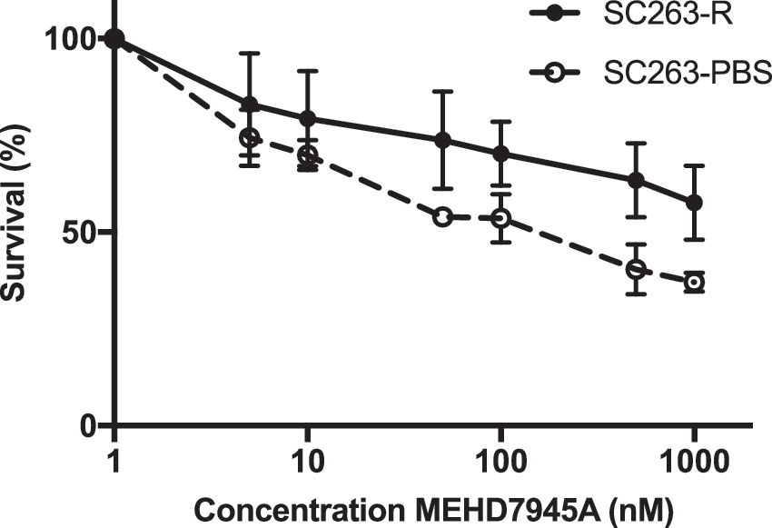
Dose–response curves of MEHD7945A for the cetuximab-sensitive and isogenic, acquired cetuximab-resistant HNSCC cell lines SC263-PBS and SC263-R after treatment for 144 hours. Only a small cytotoxic effect was observed in the acquired cetuximab-resistant SC263-R cell line. PBS, phosphate-buffered saline.
Combining MEHD7945A with cisplatin in HNSCC cell lines with different HPV status shows additive effects
To investigate the potential interaction between MEHD7945A and cisplatin, tumor cells were incubated with fixed doses of MEHD7945A for 96 hours combined with sequential treatment with 0–10 μM cisplatin for 24 hours. The fixed MEHD7945A concentrations were based on the outcome of the monotherapy experiments. The dose–response curves of the intrinsically cetuximab-resistant cell lines CAL-27 and 93-VU-147T after treatment with this combination regimen are shown in Figure 5. Sequential exposure to MEHD7945A, followed by cisplatin, revealed an additive, but not synergistic, interaction (Table 2).
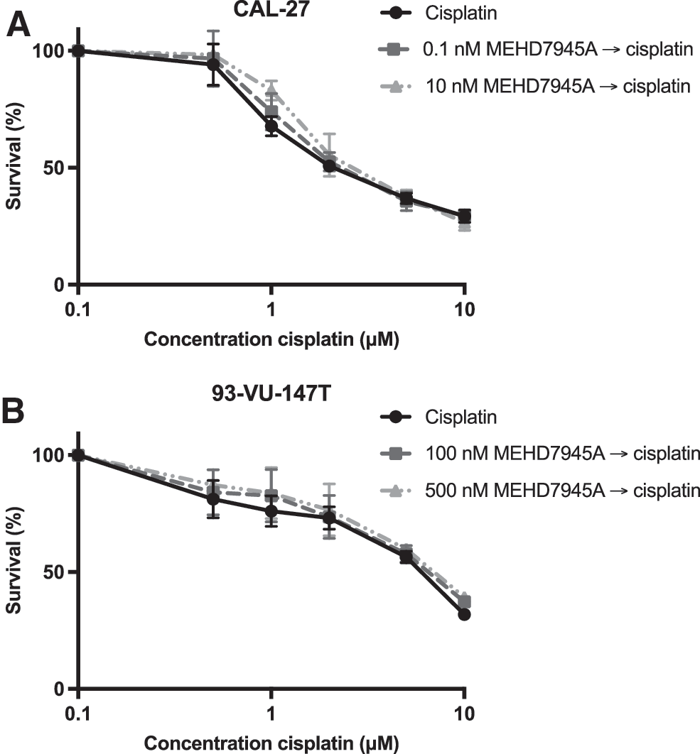
The inhibitory effect of sequential exposure to MEHD7945A, followed by cisplatin, on cell survival of HNSCC cell lines. Dose–response curves for sequential treatment with MEHD7945A, followed by cisplatin, show an additive effect in the intrinsically cetuximab-resistant cell lines CAL-27
The table gives an overview of the IC50 value of cisplatin after both monotherapy and sequential combination therapy with MEHD7945A. The average CI is provided for each combination therapy. CI <0.8, CI = 1.0 ± 0.2, and CI >1.2 indicated synergism, additivity, or antagonism, respectively.
p < 0.05: significant difference in IC50 value compared with cisplatin monotherapy.
CI, combination index; /, cannot be calculated.
Discussion
After the initial promising results of EGFR-targeted therapies, the problem of therapeutic resistance is emerging. To overcome resistance to these EGFR-targeted therapies, new treatment options are necessary. Due to extensive cross talk among HER receptors, blockade of one HER receptor can be compensated by other HER family members, which must be targeted by new therapeutic regimens. In contrast to the first-generation EGFR inhibitors, MEHD7945A inhibits EGFR as well as HER3. Consequently, the authors hypothesized that treatment with MEHD7945A might result in a distinct and more pronounced therapeutic benefit. To test this hypothesis, the authors examined the cytotoxicity of MEHD7945A in a panel of cetuximab-sensitive and (intrinsically/acquired) -resistant HNSCC cell lines.
First, the authors determined the expression of EGFR and HER3 under normal and reduced oxygen conditions in this panel of HNSCC cell lines with different sensitivity to cetuximab. It has been suggested that tumors harboring amplification or overexpression of both EGFR and HER3 may show favorable response to MEHD7945A. 30 No significant difference in EGFR and HER3 expression was found between cetuximab-sensitive and (intrinsically and acquired) -resistant HNSCC cell lines. Furthermore, it has already been established that EGFR is a key survival factor under hypoxic conditions as EGFR stimulates hypoxia-inducible factor (HIF) signaling to improve cellular survival. 21 On the other hand, HIF signaling can also activate the EGFR pathway. 31 Consistent with previous studies, the authors demonstrated that the percentage of EGFR-positive cells and the EGFR expression level significantly increased under reduced oxygen conditions. Despite a significant decrease of HER3-positive cells, no significant change in HER3 expression level under hypoxic conditions was observed.
Overall, as the majority of HNSCC cell lines demonstrated high EGFR and HER3 expression under normal and reduced oxygen conditions, they remain a valid target candidate for MEHD7945A treatment.
Second, the authors investigated the cytotoxic effect of MEHD7945A in the panel of HNSCC cell lines with different sensitivity to cetuximab. Previous in vitro and in vivo research demonstrated that MEHD7945A exhibits increased antiproliferative activity compared with monospecific HER antibodies by inhibiting MAPK and PI3K/AKT survival pathways, which are known to play an important role in regulating resistance to EGFR inhibitors. 12,19,30 Concerning HNSCC, Huang et al. demonstrated that MEHD7945A could overcome acquired resistance in one HNSCC cell line. However, studies in a panel of both cetuximab-sensitive and (intrinsically and acquired) -resistant HNSCC cell lines showed that MEHD7945A has only a limited potential to establish a clear concentration-dependent cytotoxic effect. This seemed to be true for both intrinsic and acquired resistance, despite similar expression of EGFR and HER3. For instance, MEHD7945A had no cytotoxic effect in the cell lines, LICR-HN1 and SQD9, while they demonstrated, respectively, lower and higher EGFR and HER3 expression. In addition, the cytotoxic effect of MEHD7945A in intrinsically cetuximab-resistant cell lines was attenuated under reduced oxygen conditions. In contrast, in cetuximab-sensitive HNSCC cell lines, the authors demonstrated sustained sensitivity toward treatment with MEHD7945A under hypoxic conditions. This is in line with previous preclinical results showing that the anti-EGFR therapeutics, cetuximab and erlotinib, maintain their efficacy under hypoxic conditions in cetuximab-sensitive HNSCC cell lines. 32 This sustained sensitivity to MEHD7945A might be a consequence of hypoxia-induced EGFR expression and stable HER3 expression. However, the same observation was also made in the intrinsically resistant HNSCC cell lines, but did not result in sustained sensitivity to MEHD7945A under hypoxic conditions. Overall, these results imply that the cytotoxic effect of MEHD7945A is not primarily dependent on the expression of EGFR and HER3 in HNSCC cell lines. Consequently, it remains an open question which molecular mechanisms play a key role in resistance to cetuximab as well as MEHD7945A in HNSCC.
Similarly to cetuximab, in vitro and in vivo data imply that antibody-dependent cell-mediated cytotoxicity (ADCC) is involved in the antitumor activity of MEHD7945A. 12 In this experimental setup, the authors did not take into account the contribution of ADCC to the in vitro activity of MEHD7945. This might partly explain the limited cytotoxic effect observed in this study. Therefore, further studies evaluating the antitumor activity of MEHD7945A should consider this.
It has already been established that HPV has a prognostic significance in HNSCC, while there are only limited data available on its predictive significance. 33 As such, the predictive role of HPV status concerning the efficacy of EGFR inhibition is still unclear. Pollock et al. demonstrated that the expression of HER2 and HER3 was significantly elevated in HPV-positive HNSCC and they suggested that agents targeting multiple HER receptors may be effective in HPV-positive HNSCC. 34 However, the authors did not find a significant difference in EGFR and HER3 expression between HPV-positive and -negative HNSCC cell lines. Hence, HPV did not induce overexpression of EGFR and HER3 in the cell line. In the HPV-positive cell line, MEHD7945A established a limited cytotoxic effect after drug exposure, which might indicate the limited potential for the treatment of HPV-positive HNSCC patients. However, it is important to emphasize that the use of only one HPV-positive cell line limits the authors' interpretations. Nevertheless, their data are supported by the biomarker analysis performed in the MEHGAN study, indicating that HPV-negative HNSCCs, but not HPV-positive HNSCCs, are most likely to respond to EGFR inhibition by cetuximab or MEHD7945A. 35
In addition, Juric et al. previously reported that MEHD7945A is well tolerated as a single agent administered every 2 weeks and demonstrated promising antitumor activity in HNSCC. 36 Furthermore, they initiated phase II studies in patients with HNSCC and colorectal cancer. Remarkably, recent results of the earlier mentioned MEHGAN study, a randomized phase II study comparing MEHD7945A with cetuximab in platinum-pretreated, but cetuximab-naïve, HNSCC patients, demonstrated no benefit for MEHD7945A over cetuximab in either all randomized patients or in biomarker-selected subjects. 35 These data provided clinical evidence that blockade of EGFR alone is sufficient to block EGFR-HER3 signaling. Consequently, the hypothesis that simultaneous blockade of EGFR and HER3 may improve treatment outcome in HNSCC was refuted by this clinical study. This study suggests that also in patients with acquired cetuximab resistance, a similar outcome might be expected. Nevertheless, confirmation in the clinic is needed to substantiate this.
At the moment, most cancer treatments are combinations of chemotherapeutic agents and/or radiotherapy, and it is expected that new EGFR-targeted agents will achieve their greatest efficacy in combination with traditional cytotoxic agents and/or radiotherapy. Indeed, previous research demonstrated that MEHD7945A has the potential to augment radiation response by increasing double-strand DNA breaks, blocking radiation-induced EGFR and HER3 activation, and affecting the tumor vasculature. 19,30 Furthermore, MEHD7945A enhances gemcitabine-mediated cytotoxicity in vitro and in vivo. 12 As cetuximab has been approved for the treatment of HNSCC in combination with platinum-based drugs, 8 the authors investigated the combination of MEHD7945A with cisplatin. The results did not show any synergistic interaction between MEHD7945A and cisplatin. However, recent data from a phase Ib study demonstrated encouraging activity of MEHD7945A in combination with cisplatin/5-fluorouracil (5-FU) (arm A) or carboplatin/paclitaxel (arm B) in patients with recurrent/metastatic HNSCC. 37 In this study, MEHD7945A was administered on day 1 every 3 weeks. Patients in treatment arm A received cisplatin and 5-FU, respectively, on day 1 and continuously over days 1–4 every 3 weeks, whereas patients in treatment arm B received both carboplatin and paclitaxel on day 1 every 3 weeks. However, this treatment regimen was associated with an increase in frequency and severity of select adverse events relative to historical data, suggesting potentiation of chemotherapy-related adverse events. 37
In conclusion, the results suggest that MEHD7945A has the potential to partially overcome cetuximab resistance as it was able to establish cytotoxicity in some, but not all, HNSCC cell lines resistant to cetuximab. Thus, additional blockage of HER3 does only partially overcome resistance to EGFR inhibition by cetuximab. In this study, the cytotoxicity of MEHD7945A was not dependent on the expression of EGFR and HER3 in HNSCC cell lines. This implies that other mechanisms besides HER3 expression and signaling play a pivotal role in resistance to cetuximab. Further unraveling of these molecular resistance mechanisms will lead to the development of new therapeutic strategies improving the response to EGFR blockage by cetuximab and ultimately achieving a more durable therapy for patients with HNSCC.
Footnotes
Acknowledgments
I.D.P. is funded by the University Research Fund (BOF) of the University of Antwerp. The authors would like to thank Genentech (South San Francisco, CA) for providing MEHD7945A, Erik Fransen (StatUa Center for Statistics, University of Antwerp) for his help on the statistical analysis, Hilde Lambrechts and Christophe Hermans for their help and technical advice on the performed experiments, and Mr. Floren for funding some of the equipment used in this study.
Disclosure Statement
There are no existing financial conflicts.