
Abstract
Endoplasmic reticulum (ER) stress has been reported to be associated with metastasis in many malignant tumors. PKR-like ER kinase-phosphorylated eukaryotic translation initiation factor 2α (PERK-p-eIF2α) pathway is one of the three main signal pathways in ER stress, however, its mechanism in regulating breast cancer (BC) relapse or metastasis was still not completely understood. Besides, drug resistance was an important factor influencing the effect of tumor treatment and whether PERK-p-eIF2α pathway was involved in the drug resistance to BC treatment also needs to be explored. The authors conducted survival analysis of ER stress-related genes in the The Cancer Genome Atlas (TCGA) database to find the candidate molecule and found that eIF2α was significantly correlated with relapse-free survival in BC patients, especially in the triple-negative BC (TNBC) patients. Furthermore, BC cell lines were used to study the downstream target of PERK-p-eIF2α. In this study, p-eIF2α could negatively regulate the expression of programmed death ligand 1 (PDL1) and C-X-C motif chemokine ligand 5 (CXCL5), which were important ligands of the immune cells such as T cells and myeloid-derived suppressor cells in the tumor microenvironment. Besides, p-eIF2α expression in highly metastatic human TNBC cells after treatment of carboplatin was significantly decreased. The data indicated the possible novel immune-related mechanism of PERK-p-eIF2α in regulating TNBC metastasis and drug resistance of carboplatin in highly metastatic TNBC.
Introduction
The endoplasmic reticulum (ER) stress is featured as abnormal accumulation of unfolded proteins in the cytoplasm and involved in many diseases, including diabetes, neurodegeneration, and cancer. 1 Three distinct stress sensors located at the ER membrane, inositol-requiring protein 1α (IRE1α), activating transcription factor 6 (ATF6), and PKR-like ER kinase (PERK), play important roles in the process of ER stress. 1 It was reported that activated PERK could phosphorylate eIF2, inducing an integrated stress response associated with global translational repression and selective translation of repair proteins. 2,3 Recently, researchers have demonstrated that high expression of p-eIF2α is associated with tumor progression. 4,5 However, other studies have demonstrated that p-eIF2α has a potential protective effect. 6,7 The function of PERK-p-eIF2α pathway in tumor is still contentious.
In breast cancer (BC), it was reported that p-eIF2α could predict disease-free survival in triple-negative BC (TNBC) patients. 8 However, the mechanism of p-eIF2α protective function in TNBC remains to be deeply investigated. Previous studies mainly focused on the inner part of the tumor itself. Tumor microenvironment also plays important roles in the metastasis of tumor. It was reported that increased programmed death ligand 1 (PDL1) cell expression by TNBC cells induced by PTEN loss could lead to decreased T cell proliferation and increased apoptosis. PDL1 is expressed in 20% of TNBCs, suggesting PDL1 as a therapeutic target in TNBCs. 9 Besides, tumor-associated macrophages (TAMs), 10 tumor-associated neutrophils (TANs), 11 and myeloid-derived suppressor cells (MDSCs) 12 were also involved in the process of BC metastasis. Although it was demonstrated that p-eIF2α could be a biomarker of immunogenic cell death as reviewed by Kepp et al., 13 the correlation between PERK-p-eIF2α pathway and T cells, TAMs, TANs, and MDSCs was largely unknown.
As reviewed by Kitamura et al., PDL1, colony-stimulating factor 1 (CSF1), macrophage migration inhibitory factor (MIF), CXC-chemokine ligand 5 (CXCL5), and high-mobility group protein B1 (HMGB1) were important molecules in regulating the activity of T cells, TAMs, MDSCs and TANs separately. 14 In this study, the authors explored the function of PERK-p-eIF2α in regulating the immune-related molecules in BCs and tried to find the possible mechanism of PERK-p-eIF2α in controlling BC metastasis in an immune-related way. Besides, drug resistance was common in the process of tumor treatment, and whether PERK-p-eIF2α pathway was involved in the drug resistance to BC treatment was also investigated.
Materials and Methods
Materials and patients
PKR inhibitor C16, paclitaxel, and carboplatin were purchased from Sigma Chemical Company (St. Louis, MO). eIF2α dephosphorylation inhibitor salubrinal was purchased from Santa Cruz Biotechnology. β-Actin was purchased from Santa Cruz Biotechnology. Antibodies against phospho-eIF2α (Ser51) and eIF2α were purchased from Cell Signaling Technology (CST) and Abcam Company. PDL1-PE antibody was purchased from BD company. Twenty-eight samples of TNBC patients from Binzhou Medical University Hospital were used for immunohistochemical staining. The medium follow-up was 8 years. Eight patients developed metastasis during the follow-up. The study was approved by the Ethics Committee of Binzhou Medical University, and written informed consent was signed by each patient.
Cell culture
Three breast cell lines were obtained from the cell bank of this laboratory. MCF-7 cells were grown using 1640 medium. MDA-MB-231, MDA-MB-231BO cells were cultured using F15. All media were with 10% FBS, 100 μg/mL penicillin, and 100 μg/mL streptomycin. The cells were cultured at 37°C and 5% CO2.
RNA preparation and reverse transcription-polymerase chain reaction analysis
Total RNA was extracted with TRIzol reagent (Invitrogen, Carlsbad, CA) following the manufacturer's instructions. After converting total RNA to cDNA in a reverse transcription reaction, quantitative real time polymerase chain reaction (qPCR) was used to quantify the mRNA expression levels with SYBR Green mix (Applied Biosystems, CA). The primers used in this study are summarized in Supplementary Table S1 (Supplementary Data are available online at
Western blotting
MCF-7, MDA-MB-231, and MDA-MB-231BO BC cells were harvested for the experiment. Proteins from total cell lysates were separated by 10% SDS-PAGE and transferred onto PVDF membranes (Millipore). The blots were probed with antiphospho-eIF2α (Ser51) (CST Company) and eIF2α (CST Company) followed by either anti-rabbit or anti-mouse IgG secondary antibodies conjugated to horseradish peroxidase (1:2000; Cell Signaling Technologies, Beverly, MA) and detection with the ECL system (Pierce). β-Actin protein was used as an internal control.
Immunohistochemical staining
Paraffin sections were deparaffinized in xylene and rehydrated in a graded alcohol series, boiled with 10 mmol/L of citrate buffer (pH 6) for 20 min, and preincubated in blocking solution (10% normal goat sera) for 2 hours at room temperature. The steps were performed using the Envision two-step method. A mouse anti-human monoclonal antibody against eIF2α (1:250; Abcam) and a rabbit anti-human monoclonal antibody against p-eIF2α (1:500; Abcam) were used. The expression of total eIF2α and p-eIF2α in the immunohistochemically stained specimens was evaluated by two professional pathologists concurrently and assigned scores according to the intensity of the staining and the percentage of cells stained.
Flow cytometry
Cells were incubated with the indicated PE-labeled specific PDL1 mAb (1:250) or isotype control and then analyzed on an FACSCalibur (Becton Dickinson). Data were analyzed using FlowJo software (version 9.3.2).
Processing of genomic data from The Cancer Genome Atlas project
The authors used publicly available data of The Cancer Genome Atlas (TCGA) in this study. Clinical information and mRNA expression data obtained by RNAseq of the TCGA samples were downloaded from the UCSC Cancer Browser (
Statistical analysis
The data are expressed as mean ± S.E. using GraphPad Prism software, version 5.0 (GraphPad software, San Diego, CA). Multiple comparisons were tested with two-way ANOVA followed by Bonferroni's posttest. Fisher's exact p-value was used for the comparison of metastasis events for TNBC patients with different expressions of eIF2α and p-eIF2α. p < 0.05 was considered statistically significant. Kaplan–Meier curves and log-rank test were used for survival analyses. Pearson correlation analysis was used to evaluate the correlation between two variables. All analyses were conducted using SPSS version 15 software for Windows (SPSS, Chicago, IL). p < 0.05 (two tailed) was identified as statistically significant.
Results
eIF2α mRNA level is negatively correlated with relapse-free survival in TNBC
TCGA database was used to analyze the correlation between eIF2α mRNA level and relapse-free survival (RFS) in 3951 BC patients. eIF2α mRNA level was negatively correlated with RFS in all BC patients (hazard ratio [HR] = 1.15, p = 0.013) (Fig. 1A). The median survival time is 216.66 and 191.21 months for the low expression cohort and high expression cohort, respectively. Furthermore, the prognostic function of eIF2α mRNA level in different BC molecular subtypes was investigated. It was found that there was no correlation between eIF2α mRNA level and luminal A (Fig. 1B), luminal B (Fig. 1C), and human epidermal growth factor receptor-2 (HER2)-positive (Fig. 1D) BC patients. However, for the TNBC patients, the eIF2α mRNA level was negatively correlated with RFS (HR = 1.4, p = 0.0082) (Fig. 1E).
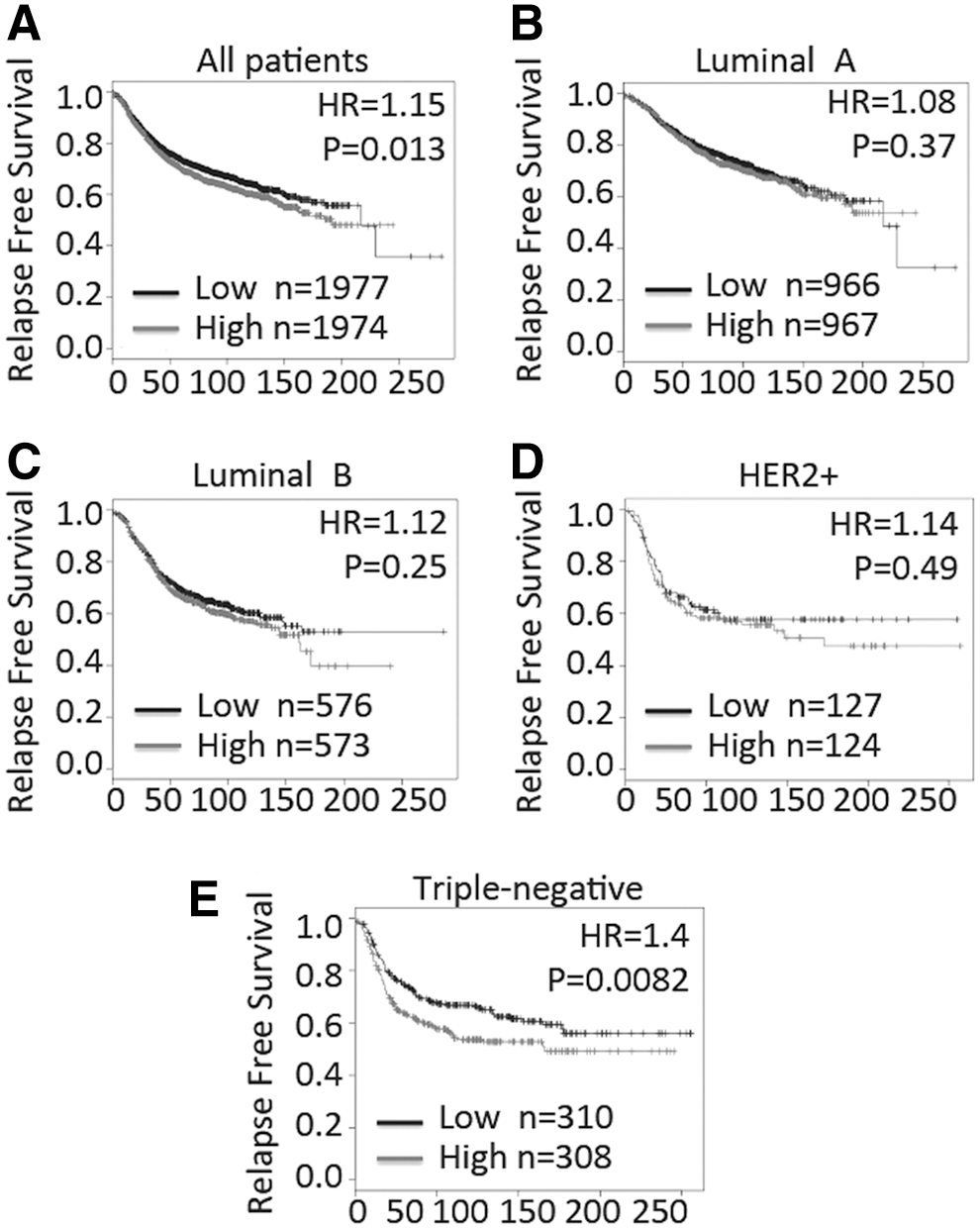
Prognostic ability of eIF2α in BC patients. Kaplan–Meier curves of estimated 20-year
p-eIF2α is negatively correlated with metastasis in TNBC
Protein level of eIF2α was further validated for the association between eIF2α and RFS in TNBC using the immunohistochemical method. The TNBC patients with metastasis found in the follow-up had higher expression of eIF2α, however, TNBC patients without metastasis had significantly lower expression of eIF2α (Fig. 2A). Expression of p-eIF2α was also checked in the immunohistochemical slides. Conversely, p-eIF2α was negatively correlated with metastasis in TNBC (Fig. 2B). Totally, 6 in 12 TNBC patients with high expression of eIF2α developed distant metastasis, however, only 2 in 16 TNBC patients with low expression developed distant metastasis (p = 0.04, Fig. 2C). As for p-eIF2α, 4 in 6 TNBC patients with low expression developed distant metastasis, however, only 4 in 22 TNBC patients with high expression developed distant metastasis (p = 0.038, Fig. 2D).
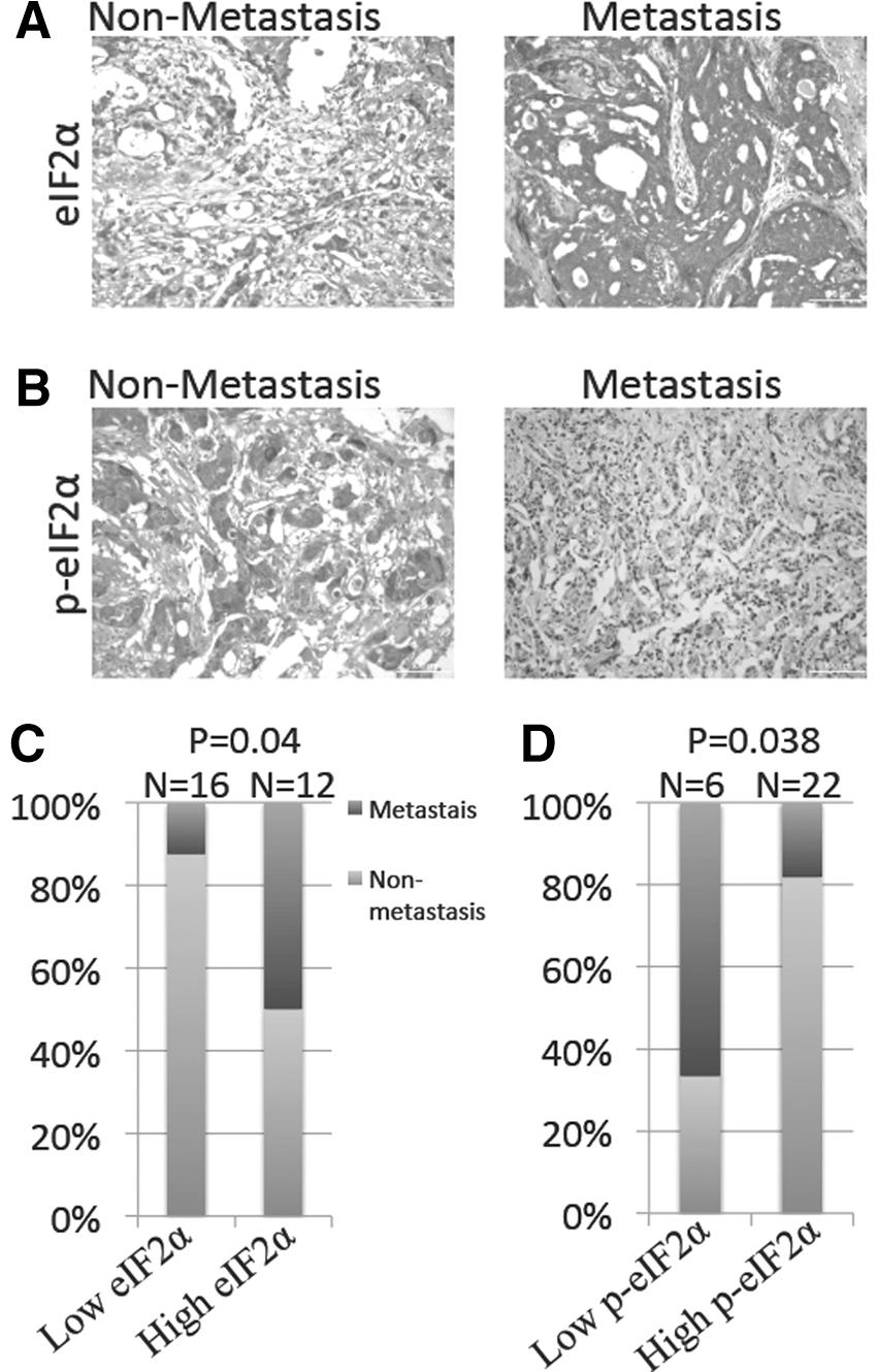
Metastasis events for TNBC patients with different expressions of eukaryotic translation initiation factor 2α(eIF2α) or p-eIF2α. Representative immunohistochemical staining of eIF2α and p-eIF2α in TNBC with or without metastasis was shown in the figure. The staining of eIF2α
p-eIF2α suppresses PDL1 expression in TNBC
The expression of PDL1 was first checked in different BC cell lines. MDA-MB-231 cell lines had significantly higher expression of PDL1 compared with CF-7 cell lines (Fig. 3A). eIF2α dephosphorylation inhibitor salubrinal and PKR inhibitor C16 were used to increase or decrease the level of p-eIF2α in MDA-MB-231 cell lines, respectively. p-eIF2α was negatively correlated with the mRNA level of PDL1, although it was not significant in the salubrinal group because the p-eIF2α was already highly expressed in MDA-MB-231 cell lines and could not be further improved by salubrinal (Fig. 3B). Protein level was further validated for the negative regulation of p-eIF2α on PDL1 and results were similar with the mRNA level (Fig. 3C). Besides, the mRNA level of PDL1 (CD274) was positively with that of PD1, which indicated that p-eIF2α expression in TNBC could influence the activity of immune cells depending on the PD1/PDL1 interaction (Fig. 3D).
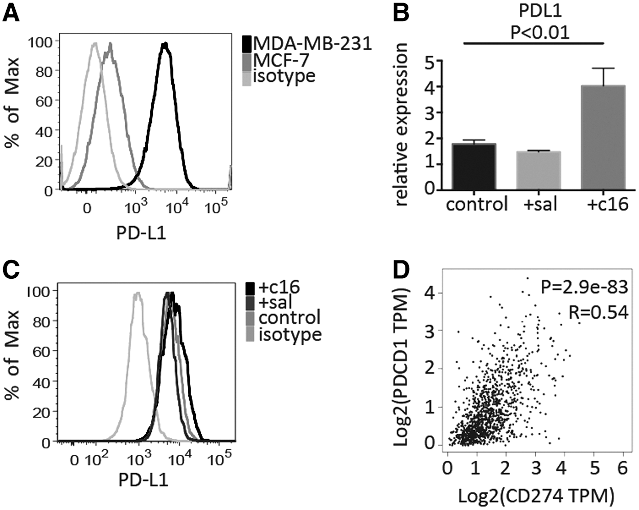
Regulation of programmed death ligand 1 (PDL1) expression by the PKR-p-eIF2α pathway.
p-eIF2α regulates CXCL5 expression in TNBC
To figure out whether p-eIF2α in TNBC could regulate the activity of immune cells in the tumor microenvironment, downstream immune-related molecules in TNBC were investigated. Expression of p-eIF2α has no correlation with MIF (Fig. 4A), HMGB1 (Fig. 4C), and CSF1 (Fig. 4D). However, it could significantly regulate CXCL5. Improved p-eIF2α significantly decreased the relative expression of CXCL5 and vice versa (Fig. 4B).
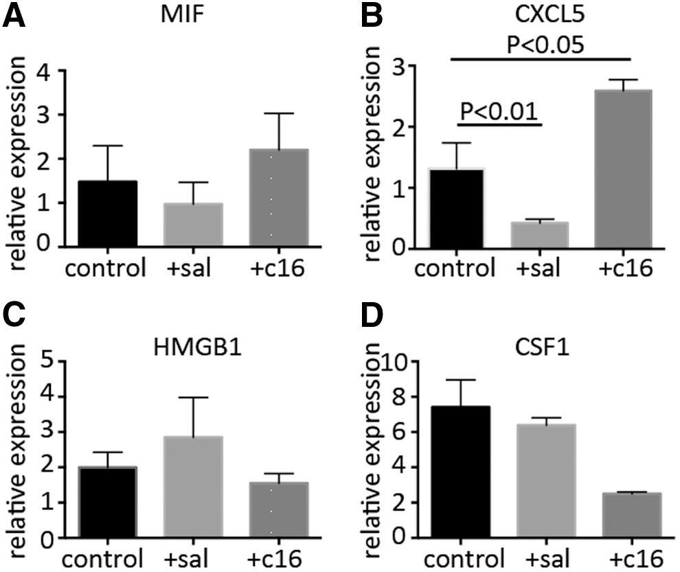
Regulation of downstream immune-related gene expression by the PKR-p-eIF2α pathway. MDA-MB-231 cell lines were separately treated with salubrinal and C16 for 48 hours. Relative expression of macrophage MIF
Carboplatin decreased expression of p-eIF2α in TNBC
To indicate the correlation between ER stress and chemotherapy treatment, carboplatin and paclitaxel were used to deal with different BC cell lines. It was found that all cell lines had increased p-eIF2α expression after adding carboplatin or paclitaxel except for the MDA-MB-231BO cell lines treated with carboplatin (Fig. 5). It indicated that TNBC with a high invasive characteristic might develop drug resistance to carboplatin depending on the PERK-p-eIF2α pathway of ER stress.
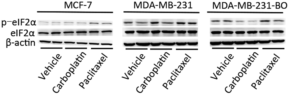
Influence of eukaryotic translation initiation factor 2α(eIF2α) and p-eIF2α expression by chemotherapy drugs in BC cell lines. MCF-7 cell lines, MDA-MB-231 cell lines, and MDA-MB-231BO (high bone metastasis) were separately treated with vehicle, carboplatin (1 μg/mL), and paclitaxel (20 μM) for 24 hours. Whole cell extracts were analyzed by western blot using indicated antibodies.
Mechanism of PERK-p-eIF2α in regulating metastasis and drug resistance in TNBC
The authors summarized the mechanism of PERK-p-eIF2α pathway in regulating metastasis and drug resistance in TNBC. ER stress in TNBC could lead to the activation of eIF2α, and subsequently, p-eIF2α could suppress the expression of PDL1 and CXCL5, which could further promote the activity of tumor-infiltrated T cells, inhibit the activity of MDSCs, and thus regulate the metastasis of TNBC. Besides, PERK-p-eIF2α pathway could possibly regulate drug resistance to carboplatin in highly metastatic TNBC (Fig. 6).
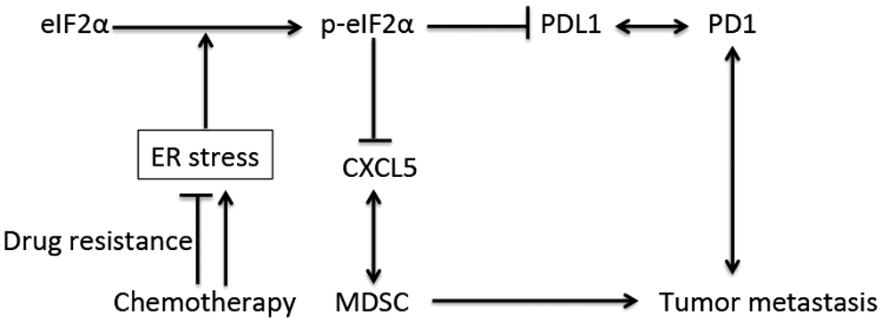
Schematic summarizing of the mechanisms by which PKR/eIF2a signaling suppresses TNBC and possible drug resistance of carboplatin in highly metastatic human TNBC. MDSCs, myeloid-derived suppressor cells.
Discussion
TNBC has the greatest need for improved therapies because of lacking of effective target and deserves more investigation in this special BC subtype. In this study, the authors found that PERK-p-eIF2α pathway of ER stress was correlated with metastasis of TNBC and further studied the possible mechanism of PERK-p-eIF2α in regulating metastasis and drug resistance in TNBC. PERK-p-eIF2α pathway could be a potent target in treating TNBC patients.
It was reported that p-eIF2α was a good prognostic factor in nonsmall-cell lung cancer 17 and TNBC. 8 Consistent with previous studies, the authors also found p-eIF2α had a protective function in TNBC. The prognostic role of eIF2α and p-eIF2α seems opposite in this study, however, it was not contradictory. p-eIF2α was the activated molecule of eIF2α and really worked in the cells. Besides, it was also indicated that eIF2α and p-eIF2α had opposite prognostic roles in TNBC in another study. 8
Programmed cell death protein 1 (PD1) is a second immune checkpoint receptor that limits T cell effector function within tissues. 18 PDL1 is the main ligand for PD1 and expressed on many tumors. 19 It was demonstrated that PDL1 was expressed in 34% of breast tumors and the expression was associated with high-risk clinicopathologic features. 20 The authors also found that TNBC had more expression of PDL1, which was consistent with the previous report. TNBC patients possibly suppressed the expression of PDL1 by p-eIF2α and thus led to the increased activity of tumor-infiltrated immune cells. Besides, it was shown that CXCL5 could recruit CD11b+GR1hi cells to the primary tumor, and depletion of these cells by LY6G-specific antibody treatment reduces dissemination of melanoma cells. 21 Another study showed that loss of TGFβ signaling in tumor cells increased CXCL5 secretion and recruited CD11b+GR1+MDSCs to the invasion front of the tumors, which could promote tumor cell invasion in vitro through the expression of MMPs. 22 p-eIF2α could inhibit the expression of CXCL5 and further restricted the MDSC infiltration in the primary tumor of TNBC, resulting in less metastasis of TNBC.
There are some limitations in this study. Although the authors found that PERK-p-eIF2α pathway could regulate PDL1 and CXCL5 expression, however, the particular mechanism how the p-eIF2α regulated expression remains to be fully explored. Besides, the deep mechanism of how the PERK-p-eIF2α pathway influenced drug resistance to carboplatin in TNBC also needs to be further investigated. Immunotherapy targeting the immune checkpoints or inhibitory immune cells plays more and more important roles in many cancers, including TNBC, and future studies should pay more attention to it in this special subtype of BC.
In conclusion, the authors found that PERK-p-eIF2α pathway could suppress metastasis in TNBC by inhibiting expression of PDL1 and CXCL5 in tumor cells. Besides, PERK-p-eIF2α pathway might also be involved in drug resistance to carboplatin in highly metastatic TNBC. This indicated a potent target in treating TNBC and might provide more choices for the clinicians in future.
Footnotes
Acknowledgment
The authors acknowledge the contribution of the laboratory members of Binzhou Medical University for their assistance with this research. This work was supported by the National Natural Science Foundation of China (NO. 81173601) and Scientific Research Staring Foundation of Binzhou Medical University (NO. BY2013KJ06 and NO. BY2015KJ23).
Disclosure Statement
No competing financial interests exist.
References
Supplementary Material
Please find the following supplemental material available below.
For Open Access articles published under a Creative Commons License, all supplemental material carries the same license as the article it is associated with.
For non-Open Access articles published, all supplemental material carries a non-exclusive license, and permission requests for re-use of supplemental material or any part of supplemental material shall be sent directly to the copyright owner as specified in the copyright notice associated with the article.