
Abstract
Objective:
Many malignant tumors grow in hypoxic condition, which is associated with tumor growth, invasion, and metastasis. MicroRNAs are of great significance in the development of multiple malignant tumors. This study cultured breast cancer cell MCF-7 under the condition of different concentrations of oxygen, to test cell proliferation and invasion, and detect miR-210 expression, aiming to analyze the influence of hypoxia on breast cancer cell behaviors as well as miR-210 expressions.
Materials and Methods:
Breast cancer cell MCF-7 was cultured under normoxia, hypoxia, or anaerobic conditions for 12, 24, or 48 hours. Cell proliferation was detected by MTT assay. Cell invasion and migration were tested by transwell assay. HIF-1α mRNA and miR-210 expressions were determined by real-time polymerase chain reaction.
Results:
MCF-7 cell proliferation was gradually increased following time extension (p < 0.05). MCF-7 cell exhibited higher proliferation, invasion, and migration activities in hypoxic and anaerobic groups compared with those in normoxic group during the same time period. HIF-1α mRNA and miR-210 were significantly upregulated in anaerobic group compared with those in other groups (p < 0.05). HIF-1α mRNA and miR-210 were obviously elevated at 12, 24, and 48 hours (p < 0.05).
Conclusion:
MCF-7 cell proliferation was increased, invasion and migration were enhanced, with upregulated expression of HIF-1α mRNA and miR-210 in the hypoxic and anaerobic group following time extension.
Introduction
Numerous malignant tumors have rapid proliferation, such as breast cancer, pancreatic cancer, and glioma, leading to a local hypoxia microenvironment. 1,2 MicroRNAs showed significant changes under ischemia/hypoxia, thus affecting cell survival, apoptosis, invasion, and other important biological processes. 3 At present, molecular targeted therapy is a hotspot on malignant tumor treatment. MiRNAs become the new direction for breast cancer early diagnosis and targeted therapy. 4 MiRNA is a type of small noncoding RNA composed of 18–23 nucleotides. They are a kind of independent transcripts that can regulate transcription factors, degrade mRNA, and regulate gene expression process. MiR-210 is commonly considered to be regulated by hypoxia and found to be upregulated in tissues and cells under the local hypoxic microenvironment. MiR-210 expression may not be affected by osmotic pressure changes or acidosis. 5,6 This study cultured breast cancer cell MCF-7 under the condition of different concentrations of oxygen to test cell proliferation and invasion, and detect miR-210 expression, aiming to analyze the influence of hypoxia on breast cancer cell behaviors and miR-210 expression.
Materials and Methods
Experimental materials
Breast cancer cell MCF-7, which was first isolated in 1970 from the breast tissue of a 69-year-old Caucasian woman and a widely studied epithelial cancer cell line derived from breast adenocarcinoma with characteristics of differentiated mammary epithelium, was provided by the experiment center in Hebei Medical University. Polymerase chain reaction (PCR) primers were supplied by Shanghai Boya Biological Technology Co., Ltd. Transwell chamber was purchased from BD.
Experimental methods
Conventional cell culture
MCF-7 cells were cultured in RPMI-1640 medium and maintained in an incubator at 37°C with 5% CO2.
Hypoxic cell culture
MCF-7 cells were cultured in RPMI-1640 medium and maintained at 37°C with 1% O2 and 5% CO2. Cell morphology was observed after 6 hours.
Anaerobic cell culture
MCF-7 cells were treated by CoCl2 at 600 μmol/L to mimic a chemical anaerobic microenvironment. Cell morphology was observed after 6 hours.
MTT assay
MCF-7 cells in logarithmic phase were seeded into a 96-well plate at a density of 8 × 10⁴/well under normoxic, hypoxic, or anaerobic conditions. After 12, 24, or 48 hours, 5 mg/mL MTT was added into each well in the plate for 4 hours. At last, DMSO was added followed by detection of the absorbance value at a wavelength of 570 nm. Three independent cultures per time point were performed for MTT assay.
Transwell assay
Cell invasion: Matrigel was diluted by serum-free medium at 1:8 for 24 hours. Then 60 μL Matrigel was put in the chamber. A total of 200 μL MCF-7 cells at a density of 106/mL were seeded on the upper chamber, while 1300 μL complete medium was added to the lower chamber. After 24 hours, the cells that passed the membrane were fixed with ethanol and stained for observation.
Cell migration: A total of 200 μL MCF-7 cells at 106/mL were seeded on the upper chamber, while 1300 μL complete medium was added to the lower chamber. After incubation for 24 hours, the cells that passed the membrane were fixed with ethanol, stained with crystal violet, and observed under the microscope.
Real-time PCR
The cells were collected to extract total RNA. Next, the RNA was reversely transcripted to cDNA according to the manual. PCR was performed under the condition of 95°C for 30 seconds, followed by 40 cycles of 95°C for 5 seconds, 60°C for 34 seconds, and 95°C for 15 seconds at last. The primers used are listed in Table 1. The expression of target genes was quantified as a fold change relative to internal control (GAPDH).
Data analysis
SPSS 17.0 software was adopted for data analysis. Measurement data are presented as mean ± standard deviation and compared by t test. Enumeration data were analyzed by analysis of variance. p < 0.05 was considered statistical significance.
Results
MCF-7 cell proliferation under normoxic, hypoxic, or anaerobic conditions
MCF-7 cell proliferation was gradually increased following culture time extension (p < 0.05). MCF-7 cell exhibited a higher proliferation activity in hypoxic and anaerobic groups compared with that in normoxic group during the same time period (p < 0.05) (Fig. 1).

MCF-7 cell proliferation under normoxic, hypoxic, or anaerobic conditions. *p < 0.05, versus normoxic group. # p < 0.05, versus hypoxic group. & p < 0.05, versus 12 hours. @ p < 0.05, versus 24 hours.
MCF-7 cell invasion and migration under normoxic, hypoxic, or anaerobic conditions
MCF-7 cell invasion and migration were obviously enhanced in hypoxic and anaerobic groups compared with those in normoxic group (p < 0.05) (Fig. 2).
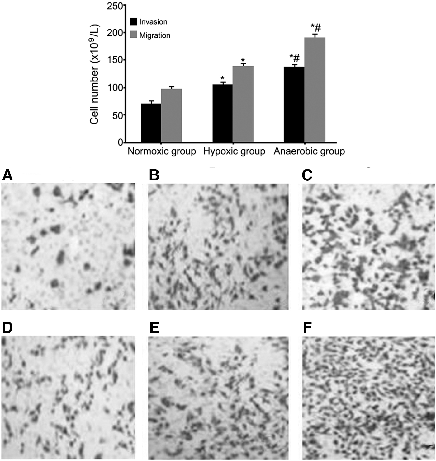
MCF-7 cell invasion and migration under normoxic, hypoxic, or anaerobic conditions. *p < 0.05, versus normoxic group. #
p < 0.05, versus hypoxic group.
HIF-1α mRNA and miR-210 expression in MCF-7 cells under normoxic, hypoxic, or anaerobic conditions
HIF-1α mRNA and miR-210 expression was significantly upregulated in hypoxic and anaerobic groups compared with those in normoxic group at 12, 24, and 48 hours (p < 0.05). In addition, HIF-1α mRNA and miR-210 expression was markedly enhanced following time extension (p < 0.05) (Fig. 3).
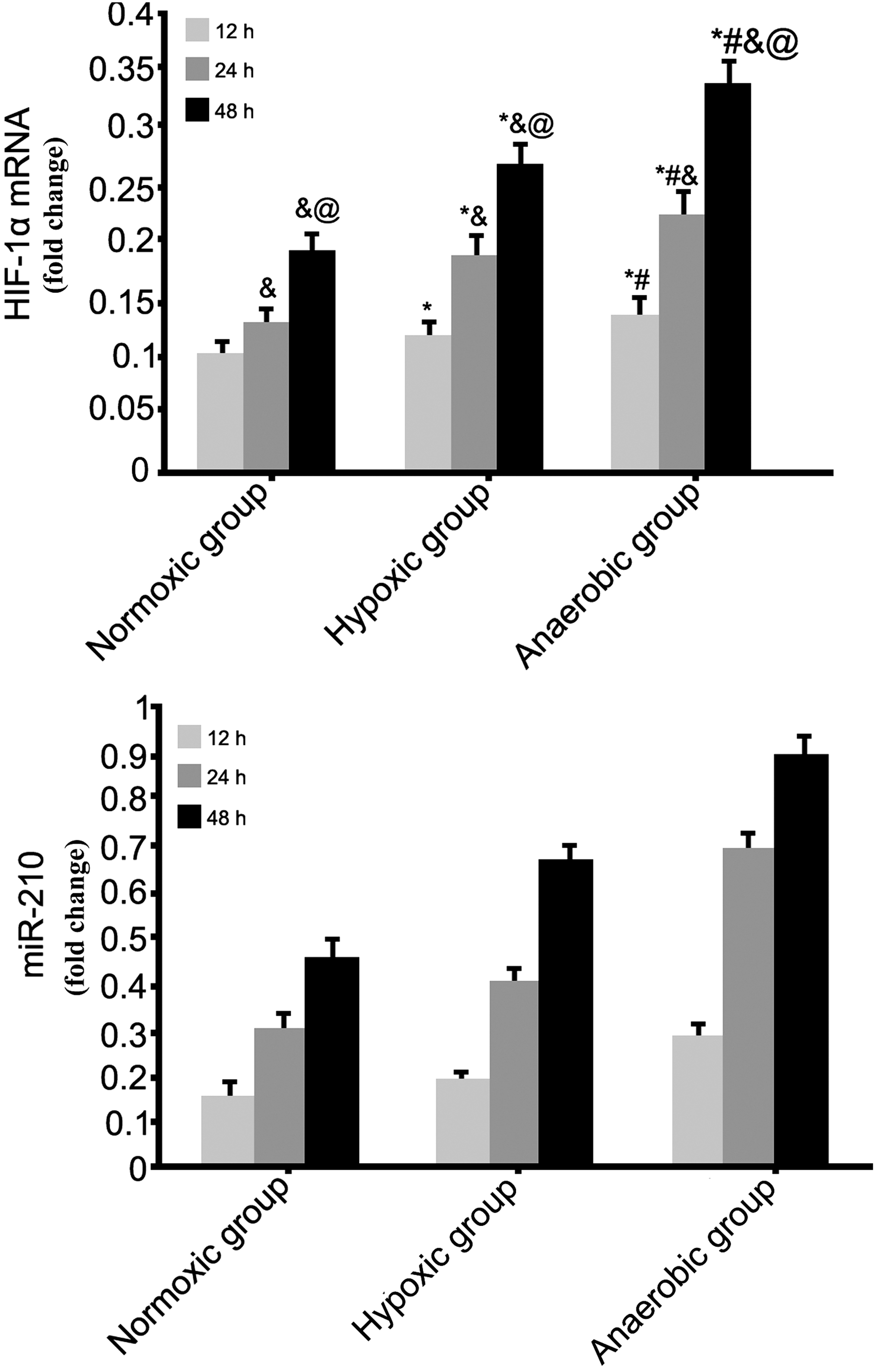
HIF-1α mRNA and miR-210 expression in MCF-7 cells under normoxic, hypoxic, or anaerobic conditions. A, HIF-1α mRNA expression. B, miR-210 expression. *p < 0.05, versus normoxic group. # p < 0.05, versus hypoxic group. & p < 0.05, versus 12 hours. @ p < 0.05, versus 24 hours. The numerical values in the y-axis represented fold changes relative to the internal control.
Discussion
Tumor invasion and metastasis is the process of the cancer cell detaching from the original site and reaching remote organs, which is regulated by multiple steps and multiple factors. 7 Cancer cells need a large amount of oxygen and nutrients in the process of invasion and metastasis. Hypoxia may lead to changes of a variety of miRNA expressions. Original miRNAs existing in introns are formed through gene transcription. 8 It was suggested that most malignant tumor cells had miRNA abnormal expressions. 9,10 Basic study showed that miR-10b, miR-520c, and miR-155 upregulation may promote breast cancer cell invasion and migration, while miR-335, miR-126, and miR-205 elevation can suppress cell migration. 11 –15 As a specific hypoxia-related miRNA, miR-210 is expressed in a low level under normoxic condition. It was found that miR-210 expression was upregulated in hypoxic environment in renal cancer. 16 MiR-210 is characterized as rapid response and concentration dependency in hypoxia. Malignant tumor cells originated from different sources and showed significant upregulation of miR-210 in hypoxic microenvironment with negative correlation with hypoxia severity. 17 This research cultured breast cancer cell MCF-7 under the condition of different concentrations of oxygen and tested cell proliferation, invasion, migration, and miR-210 expression, aiming to explore the impact of hypoxia on breast cancer cell behaviors and miR-210 expression.
In this study, MCF-7 proliferation was increased following time extension in all the anaerobic, hypoxic, and normoxic conditions. Compared with normoxic group, MCF-7 cell proliferation in hypoxic and anaerobic groups was significantly enhanced under the same time period. Moreover, it showed more apparent changes in anaerobic group than hypoxic group, suggesting that MCF-7 cell proliferation was increased in hypoxic and even anaerobic conditions in a time-dependent manner. Next, the authors further analyzed MCF-7 invasion and migration under hypoxic environment and found that they were markedly enhanced in hypoxic group and anaerobic group compared with normoxic group. It revealed that MCF-7 cell invasion and migration were enhanced under the hypoxic and even anaerobic local microenvironment. Hypoxic microenvironment is a common phenomenon closely associated with tumor excessive growth, causing hypoxic region in local tissues, requiring blood supply and nutrients, resulting in excessive formation of new vessels with abnormal structure and function. On this occasion, hypoxic microenvironment further changes some of the tumor biological characteristics, such as genetic instability, resistance to radiotherapy and chemotherapy, or even local invasion and distant metastasis. 18
This study further investigated the impact of hypoxic or even anaerobic microenvironment on miR-210 expression in MCF-7 cells. HIF-1α mRNA and miR-210 expression was significantly upregulated in hypoxic and anaerobic groups compared with normoxic group at the same time point. In addition, HIF-1α mRNA and miR-210 expression was markedly enhanced following time extension. It indicated that HIF-1α mRNA and miR-210 levels were elevated in MCF-7 cells under hypoxic or anaerobic condition. HIF-1α level was low under enough oxygen supply, while it may quickly increase under severe hypoxic or anaerobic condition to regulate target gene expression and cell response to hypoxia signaling transduction. 19 Fasanaro, et al. constructed the hypoxia model on umbilical vein endothelial cells and osteosarcoma cells for 4 hours and found miR-210 expression was elevated, while it slowly declined after normal oxygen supply. 20 MiR-210 expression was obviously increased in hypoxic or even anaerobic microenvironment, whereas reduced after oxygen recovery. Studies on pancreatic cancer and lung cancer demonstrated that miR-210 elevation was associated with malignant tumor invasion, migration, and patient's prognosis. 21 MiR-210 expression was upregulated in local hypoxic microenvironment for the same type of malignant tumor. However, miR-210 may gradually keep on high level instead of continuous elevation once the tumor tissue keeps in hypoxic or even anaerobic condition for a long time. It may be caused by the tumor protection mechanism activated following time extension, thus activating Drosha and Dicer enzymes, leading to miR-210 abnormal expression. 22
Conclusion
MCF-7 cell proliferation was increased, invasion and migration were enhanced, with upregulated expression of HIF-1α mRNA and miR-210 in hypoxic and anaerobic group following time extension, suggesting that the HIF-1α signaling pathway and miR-210 might be involved in the pathogenesis of breast cancer invasion and metastasis, and therapeutically targeting them might be a novel strategy for the treatment of breast cancer.
Footnotes
Disclosure Statement
No competing financial interests exist.