
Abstract
Background:
Gastric carcinoma is the most popular cancer worldwide. Anoctamin-1 is a calcium-activated channel and highly expressed in various tumors. A previous study indicated that suppressed Anoctamin-1 expression decreased cancer cell proliferation or migration. As a signal transduction and transcription activator, STAT6 is a novel agonist for Anoctamin-1 promoter. However, its role in tumor cell proliferation or migration remains unclear. Therefore, this study aimed to suppress STAT6 and Anoctamin-1 protein expression in gastric cancer cells to test the inhibitory effects on gastric cancer cell migration or invasion.
Materials and Methods:
MTT colorimetry was used to test cell proliferation. Western blot was used to measure STAT6 and Anoctamin-1 expression before and after small interfering RNA (siRNA) treatment. A scratch assay was performed to measure cell migration, followed by Transwell chamber assay analysis of cell invasion.
Results:
After STAT6 siRNA interference, the expression of STAT6 and Anoctamin-1 was significantly decreased in the gastric carcinoma cell line. Anoctamin-1 siRNA interference only decreased its protein expression, but not STAT6 protein expression. Interference of STAT6 or Anoctamin-1 reduced their protein expression and inhibited proliferation, migration, or invasion of gastric cancer cells.
Conclusions:
Inhibition of STAT6/Anoctamin-1 activation decreased proliferation, migration, or invasion of gastric cancer cells, suggesting that the STAT6/Anoctamin-1 pathway might be a novel target for treating gastric cancer.
Introduction
Gastric cancer is the most common malignant tumor in the digestive tract. With lifestyle transition, patients with gastric cancer show a younger trend, with risk factors, including unhealthy habitats and environmental pollution. 1 The investigation of potential treatment target of gastric cancer is thus of critical importance.
Anoctamin-1 is an ion channel activated by calcium ion, or called TMEM16A, which is encoded by the ANO1 gene. 2 A previous study showed the expression of Anoctamin-1 in gastrointestinal smooth muscle and epithelium, with activation of the chloride channel by voltage-sensitive calcium. 3 Moreover, Anoctamin-1 is highly expressed in Cajal mesenchymal cells in the gastrointestinal tract, with close correlation of its expression level with the activity of Cajal mesenchymal cells. 4 A recent study showed significantly elevated expression of Anoctamin-1 protein in various tumors, including esophageal squamous carcinoma 5 and breast cancer. 6 So far, no study has reported regarding the expression of Anoctamin-1 in the gastric cancer cell line. Another study indicated decreased proliferation or migration of cancer cells after Anoctamin-1 expression was suppressed. However, the exact role remains poorly understood.
STAT6 is a member of the signal transduction and transcription activator family and can respond to a series of cytokines and growth factors. After phosphorylation by receptor-related kinase, it can form dimers and subsequently translocate into the nucleus, where it can activate gene transcription. Early study showed that STAT6 could induce Bcl-2 and Bcl-xl expression, thus showing antiapoptotic activity. 7 Moreover, it is a newly discovered agonist of Anoctamin-1 promoter. 8 Whether it is involved in Anoctamin-1-induced tumor cell proliferation or migration has not been studied.
Based on previous studies previously mentioned, the authors proposed the upregulation of Anoctamin-1 in gastric cancer cells, which might be under STAT6 regulation. Moreover, the STAT6/Anoctamin-1 pathway can regulate cancer cell proliferation, migration, and invasion. This study thus aimed to investigate the role of the STAT6/Anoctamin-1 pathway on invasion or migration of gastric cancer cells.
Materials and Methods
Drugs and reagents
The human gastric cancer cell line MGC-803 was obtained from Cell Bank, Chinese Science Academy (China). MTT reagent was purchased from Sigma (China). Dulbecco's modified Eagle's medium (DMEM), fetal bovine serum, and dual antibiotics were purchased from Gibco (USA). Matrigel was purchased from BD (USA). Antirabbit STAT6 and Anoctamin-1 polyclonal antibodies were purchased from Abcam (China). Goat antirabbit IgG (H+L) and anti-β-actin antibodies were purchased from Proteintech (China). Transwell chamber was purchased from Costar Cambridge (USA). Si-STAT6 and Anoctamin-1 and si-control (NC) were designed and synthesized by Sigma (USA).
Cell culture and treatment
The human gastric cancer cell line MGC-803 was kept in DMEM containing 10% serum, 100 U/mL penicillin, and 100 μg/mL streptomycin in a 37°C chamber with 5% CO2. Cell passage was changed for fresh medium each day.
RNA transfection
Small interfering RNA (siRNA) was synthesized by Sigma. One day before transfection, MGC-803 cells were digested and resuspended in a 24-well plate. siRNA transfection buffer was prepared by adding 1.25 μL siRNA stock solution (20 μM) into 100 μL Opti-MEM. One microliter of Lipo2000 was solved in serum-free DMEM. After 5 minutes of mixing, Lipo2000 and siRNA solutions were mixed for 20-minute incubation. The mixture was eventually added into a 24-well plate for a certain period of incubation. After 4 hours, normal growth medium was added for observing transfection efficiency under the fluorescence microscope.
MTT assay for tumor proliferation by STAT6 and Anoctamin-1
Human gastric cancer cells MGC-803 at the log growth phase were digested and resuspended into single-cell suspension in serum-containing DMEM. Cells were adjusted into a certain concentration and inoculated into a 96-well plate at 3000–5000 cells per well. After attached growth, siRNA for STAT6 or Anoctamin-1 (50 nM) was added. After 6 hours of transfection, normal culture medium was applied for 18 hours of continuous incubation. After adding 10 μL MTT solution for 4 hours of incubation, a violet crystal complex was observed under the microscope. The upper culture medium was extracted. Each well was mixed with 150 μL Tris buffer for vortex for resolving. After 12–15 hours, the absorbance value at 490 nm wavelength was measured for calculating cell activity. Eight paralleled wells were set in each group.
Scratch assay for cell migration potency
Gastric cancer MGC-803 cells at the log growth phase were rinsed three times with phosphate-buffered saline (PBS) and digested in 0.25% trypsin for resuspension in serum-containing DMEM. One milliliter of cell suspension was inoculated into a six-well plate, followed by transfection when cells reached 80% confluence. Normal culture medium was then used until complete confluence. Parallel lines were drawn on the culture plate using a sterile pipette for removing cells. After rinsing three times in PBS, normal culture medium or different concentrations of drugs were used. After 24-hour observation, an inverted microscope was used to check the effect of siRNA knockdown of STAT6 and Anoctamin-1 expression on cell migration.
Transwell assay for cell invasion potency
Matrigel-based gel was paved on the membrane of the Transwell chamber inside a 24-well plate. After incubation at 37°C for 30 minutes, cells were digested by 0.25% trypsin and prepared for single-cell suspension. Two hundred microliters of cell suspension and 500 μL chemokines were added into the upper and lower chamber, respectively. After knock down of protein expression of STAT6 or Anoctamin-1 by siRNA, the culture plate containing chambers was cultured for 24 hours. Unpenetrated cells were removed by wet cotton, followed by hematoxylin staining, rinsing, and coverslip mounting. Under an inverted microscope, three fields were randomly selected from each Transwell chamber. Cell invasion potency was evaluated by the number of cells for membrane penetration.
STAT6 and Anoctamin-1 expression in gastric cell lines by Western blot
MGC-803 cells were treated with siRNA and digested in trypsin, followed by adding 100 μL lysis buffer (containing proteinase inhibitor, PMSF, and phosphatase inhibitor) for 15 minutes of iced incubation. After complete lysis, the mixture was centrifuged for 10 minutes at 14,000 g. The supernatant was obtained and quantified for protein content using the BCA method. All samples were adjusted to equal concentration using saline and mixed with an equal volume of loading buffer for 5-minute boiling. Fifty-microgram samples were then separated in sodium dodecyl sulfate–polyacrylamide gel electrophoresis and transferred to a PVDF membrane by the semidry approach. The membrane was blocked at 5% defatted milk powder at 37°C for 1 hour. Primary antibodies for STAT6, Anoctamin-1, and β-actin were added for a 4°C overnight incubation (in 1:1 000 dilution). On the second day, the membrane was rinsed in TTBS three times, followed by secondary antibody incubation for 1 hour at 37°C. ECL substrate was added for development.
Statistical methods
All experimental data were collected from at least three times of independent tests. Data are presented as mean ± standard deviation. One-way analysis of variance was used for comparison among multiple groups, while Student's t-test was used for between two groups. SNK test was used for paired comparison. All studies were carried out in a two-sided manner. Statistical significance was defined when p < 0.05.
Results
Expression of STAT6 and Anoctamin-1 in the gastric cancer cell line
siRNA interference was first used to knock down the expression of STAT6 and Anoctamin-1, which were examined by Western blot. After STAT6 siRNA transfection, both STAT6 and Anoctamin-1 expression levels were downregulated with significant difference compared with the untreated group (Fig. 1A, B). Anoctamin-1 siRNA treatment barely changed STAT6 expression, but remarkably decreased Anoctamin-1 expression with significant difference compared with the normal interference group. All these results demonstrated a regulatory effect of STAT6 on Anoctamin-1 expression.
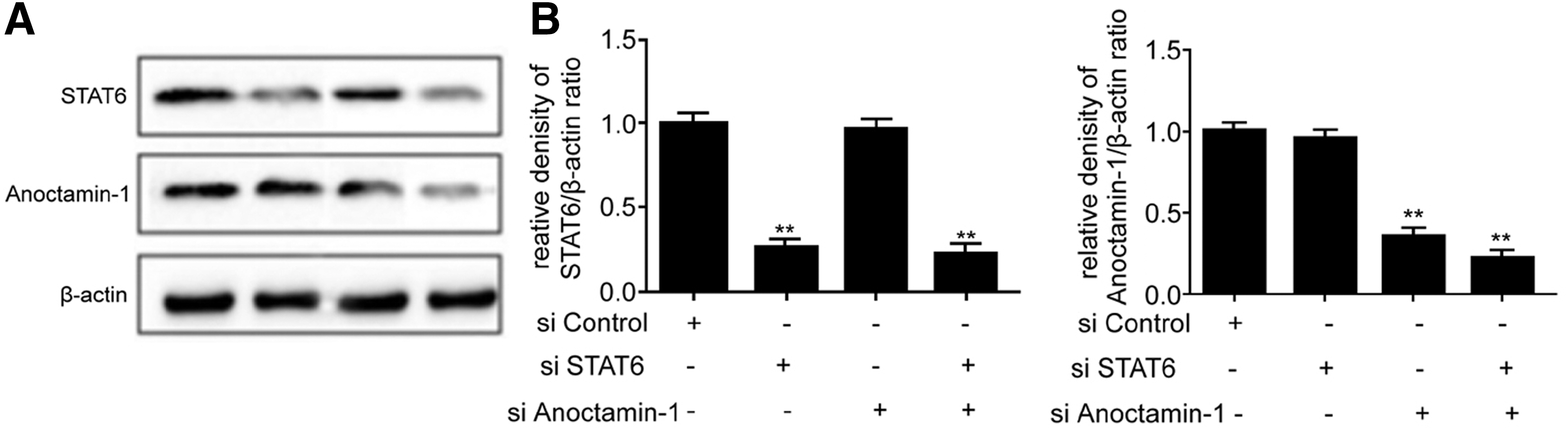
Expression of STAT6 and Anoctamin-1 in the gastric cancer cell line. **p < 0.01 compared with the si-control group.
Effects of STAT6 and Anoctamin-1 on gastric cancer cell proliferation by MTT approach
MTT is a classical way to evaluate cell viability. 9 In this study, siRNA approach was used to knock down the expression level of STAT6 and Anoctamin-1, followed by observation of their effect on cell proliferation. Anoctamin-1 and STAT6 siRNA treatment significantly decreased cell proliferation compared with si-NC control (Fig. 2). In STAT6 and Anoctamin-1 double knockdown group, cell proliferation potency was further inhibited.
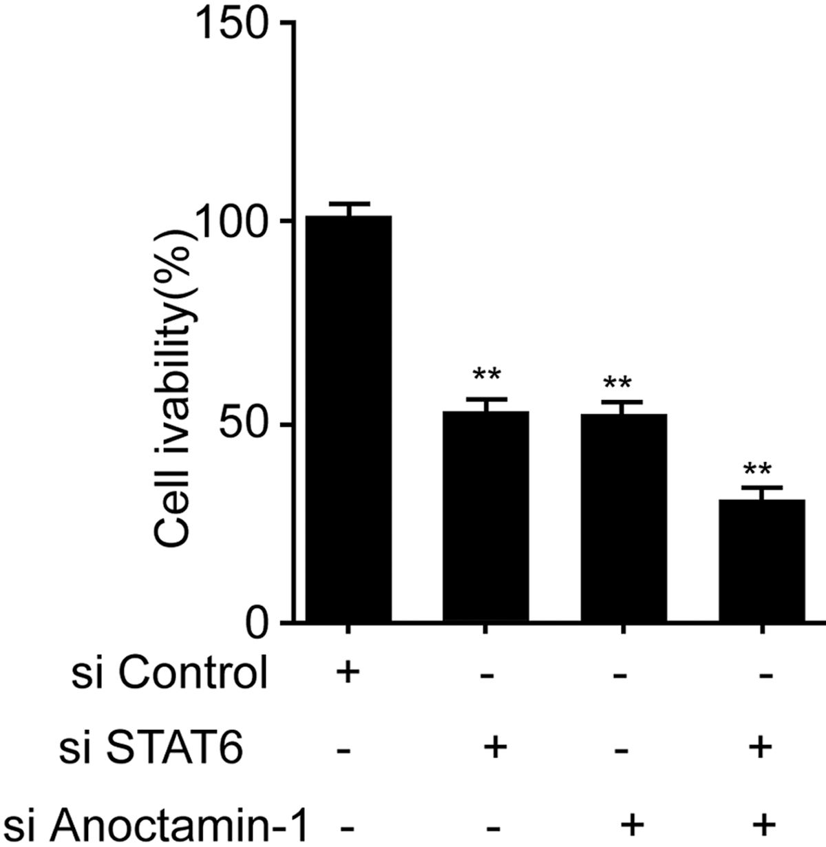
Effects of STAT6 and Anoctamin-1 on gastric cancer cells. **p < 0.01 compared with the si-control group.
Scratch assay for the effect of STAT6 and Anoctamin-1 on gastric cancer cell migration
Cell migration ability was tested by scratch assay. 10 The authors measured cell migration at 24 hours after interference by STAT6 and Anoctamin-1 siRNA. As shown in Figure 3, compared with the control group, STAT6 or ANO2 siRNA transfection suppressed their expression level and weakened cell migration. Dual knockdown of STAT6 and Anoctamin-1 further decreased the migration ability of cells.

Effects of STAT6 and Anoctamin-1 for gastric cancer cell migration.
Transwell assay for effects of STAT6 and Anoctamin-1 on invasion of gastric cancer cells
The Transwell chamber with matrix gel pavement was used to evaluate invasion of gastric cancer cells. Twenty-four hours after interference by STAT6 or Anoctamin-1 siRNA, cell invasion was tested. As shown in Figure 4, after knocking down STAT6 or Anoctamin-1 expression, cell invasion potency was significantly reduced, with significant difference compared with the control group. In the STAT6 and Anoctamin-1 dual knockout group, cell invasion was further suppressed.

Effects of STAT6 and Anoctamin-1 on invasion potency of gastric cancer cells. **p < 0.01 compared with the si-control group.
Discussion
Gastric cancer is the most common malignant tumor in the digestive tract. 11 A previous study indicated significant correlation between disease occurrence and a unhealthy life habitat or environmental pollution. 12 Currently, the 5-year survival rate of gastric cancer in China is only 10%, making early diagnosis a critical challenge in the clinic. Determination of related targets involved in gastric cancer pathogenesis and treatment is thus of critical importance.
Tumor metastasis is a complicated process, including detachment of tumor cells from the original position, followed by penetration across matrix tissues and entry into the circulation system through vascular walls. 13 Cell proliferation can represent tumor cell growth, while migration and invasion are closely associated with cancer metastasis. 14 The authors employed the MTT approach to observe cell proliferation, focusing on inhibitory effects of STAT6 and Anoctamin-1 on tumor growth. The authors also used the scratch assay to observe cell migration potency, followed by Transwell chamber assay to measure effects of the STAT6 and Anoctamin-1 pathway on tumor invasion, thus mimicking the tumor metastasis process.
STAT6 is a member of the signal transduction and transcription activator family. It can form dimers after being phosphorylated by receptor-related kinase, in response to a series of cytokines and growth factors, and then translocate into the nucleus. In various human tumors, STAT6 activity was significantly elevated. 15,16 Previous studies showed overexpression of interleukin 4 (IL-4) and its receptor in various epithelial-derived tumors, making them promising targets for treating metastatic tumors. 7 IL-4 is an effective agonist for STAT6. 17,18 Previous literatures showed the regulatory role of STAT6 activity in cell cycler regulation and cancer metastatic genes, and STAT6 transduction pathway activation was related with tumor cell growth, apoptosis, and metastasis. Moreover, it is a newly discovered agonist for Anoctamin-1 promoter. However, the role of Anoctamin-1 in STAT6-activated tumor cell proliferation or migration has not been reported. Anoctamin-1 is a calcium-activated chloride channel and is firstly discovered in gastrointestinal smooth muscle cells and Cajal mesenchymal cells, functioning as a critical protein for slowing wave potential. 19 Recent studies showed overexpression of Anoctamin-1 in various tumors. 20 Other reports indicated that Anoctamin-1 could work as a sensitive marker for gastrointestinal matrix carcinoma as well as a negative prediction factor for survival rate. 21,22 Inhibition of Anoctamin-1 expression can decrease proliferation or migration of cancer cells.
In this study, the siRNA approach was used to knock down protein expression of STAT6 and Anoctamin-1, followed by analysis of proliferation, invasion, and migration of gastric cancer cells, to investigate the effect of the STAT6/Anoctamin-1 signal pathway on cell proliferation, invasion, and migration. The results showed that STAT6 knockdown inhibited Anoctamin-1 expression and suppressed proliferation/invasion and migration of gastric cancer cells. However, knockdown of Anoctamin-1 had no effects on STAT6 expression, but inhibited gastric cancer cell invasion or migration. These results indicated that STAT6 could affect cell behaviors through mediating Anoctamin-1 expression, which, however, had no regulatory effects on STAT6 expression.
This study, for the first time, illustrates the upstream–downstream relationship between STAT6 and Anoctamin-1 and suppressed tumor cell growth or metastasis by inhibition of STAT6/Anoctamin-1 activation. However, the exact role of STAT6/Anoctamin-1 in gastric cancer progression in vivo was not investigated in the present study and requires further studies.
Conclusions
Inhibition of STAT6/Anoctamin-1 activation suppressed proliferation/migration and invasion potency of gastric cancer cells, suggesting that the STAT6/Anoctamin-1 pathway might be a novel therapeutic target for treating gastric cancer.
Footnotes
Acknowledgments
This work was supported by the Guangxi Scientific Research and Technology Development projects (No. 14122007-44) and the Nanning QinXiu District Scientific Research and Technology Development projects (No. 2015S08).
Disclosure Statement
There are no existing financial conflicts.