
Abstract
Aim:
The aim of the present investigation was to examine the suitability of 99mTc–N-HYNIC–BZMB as a specific vascular endothelial growth factor (VEGF)-targeting agent. Bevacizumab is a recombinant humanized monoclonal antibody that inhibits VEGF.
Methods:
N-hydroxysuccinimide-2-hydrazinonicotinic acid (N-HYNIC) was conjugated to BZMB, followed by labeling with 99mTc using N-[Tris(hydroxymethyl)methyl] glycine (tricine), ethylenediamine-N,N′-diacetic acid (EDDA), and nicotinic acid as coligands. 99mTc-labeled BZMB was characterized in terms of 99mTcO4, radiocolloids, and labeled N-HYNIC–BZMB using thin-layer chromatography and HPLC. Poor metastatic SKOV-3 and high metastatic SKOV-3.ip1 human ovarian cancer cell lines were used for in vitro binding uptake of 99mTc–N-HYNIC–BZMB. Biodistribution and scintigraphy accuracy were examined in human ovarian tumor xenografts in rats and rabbits.
Results:
99mTc–N-HYNIC–BZMB prepared by using a mixture of tricine and EDDA demonstrated relatively high radiochemical purity (more than 98%). In L-cysteine and serum, it exhibited a stable behavior up to 16 h. In vitro binding uptake indicated that it targets high metastatic SKOV-3.ip1 tumors. Biodistribution in human ovarian tumor xenografts in rats confirmed a significant uptake in SKOV-3.ip1 tumors (5.69% ± 1.86%, 4 h). Scintigraphic accuracy in human ovarian tumor xenografts in rabbits validated its suitability as a high metastatic SKOV-3.ip1 radiotracer.
Conclusion:
High radiochemical purity, stability in saline and serum, biodistribution, and scintigraphy of 99mTc-N-HYNIC–BZMB in human ovarian tumor xenografts in rats and rabbits confirmed its suitability as a potential radiotracer for imaging high metastatic SKOV-3.ip1 sites.
Introduction
Angiogenesis is a constant process necessary for the growth and metastasis of various tumors. It is a complex process through which synthesis of new blood vessels is initiated from active vasculature for the supply of energy-rich compounds, including oxygen, to various tissues. Nevertheless, its probable associated undesirable effects for human health are the prime concerns of clinicians/investigators. 1 –3 An essential factor of angiogenesis is vascular endothelial growth factor (VEGF), which consists of at least four splice variants containing 121, 165, 189, and 206 amino acids. 4 VEGF has been widely investigated as one of the essential factors involved in the progression of pathophysiological angiogenesis. In a variety of human tumors, overexpressing of VEGF renders it a suitable target for cancer diagnosis. 5 This, in turn, has contributed to development of anti-VEGF agents that include monoclonal antibodies (mAbs) or receptor hybrids and tyrosine kinase inhibitors. 6
Anti-VEGF antibodies (Abs) are specifically attached to free circulating VEGF for blocking attachment to its receptors. So far, the FDA has approved bevacizumab (BZMB) as an anti-VEGF Ab for human treatment. 7,8 BZMB is a recombinant humanized mAb of mouse origin that binds to almost all variants of VEGF to block ligand receptor communications. 9 It also blocks VEGF-induced proliferation of endothelial cells and angiogenesis. 10 It selectively binds to circulating VEGF, inhibiting the binding of VEGF to its receptors, and has shown promising results in a variety of malignancies. 11
Radiolabeled bioactive molecules have shown encouraging results in identification and treatment of various tumors. Technetium-99m (99mTc), indium-111 (111In), and zirconium-89 (89Z)-labeled BZMB have been reported for noninvasive in vivo VEGF localization and quantification. 12 –14 However, 111In and 89Z are both produced by cyclotron, presenting multiple limitations. In spite of the expansion in positron emission tomography, the investigators depend much on 99mTc-labeled radiotracers due to their radiobiological properties, low cost, and easy availability in the form of 99mTc/99Mo generator. 15
At the beginning of the 1990s, development of various strategies for labeling of biomolecules with 99mTc, including labeling by employing the affinity of native or reduced cysteine in proteins and various bifunctional chelators, has been reported. 16,17 2-Hydrazinonicotinic acid (HYNIC) (Fig. 1A) has been one of the most promising, accepted, and effective bifunctional chelators intended for labeling of biomolecules with 99mTc. It is coupled to biomolecules through amide linkage formed by the amide terminal with the ester derivative of HYNIC, the N-hydroxysuccinimide (N) ester (Fig. 1B). 18

In the present investigation, N-hydroxysuccinimide-HYNIC (N-HYNIC) has been conjugated to BZMB, followed by labeling with 99mTc using N-[Tris(hydroxymethyl)methyl] glycine (tricine), ethylenediamine-N,N′-diacetic acid (EDDA), and nicotinic acid as coligands. The suitability of 99mTc–N-HYNIC–BZMB as a specific VEGF-targeting agent was further evaluated in terms of radiochemical purity, stability in cysteine and serum, in vitro binding to highly metastatic SKOV-3.ip1 and poorly metastatic SKOV-3 human ovarian cancer cell lines, biodistribution, and scintigraphy in human ovarian tumor xenografts in rats and rabbits.
Experiments
Materials
Bevacizumab (BZMB) was obtained from Roche, Inc. (Switzerland). N-HYNIC was from Synchem UG & Co. (Germany), and N-[Tris(hydroxymethyl)methyl]glycine (tricine), EDDA, nicotinic acid, and all other chemicals were purchased from Sigma-Aldrich. Rats and rabbits were obtained from the Veterinary Research Institute (Pakistan), and HPLC from Shimadzu, well counter and scalar count rate meter from Ludlum, and dose calibrator from Capintech were used for radioactivity measurement and dose adjustment. A gamma camera from GEADE Nuclearmedizine system was used for animal scintigraphy.
Methods
Bioconjugation of bevacizumab
Bioconjugation of N-HYNIC with bevacizumab (BZMB) was performed using an already reported method. 19 Briefly, 5 mL (10 mg) of BZMB and N-HYNIC (ester) was conjugated for 1 hour at 27°C using Na2CO3 (0.1 M), pH 9.5–9.7, with 3–5 ester molecules per BZMB. The excess of the unconjugated ester was discarded by washing and ultrafiltration using phosphate buffer (PB: 0.05 M, pH 7.4).
Labeling of conjugated BZMB
In three (2-mL) round bottom, polypropylene corning vials, 0.5 mL (10 μg/mL in distilled water) of conjugated BZMB was elaborated separately. The mixture of vial (I) was incubated with 0.5 mL (20 mg/mL in distilled water) of tricine (N-[Tris(hydroxymethyl)methyl]glycine) at 100°C for 25 min, the contents of vial (II) with 0.25 mL (20 mg/mL in 0.3 M sodium dihydrogen phosphate) of tricine and 0.25 mL (10 mg/mL in 0.1 M NaOH) of EDDA, and the contents of vial (III) with 0.4 mL (100 mg/mL in water) of tricine solution and 0.1 mL (90 mg/mL in water) of nicotinic acid as coligands. Subsequently, 0.5 mL (10 mCi/mL) of freshly eluted sodium pertechnetate and 10 μL (1 mg/mL in 0.1 N HCl) of tin (II) solution were added to vials (I), (II), and (III), separately.
99mTc–N-HYNIC–BZMB was characterized in terms of free technetium-99m (99mTcO4), radiocolloids, and labeled conjugated BZMB using thin-layer chromatography (TLC) and HPLC. In TLC, acetone, sodium citrate, and methanol: ammonium acetate (1:1 v/v) were used as mobile phases for separation of 99mTcO4, 99mTc-coligand, 99mTcO4, and radiocolloids, respectively.
HPLC was performed with Shimadzu equipment with a UV photodiode detector, flow scintillation counter, binary pump, online degasser, and TSKgel SuperSW (4 × 6 mm) column. Five microliters of 99mTc–N-HYNIC–BZMB was injected into the stationary phase (column), followed by elution with 0.1% TFA/water (solvent A) and 0.1% TFA/acetonitrile (ACN) (solvent B) with a flow rate of 1 mL/minute. The detailed A-B gradient conditions were 100% solvent A for 0–2 min, modified to 50% solvent A over 10 min, and sustained for 10 min. Thereafter, it was modified to 30% solvent A over 3 min and finally to 100% solvent A over 4 min.
Stability in cysteine and serum
At room temperature (27°C), 99mTc–N-HYNIC–BZMB was incubated with 1 mL (1 mg/mL) of L-cysteine. The stability of 99mTc–N-HYNIC–BZMB in L-cysteine solution was determined by TLC at different intervals up to 16 h, after incubation.
At 37°C, 99mTc–N-HYNIC–BZMB and human serum albumin obtained from a normal donor were incubated up to 16 h. Thereafter, 0.25 mL of the incubated mixture was precipitated with 0.75 mL of ACN/ethyl alcohol (EtOH) (v/v = 1:1), followed by centrifugation at 5000 rpm for 2 min. The mixture was filtered, and stability was determined with TLC and HPLC.
Partition coefficient
Equimolar amounts of 99mTc–N-HYNIC–BZMB, octanol, and PB in saline were poured in an Eppendorf vial. The mixture was vortexed for 10 min, followed by centrifugation at 3000 rpm for 15 min. At different intervals, aliquots were withdrawn and counted for activity using a single well counter interface with a scalar count rate meter. Log p was calculated with the following formula: p = counts per minute (CPM) in octanol—CPM in the background/CPM in the buffer—CPM in the background.
Cell culture and in vitro binding
Human ovarian cancer cells lines (poor metastatic SKOV-3 and high metastatic SKOV-3.ip1) in Dulbecco's modified Eagle's medium were cultured with 5 gm/mL D-glucose and 10% fetal calf serum. Before inoculation, SKOV-3 and SKOV-3.ip1 were obtained with trypsinization, followed by resuspension in medium and Matrigel.
In vitro binding of 99mTc–N-HYNIC–BZMB was assessed using an already reported method. 20 Briefly, 106 cells (human ovarian cancer cell line: SKOV-3) were incubated in triplicate with increasing concentrations of labeled conjugates from 1 to 2000 ng, shelved in 0.5% BSA in 550 μL of PBS (pH 7.4) at 37°C for 2 h. To determine binding specificity, the overall procedure was replicated with a higher concentration of BZMB than the labeled conjugate. The mixture was centrifuged at 3000 rpm for 5 min, followed by washing with 0.5% BSA in PB (pH 7.4). The uptake (activity) in SKOV-3 was measured with a single well counter interface with scalar count rate meter. The same procedure was repeated for SKOV-3.ip1.
Biodistribution in Wistar rats
Biodistribution of 99mTc–N-HYNIC–BZMB was performed using Wistar rats (WRs) (weight: 250–350 g, age: 56 days). The WRs used in the present investigation were treated in accordance with the procedure (NMRL part-I) and approval of the institutional Ethics Committee (EC). Twelve WRs were equally divided into three groups, that is, A, B, and C (n = 4); 0.2 mL of SKOV-3.ip1 cells (1 × 10⁶ cells per WR) and Matrigel were subcutaneously injected into the axial fat pad of the WRs of group A for tumor creation. The experimental animals were kept in a temperature and humidity control room and fed with food and water ad libitum. After 2–3 weeks of inoculation or when a palpable tumor is observed, 0.1 mL (18 MBq) of freshly prepared 99mTc–N-HYNIC–BZMB was injected into animals of group A. Then, the animals were sacrificed as per guidelines and approval of EC. Thereafter, 100 mg of blood, tumor, liver, spleen, kidney, and muscle samples was accurately weighed for determination of activity using a well counter. The above procedure was repeated for WRs of group B, with minor modification by injecting 0.2 mL of SKOV-3 cells (1 × 10⁶ cells per WR) instead of SKOV-3.ip1 cells. Tumors were created in group C WRs employing exactly the above procedure. However, 30 min before the administration of 99mTc–N-HYNIC–BZMB, unlabeled BZMB was injected intravenously (I.V.).
Animal scintigraphy
Scintigraphic accuracy of 99mTc–N-HYNIC–BZMB was performed using Angora rabbits (ARs) (weight: 3–4 kg). Ovarian cancer was created artificially by subcutaneously injecting 0.2 mL of SKOV-3.ip1 cells (1 × 10⁶ cells per AR) with Matrigel. After 2–3 weeks of inoculation and appearance of a palpable mass, 0.2 mL (37 MBq) of 99mTc–N-HYNIC–BZMB was I.V. administered through the ear vein. After that the anesthetized AR was placed prone under the gamma camera equipped with a low-energy, high-resolution general-purpose collimator. Whole-body planar images were obtained at 15, 30, 60, and 90 min with 20% window over 140 KeV energy peak. After acquisition of 200,000–300,000 counts, the images of ARs were digitally stored in a 256 × 256 matrix.
Statistical analysis
Data are presented as mean ± standard deviation. Statistical analysis was performed using the Mann–Whitney test.
Results
Labeling and characterization
The labeling of conjugated BZMB with technetium-99m (99mTc) was examined using N-[Tris(hydroxymethyl)methyl] glycine (tricine), EDDA, and nicotinic acid as coligands. The 99mTc–N-HYNIC–BZMB prepared with different coligands was characterized in terms of free 99mTc, radiocolloids, and labeled BZMB bioconjugates using TLC and HPLC. It was observed that labeling with 0.5 mL (20 mg/mL in distilled water) of tricine as a coligand yielded 99mTc–N-HYNIC–BZMB with maximum purity of 92.25% ± 1.00% (n = 15) after 1 minute of reconstitution, which decreased to 80.10% ± 0.95% (n = 15) after 6 h. However, the 99mTc–N-HYNIC–BZMB, prepared by adding 0.25 mL (20 mg/mL in 0.3 M sodium dihydrogen phosphate) of tricine and 0.25 mL (10 mg/mL in 0.1 M NaOH) of EDDA, showed relatively higher purity (97.50% ± 0.75%, n = 15) compared with tricine and nicotinic acid as coligands, as shown in Figure 2.
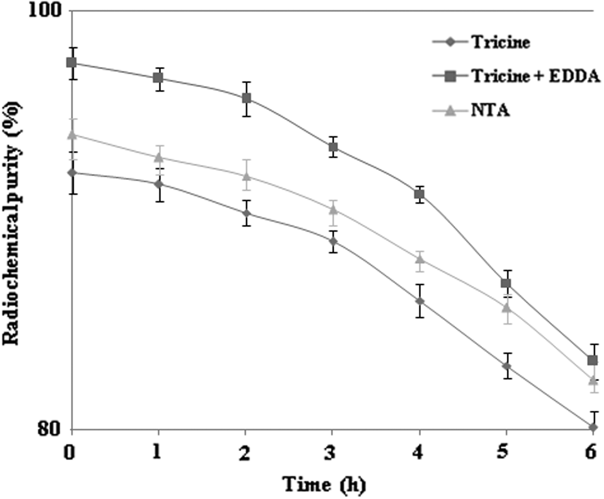
Radiochemical purity of 99mTc–N-HYNIC–BZMB using different concentrations of coligands. NTA, nicotinic acid.
Stability and partition coefficient
99mTc–N-HYNIC–BZMB prepared by using three different coligands has indicated a stable profile in L-cysteine and in serum at different intervals. At room temperature, freshly prepared 99mTc–N-HYNIC–BZMB has exhibited stable behavior in cysteine with an overall decay of 40.90% up to 16 h. In serum at 37°C, a similar trailing pattern with 26.95% decay was seen. A combined stability profile of 99mTc–N-HYNIC–BZMB in cysteine and serum is shown in Figure 3. The partition coefficient (log p) value measured in n-octanol and PB at pH 7.4 was −2.211 ± 0.032, indicating lipholicity.
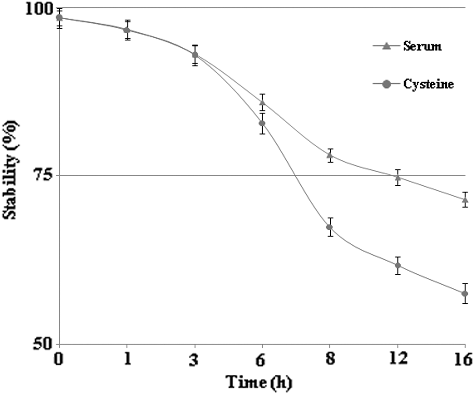
In vitro stability of labeled BZMB in cysteine and serum.
In vitro binding
In vitro binding assay of 99mTc–N-HYNIC–BZMB with poor metastatic SKOV-3 and high metastatic SKOV-3.ip1 demonstrated that the labeled conjugate maintained its integrity to target highly metastatic cancer cell lines compared with less metastatic cancer cell lines. A decrease (∼75%) in the in vitro binding capacity of labeled conjugate was observed with the addition of excess unlabeled BZMB compared with labeled BZMB. The in vitro binding capacity of 99mTc–N-HYNIC–BZMB with SKOV-3 and SKOV-3.ip1 is shown in Figure 4.
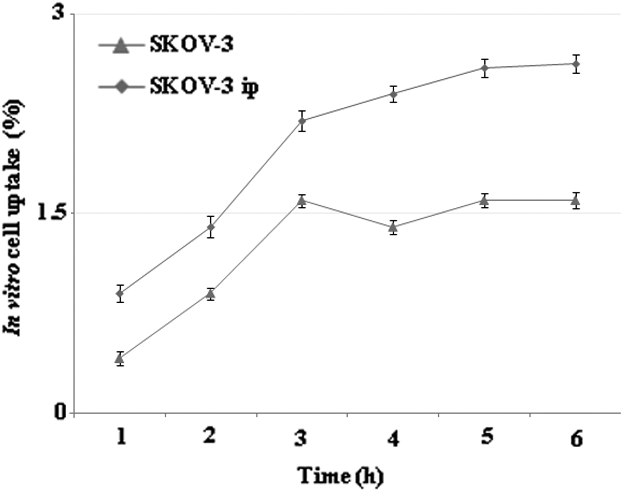
In vitro binding capacity of 99mTc–N-HYNIC–BZMB with SKOV-3 and SKOV-3.ip1.
Biodistribution
Biodistribution activities of 99mTc–N-HYNIC–BZMB in group A, B, and C WRs are summarized in Table 1. Initially, it was observed that the level of labeled conjugated BZMB was much higher in blood in all groups compared with the stomach, muscles, bones, and intestines. In all other tissues, except tumors, relatively low uptake was observed in all animals and minor uptake in the stomach confirmed in vivo stability of 99mTc–N-HYNIC–BZMB. The level of activity in the liver decreased to a minimum level of 3.79% ± 1.40%, 3.82% ± 1.24%, and 3.97% ± 1.44% after 4 h in WRs of groups A, B, and C, respectively. In kidneys, a similar trailing pattern was observed, and the retention of activity was 2.65% ± 0.99%, 2.72% ± 1.12%, and 2.86% ± 1.00%, respectively, after 4 h. Disappearance of activity from the liver and kidneys indicated clearance of the labeled conjugated BZMB from hepatobiliary and urinary systems. The amount of activity detected in the tumor was initially 4.83% ± 0.50%, which went up to 5.69% ± 1.86% after 4 h, in group A. However, in group C, the activity detected in the tumor was 1.65% ± 0.22% after 4 h. The reduced activity observed in group C animals was due to administration of unlabeled BZMB. The target (tumor)-to-nontarget (blood) (T/NT) ratio determined at different intervals was 0.39, 0.56, and 1.48. The tumor-to-blood and tumor-to-muscle ratios reported by the authors are in close correlation with the work reported by other groups. 13,14
PI, post injection; WR, Wistar rat.
Rabbit scintigraphy
The whole-body planar images obtained at 15, 30, 60, and 90 min after intravenous administration of 99mTc–N-HYNIC–BZMB are shown in Figure 5A–D. The clear visuals of kidneys, bladder, and liver validate that the labeled BZMB was excreted through hepatobiliary and urinary routes. Scintigraphically, the target site (SKOV-3.ip1) was not clear after 30 min of I.V., but could be seen clearly after 60 and 90 min.

Whole-body scintigraphy of male AR model
Discussion
BZMB is a recombinant humanized mAb against VEGF that is intended to inhibit VEGF function in vascular endothelial cells and thereby control tumor angiogenesis, on which tumors mainly depend for growth and metastasis. 19,20 The effectiveness of BZMB posed it as a promising drug for therapeutic treatment of lung, renal, ovarian, and brain cancers. 8 As a radioimmunoimaging (RII) and radioimmunotherapeutic (RIT) agent, its tagging with 99mTc, 188Re, 177Lu, 89Zr, and 111In has been reported using different methodologies. 21 –24 Recent findings signified the potential of labeled BZMB as a specific RII and RIT agent. However, the associated problems such as appearance of unwanted radionuclide species were not investigated further.
In this study, BZMB was coupled with N-HYNIC, followed by labeling with 99mTc using three different coligands, with the prime intention to design an ideal RII agent. As HYNIC cannot complete the coordination sphere of 99mTc by itself, tricine, tricine and EDDA (mixture), and nicotinic acid were used as coligands. With the exception of mixed tricine and EDDA, the rest of the coligands relatively present an unstable profile. There are reports in the literature that different coligands can significantly alter the radiobiological characteristics of labeled HYNIC conjugates. 12,25 99mTc–N-HYNIC–BZMB was synthesized using the mixture of tricine and EDDA, thus showing relatively high purity and in vitro stability in cysteine and serum compared with tricine and nicotinic acid. In an instant trial, high HYNIC-to-BZMB ratios were avoided so as to protect the antigen binding capacity of BZMB from reduction.
In vitro binding assay of 99mTc–N-HYNIC–BZMB with poor metastatic SKOV-3 and high metastatic SKOV-3.ip1 showed targeted in vitro binding. However, labeled BZMB maintains high in vitro binding affinity with SKOV-3.ip1, but not with SKOV-3. Lowering of specific uptake by adding unlabeled BZMB indicated specificity of BZMB to VEGF.
Biodistribution of 99mTc–N-HYNIC–BZMB in model rats indicated high in vivo uptake in the tumor compared with other organs and was reported as slightly high by others. 26,27 Whole-body planar images with labeled BZMB using tricine and EDDA as mixed coligands in human ovarian tumor xenografts in rabbits indicated that it could be a choice for in vivo tracing of high metastatic sites. Beside blood, normal activity distribution was seen at different time points. Uptake in the tumor and kidney was high compared with the other reported 99mTc-labeled HYNIC conjugates. 19,20,28 –30 More efforts are required to minimize the level of activity in blood so as to lessen the hyperglance of background in targeted scintigraphy.
Conclusion
In this study, radiolabeling of conjugated BZMB with 99mTc was assessed as a specific ovarian tumor-imaging agent. Radiobiological characteristics and scintigraphy of 99mTc–N-HYNIC–BZMB prepared by using a mixture of coligands demonstrated its potential to trace in vivo high metastatic sites. However, decrease in higher accumulation of 99mTc–N-HYNIC–BZMB in some nontarget organs needs further studies.
Footnotes
Disclosure Statement
No competing financial interests exist.