
Abstract
Aim:
To compare the uptake of 11C-deuterodeprenyl (11C-DED) and 11C-methionine (11C-MET) in three human glioma cell lines and study the relationship with glial fibrillary acid protein (GFAP) and monoamine oxidase B (MAO B) expression. 11C-DED is used in positron emission tomography imaging as a marker of astrocytosis in various central nervous system pathologies. It binds irreversibly to MAO B, a glial dimeric enzyme with increased activity in some neurological pathologies.
Materials and Methods:
Binding and internalization studies of 11C-MET and 11C-DED were performed in astrocytoma grade III, glioblastoma grade IV, and radio-resistant glioblastoma grade IV cells. Immunofluorescence was used.
Results:
11C-MET specific activity bound to membrane was 9.0%–11.1% and that internalized was 88.9%–91.0%. 11C-DED specific activity bound to membrane was 34.8%–58.0% and that internalized was 38.7%–65.2%. Immunocytochemistry revealed GFAP and MAO B expression.
Conclusions:
The expression of MAO B measured by 11C-DED uptake or immunocytochemistry was not significantly different in grade III or IV cells. The GFAP signal was higher for grade IV compared to grade III. 11C-MET uptake was high in all the tumor cells. 11C-DED is a dopamine analogue and the transport across cell membranes is expected to be mediated by DAT receptors present in astrocytes. Reactive astrocytes surround tumor lesions; so the authors suggest that the 11C-DED uptake might be caused by the reactive astrocytosis and not by MAO B expression in tumor cells.
Introduction
Astrogliomas are highly heterogeneous tumors, classified by WHO as grade I, II, III (anaplastic astrocytoma), and IV (glioblastoma).
Positron emission tomography (PET) imaging is a minimally invasive technique, which employs radioactive tracers to target cellular proliferation, hypoxia, and metabolism. Several radiotracers targeting amino acid transport, protein synthesis, and tumor hypoxia have been suggested as useful for glioma grading. 1,2
Amino acid cellular uptake is increased in gliomas and many amino acid analogues are used as targets for imaging. 11C-methionine (11C-MET) is the best characterized amino acid radiotracer for imaging of brain tumors. 3 –5 11 C-MET uptake correlates with tumor grade 6 –9 to some extent, but there is a high rate of variability and overlapping among different stages, which does not allow its use for accurate noninvasive grading. 10 Nevertheless, many imaging studies report an increase in 11C-MET uptake in high-grade gliomas compared to low-grade gliomas. 1,8,11
11C-deuterodeprenyl (11C-DED) is used in PET imaging as a marker of astrocytosis in various central nervous system (CNS) pathologies. It binds irreversibly to monoamine oxidase B (MAO B), a glial dimeric enzyme with increased activity in some neurological pathologies such as Alzheimer's disease, Creutzfeldt-Jakob disease, temporal lobe epilepsy, Parkinson, and amyotrophic lateral sclerosis. 12 –16 MAO B function is to degrade amines like dopamine in mice and human astrocytes. 17,18 The catalytic activity of MAO B resulting in peroxide production and oxidative damage is another process related to glioma grade. 19,20 The enzyme activity was found to be higher in astrogliomas when compared to meningiomas or postmortem tissue samples from individuals without CNS disease. However, no significant difference in MAO B activity was found among the different astrocytoma grade tissue samples. 21,22 In other studies, the enzyme level of protein expression was higher in gliomas compared to normal astrocytes and other cells of the brain. 23,24 In this context, the study of 11C-DED uptake by human glioma cells is of interest because of its potential use as a marker of tumor gradation.
Glial fibrillary acid protein (GFAP) is an intermediate filament protein present in astrocytes, radial glia, and stellate cells. It is commonly used for the detection of reactive astrocytes and the identification and diagnosis of astrocytic tumors. 22,25 Astrocytomas express other astrocyte intermediate filament proteins such as nestin, vimentin, and synemin. 26 Overexpression and rearrangement of all cytoskeletal proteins are now believed to be the response to cellular stress. High-invasive astrocytomas have shown to express higher levels of nestin and GFAPδ, the isoform most expressed in stem and glioma cells. Stoichiometry of intermediate filament proteins in grade IV glioma is different from that in nontumor astrocytes. 27 –29 However, in grade IV gliomas, a decrease in GFAP expression has been found and attributed to the dedifferentiated state of these cells. 30 –32 Accordingly, detection of GFAP is not a necessary condition to classify grade IV glioma because they are histologically characterized as GFAP-negative tumors. Therefore, GFAP by itself is not a sufficient marker to track glioma development.
Glioma cell lines are homogeneous models that allow a more accurate knowledge of the tumor cell biology. In vitro cell-based studies are useful to analyze molecular mechanisms of disease and for the identification of possible markers to be tested in vivo in animal models.
Differences in uptake between the cell lines could result in the possibility to grade these tumors in patients; therefore; the aim of this work was to compare the uptake of 11C-DED and 11C-MET in glioma grade III and IV cell lines. Immunocytochemistry was performed to relate the MAO B and the GFAP expression with the 11C-DED uptake.
Materials
All chemicals and solvents were of analytical grade (Merck, Sigma-Aldrich, Carlo Erba). The following reagents were used for the synthesis and quality control: l-deprenyl-D2 hydrochloride (DED.HCl, analytical standard; Pharma Synth AS, Estonia) and desmethyl-L-deprenyl-D2 hydrochloride (nor-DED.HCl, radiosynthetic precursor; Pharma Synth AS), and l-homocysteine thiolactone hydrochloride (radiosynthetic precursor; ABX) and DL-methionine (analytical standard; Sigma Aldrich). Sep-Pak C18 light cartridge and 0.2 μm hydrophilic sterilizing filters were purchased from Waters. The following items were used for chromatography: an HPLC semipreparative column (M-N Nucleosil Nautilus® C18 100-5VP 250 × 10 mm) and an HPLC analytical column (M-N EC 250/4.6 mm Nucleodur 100-5 C18ec).
Instruments
11C-DED and 11C-MET radiosynthesis were performed using the TRACERlab FXC PRO module (GE Healthcare). Both syntheses used 11C-CO2 produced in a PET Trace 16.5 MeV cyclotron (GE Healthcare). A high-performance target was used for 11C-CO2 production. Target content was a mixture of N2 and 1.0% O2 (Praxair). HPLC analyses were performed with a Shimadzu UFLC equipped with diode array and gamma detectors.
A Gamma Counter (Ortec) was used to perform activity measurements.
A Confocal Microscope (Olympus FV300) was employed to image cell cultures immunostained against MAO B and GFAP.
Cell lines
Cells were obtained from the American Type Culture Collection Cell: LN229 (ATCC®CRL-2611™) glioblastoma multiforme Grade IV, M059K (ATCC®CRL-2365™) glioblastoma multiforme Grade IV radio resistant, and SW1783 (ATCC®HTB-13™) astrocytoma Grade III.
Antibodies
The following primary antibodies were used: anti-MAO B (Abcam 88510) and anti-GFAP (Sigma G9363). The following secondary antibodies were used: Alexa Fluor 546 goat anti-mouse IgG (Invitrogen A21123) and Alexa Fluor 488 goat anti-rabbit IgG (Invitrogen A21206), respectively.
Methods
Radiolabeling
11C-DED was synthesized according to the method previously reported by this group. 33 11 C-CH3OTf was bubbled into the organic precursor solution to react with 1.0 mg of precursor nor-DED.HCl in 0.35 mL butanone (MEK), 1 minute at 80°C. Purification was performed using a semipreparative HPLC (MeCN: AcONH4 0.1 M; 60:40; v/v) and a second purification step through C18-SPE cartridge (Sep-Pak® C18 light). The product was eluted with 1 mL of absolute ethanol and it was finally formulated in a 0.9% NaCl solution and filter sterilized.
11C-MET was synthesized according to the solid-phase 11C-methylation of the precursor l-homocysteine thiolactone on a C18 column, using 11C-CH3I as the methylating agent. 34 11 C-CH3I was transferred under a stream of helium into a C18-SPE cartridge (Sep-Pak C18 light). The cartridge was previously loaded with a solution of l-homocysteine thiolactone hydrochloride (2 mg) dissolved in 1 mL of NaOH:ethanol (50/50; v/v). 11C-MET was eluted with 5.5 mL of NaH2PO4 0.05 M buffer and collected in a vial containing 4.4 mL of 0.9% NaCl solution and filter sterilized.
Radiochemical purity was determined using radio-HPLC on a C18 column. The mobile phase for 11C-DED analysis consisted of TFA 0.1% and MeCN (75:25; v/v), with a flow rate of 1.5 mL/min. The mobile phase for 11C-MET analysis consisted of KH2PO4 1.4 g/L (A) and MeCN (B) as mobile phase with the following gradient: 0–11 minutes. 1%–3% (B) and a flow rate of 1.0 mL/min.
Biological studies
Cells were cultured in Dulbecco's modified Eagle's medium high glucose (DMEM; Capricorn Scientific) supplemented with 10% (v/v) fetal bovine serum (FBS; Gibco), antibiotic/antimycotic solution.
All cells were grown to confluence in cell culture T-flasks in a humidified atmosphere of 5% CO2/95% air at 37°C. The cells were passaged when required.
Binding and internalization assays
These assays were performed as previously described. 35 Each cell line was used in at least three independent experiments. Briefly, cells were seeded at 300,000/well in six-well plates 2 days before the experiment (one to three plates each time). The day of the experiment, the cells were washed twice with ice-cold phosphate-buffered saline (PBS) and 1.2 mL/well DMEM/1% FBS was replaced. Half of the wells were blocked with 150 μL cold methionine or cold deprenyl at high excess and 150 μL PBS/bovine serum albumin (BSA) 0.5% was added to nonblocked wells. Next, all the wells were added 150 μL of the respective radiotracer (1–2.5 MBq/well), followed by 40 minutes of incubation at 37°C.
Incubation was stopped by addition of ice-cold PBS. Then, two 5-minute washes with 1 mL ice-cold glycine acid buffer (0.05 M glycine/0.1 M NaCl, pH 3) were performed and the radiotracer membrane-bound fraction was collected. After that, the cells were lysed with two washes of 1 mL 0.1 N NaOH and the internalized activity was measured in a Gamma counter. Specific membrane bound and specific internalized activity was calculated by subtraction of nonspecific activity measured in the blocked wells to the total activity measured in the nonblocked wells.
Immunocytochemistry
Each cell line was directly seeded on 35 mm Petri dishes and cultured until 80% confluence. Then, cells were washed with PBS and fixed with 4% paraformaldehyde (in 10 mM PBS, pH 7.2) during 30 minutes at room temperature. Permeabilization was performed with Triton 0.1% for 20 minutes. After that, two 5-minute PBS washes were done, followed by a 1-hour incubation with 5% BSA blocking solution. Then, cells were incubated overnight at 4°C in a wet chamber with a mouse anti-MAO B antibody (1:100 dilution) together with a rabbit anti-GFAP (1:400 dilution) antibody. The next day, primary antibodies were removed, cells washed, and the secondary antibodies (1:800 anti-mouse Alexa Fluor 546 and 1:800 anti-rabbit Alexa Fluor 488) added and incubated for 90 minutes at room temperature with mild agitation. After that, cells were rinsed with PBS several times and then mounted with 20 μL of 50% glycerol containing 1 μg/mL of Hoechst 33342. 2048 × 2048 images were obtained in a confocal microscope attached with 405, 488, and 543 nm lasers. All acquisition parameters were maintained when imaging all cell lines. Fluorescence quantification was performed with Image J software.
Results and Discussion
11C-DED and 11C-MET were obtained with a radiochemical purity higher than 90%, as measured by radio-HPLC. Both radiotracer retention times were similar to their respective reference cold standard.
11C-MET was used as the reference radiopharmaceutical due to previous reports about the increased amino acid metabolism in high-grade gliomas. 1 No significant difference was observed between the membrane-bound activity in nonblocked and blocked cells (nonblocked/blocked ratio: 1.1–1.4). For the internalized activity, the ratio between nonblocked and blocked cells was from 1.8 to 2.6 (Fig. 1). Nonspecific bound and nonspecific internalized activity—activity measured in blocked wells—was subtracted from the total activity bound or internalized in nonblocked wells to correct the data and estimate the activity specifically bound and internalized. The specific activity bound and internalized was calculated for each cell line (Fig. 2). The specific internalized activity was 8–10 times the specific membrane-bound activity (Table 1).
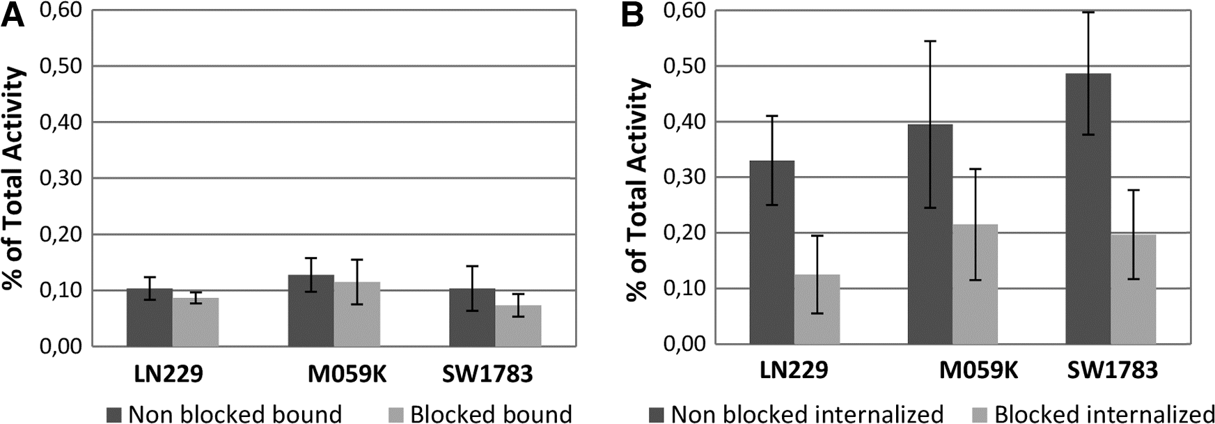
11C-MET uptake of blocked and nonblocked:

11C-MET bound and internalized specific activity, obtained as the total activity measured in nonblocked wells minus the activity measured in the blocked wells. The results are expressed as mean ± SD.
11C-Methionine Nonblocked/Blocked Ratio
All three cell lines internalized the radiopharmaceutical at a similar rate. No difference in the 11C-MET uptake in relationship with tumor grade was observed, reinforcing the information that this radiopharmaceutical cannot differentiate grade III from grade IV gliomas. 9 The cells were effectively blocked by cold methionine, confirming the specificity of 11C-MET (Fig. 1B).
11C-DED membrane-bound activity in nonblocked wells was 1.8–2.3 times higher than that in blocked ones, confirming also the specificity of this radiopharmaceutical (Fig. 3 and Table 2).
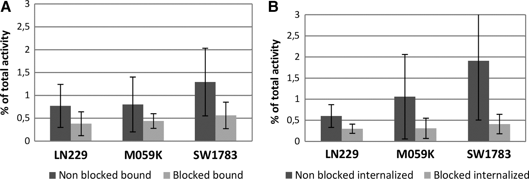
11C-DED uptake of blocked and nonblocked
11C-Deuterodeprenyl Nonblocked/Blocked Ratio
The internalized activity in nonblocked wells was 2.0–4.7 times higher than the one observed in blocked wells. These data revealed the high specificity of the radiopharmaceutical for its molecular target MAO B.
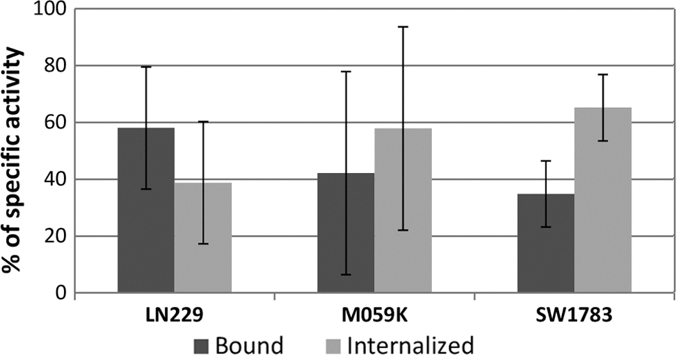
The specific internalized and bound to membrane activity was calculated in the same way as for 11C-MET. The ratio between specific internalized activity/specific bound activity was 0.6–1.9 (Fig. 4, Table 2). These results did not demonstrate significant differences in the 11C-DED specific activity bound or internalized in all cell lines, considering the high variability interassay.
11C-DED bound and internalized specific activity, obtained as the total activity measured in nonblocked wells minus the activity measured in the blocked wells. The results are expressed as mean ± SD.
Published data are controversial concerning the MAO B activity in astrogliomas. It has been reported that MAO B “level” per cell is higher in gliomas with respect to normal astrocytes. 23,24 Gabilondo et al. and Callado et al. have reported an increase in the MAO B activity, in glioma tissue samples compared to menigiomas or nontumoral tissue. 21,22 The authors tried to detect differences in MAO B expression in the above-described cell lines.
None of the three cell lines showed a difference in the uptake of 11C-DED, indicating comparable expression of MAO B in the grade III and IV cells. The authors could not verify an increase of enzyme expression depending on grade. 11C-DED has been described as a marker for reactive astrocytosis. Reactive astrocytes surround tumor lesions 36 ; so the authors suggest that the 11C-DED uptake might be caused by the reactive astrocytosis and not by MAO B expression in tumor cells.
The fact that 11C-MET internalization assays revealed low intravariability and inter variability, compared with the 11C-DED results, raises further questions to consider. L-methionine influx across the cell membrane is mediated by the Na+-independent LAT 1 system, which has a high affinity for neutral amino acids, including methionine. In vivo studies have shown that LAT1 is overexpressed in gliomas. 37 PET studies with 11C-MET differentiate low-grade from high-grade astrogliomas because an increased amino acid uptake is required for protein synthesis. 11 11 C-DED is a dopamine analogue and the transport across cell membranes is expected to be mediated by DAT receptors. However, this mechanism is not well studied in astrocytes. Naganuma et al. have proposed the existence of a low-affinity monoamine transporter system in a human astrocytoma cell line. 38 Besides, 11C-DED is a MAO B inhibitor probably located in the mitochondrial intermembrane, but this has only been reported in rat liver tissue. 19 11 C-DED uptake by astroglioma cells seems to be less direct compared with the 11C-MET uptake. Part of the radiopharmaceutical remained bound to membrane and a part reached the cytoplasm.
Immunocytochemistry
Immunocytochemistry was performed to confirm the presence of the astrocyte molecular marker GFAP and the enzyme MAO B (Fig. 5). The three cell lines were positive for both markers. The GFAP signal was higher for grade IV and radio-resistant glioblastoma cell lines. MAO B signal was lower, but similar in all cell lines (Fig. 6).
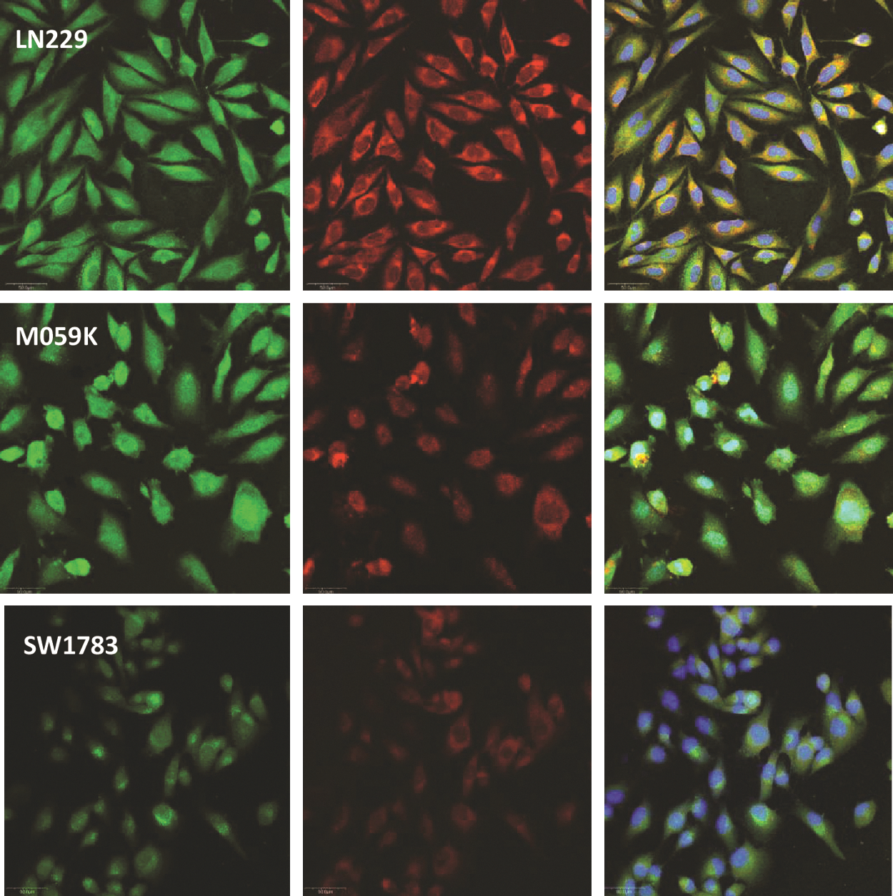
Confocal photographs showing the expression of GFAP (green) and MAO B (red) in the glioma cell lines analyzed by immunocytochemistry. Images on the right show both markers merged together with the nuclear fluorescent marker Hoechst. Calibration = 50 μm. GFAP, glial fibrillary acid protein; MAO B, monoamine oxidase B.
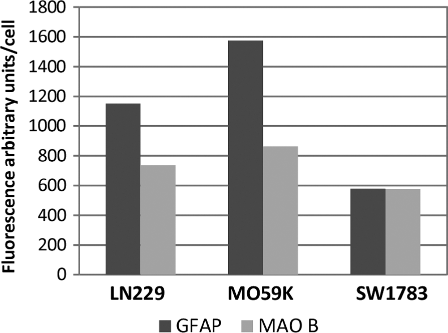
Immunocytochemistry quantification. Fluorescence in arbitrary units/number of cells for each cell line.
Conclusions
The authors present herein a comparison of the uptake of two known PET tracers (11C-DED and 11C-MET) applied to three different human glioma cell monolayers. GFAP and MAO B positivity were determined by immunocytochemistry. 11C-DED and 11C-MET uptake did not reveal differences between grade III and IV gliomas cells. MAO B expression was also similar in all cell lines assayed. GFAP signal was higher for grade IV and rRG compared to grade III. These results suggest that the high variability in uptake of 11C-DED in patients with gliomas might be caused by the reactive astrocytosis present in these lesions more than an increased uptake in tumor cells.
Footnotes
Authors' Contributions
Elena Vasilskis: participated in the study design, performed the experiments, and drafted the article; Ingrid Kreimerman: synthesis and quality control of the radiopharmaceutical and contributed to drafting the article in relation to these sections; Silvia Olivera: collaborated with the immunocytochemistry studies and contributed with the revision of that section; Eduardo Savio and Henry Engler: participated in the study design, discussion of the results, and revision of the article.
Disclosure Statement
There are no existing financial conflicts.