
Abstract
Background and Objective:
There is a high incidence of nasopharyngeal carcinoma (NPC), malignant head and neck tumors, in southern China. Radioresistance is the main cause affecting the efficacy of NPC treatments. The POLG gene particularly plays an important role in radiation-induced damage repair. In this study, the authors established RNAi CNE-1 and CNE-2 knockdown in two NPC cell lines to observe whether this gene affects the radiosensitivity of NPC cells.
Materials and Methods:
Four short hairpin RNA (shRNA) expression plasmids targeting POLG gene were constructed and transfected into the NPC cell lines CNE-1 and CNE-2. Screening was performed to evaluate the stable expression of cloned cells, which were named CNE-1/POLG-shRNA1, CNE-1/POLG-shRNA2, CNE-2/POLG-shRNA1, and CNE-2/POLG-shRNA2. The negative controls CNE-1/Neg-shRNA and CNE-2/Neg-shRNA were additionally used. The MTT method, flow cytometry, clone formation analysis, cell migration, and other experimental methods were employed to verify changes in the radiosensitivity of the NPC cells.
Results:
Fluorescent quantitative PCR and Western blot confirmed the downregulation of the PLOG gene through diminished PLOG messenger RNA and protein levels. Consequently, the authors report the stable knockdown of the POLG gene in an NPC model. Dose-dependent radiation exposure of POLG inhibited NPC cell growth and increased apoptosis compared with control cells (p < 0.01), as demonstrated through colony formation assay and flow cytometry. Functional assays indicated that knockdown of the POLG in CNE-1 and CNE-2 cells remarkably reduced cell viability and proliferation. Specifically, POLG knockdown led to G1 phase arrest and apoptosis.
Conclusions:
Overall, the authors conclude that POLG downregulation alters the radiosensitivity of NPC cells, indicating that the gene is likely involved in conferring the radiation response of the cells. In addition, findings in this study suggest a novel role for POLG as a potential predictive marker for NPC radiotherapy efficiency. POLG gene can be used as a potential clinical target to effectively improve the radiosensitivity of NPC.
Introduction
There is a high incidence of nasopharyngeal carcinoma (NPC), malignant head and neck tumors, in southern China. Radiotherapy is the current standard therapy, 1 although tumor resistance to this treatment does occur and can reduce the 5-year patient survival rate dramatically. 2 Owing to this, investigation is urgently needed to elucidate the mechanisms of NPC radioresistance. Mitochondrial DNA polymerase γ, POLG, exists exclusively in the vertebrate and is involved in mitochondrial DNA (mtDNA) replication and repair. MtDNA damage repair plays an important role in maintaining the integrity of mitochondrial function. The POLG gene plays an important role in the repair of radiation-induced damage. The purpose of this research was to elucidate the role of POLG gene in radiation resistance of NPC. The regulatory mechanisms of POLG were additionally explored.
Materials and Methods
Cell culture and cell lines
Two human NPC cell lines, CNE-1 and CNE-2, came from the Fujian Province Tumor Research Institute. Cells were cultured in RPMI-1640 Medium (Hyclone), which contained 10% of FBS (PANSera) and 0.5% of penicillin–streptomycin sulfate.
Short hairpin RNA transfection
Transfection was performed as described in the literature. 3 POLG short hairpin RNA (shRNA) was created and transfected into NPC cells to inhibit POLG expression. POLG shRNA, or scramble control shRNA (Genechem, Shanghai, China), was transfected using Lipofectamine 2000 (Invitrogen) as a carrier at a 1:1 ratio.
Quantitative real-time PCR
Real-time PCR (RT-PCR) was performed as the method of the article. 3 Quantitative RT-PCR (qRT-PCR) was used to detect POLG expression. Cells were washed twice in cold phosphate buffered saline before being harvested with TRizol (Sangon, Shanghai, China). A NanoDrop Lite spectrophotometer (Thermo Scientific) was used to measure the total RNA concentration and purity.
For POLG detection, reverse transcription was performed using an ImProm-II Transcription System Kit (Promega). PCR mixtures containing SYBR Green PCR Master Mix (Promega) and gene expression assays were used according to the manufacturer's protocols (ABI 7500; Applied Biosystems) with the 7500 system software. qRT-PCR was performed on the ABI 7500 Real-Time PCR detection system (ABI) and the procedure was performed at 95°C for 5 min followed by 40 cycles at 95°C (15 s) and annealing/extension at 60°C (1 min). All reactions were performed in triplicates. Data were analyzed using the 2−ΔΔCt method for evaluation of relative gene expression change. GAPDH served to normalize POLG values.
Western blotting
The proteins were extracted, the expression levels of POLG, E-cadherin, N-cadherin, and apoptosis marker were detected by Western blot as described previously. 4 The primary antibodies used were rabbit monoclonal to DNA polymerase-γ (1:1000; abcam, USA), rabbit monoclonal to E-cadherin (1:1000; Cell Signaling, USA), rabbit monoclonal to N-cadherin (1:1000; Cell Signaling), and Cleaved Caspase Antibody Sample Kit (1:1000, Cell Signaling). GAPDH (1:1000; Biolegend, USA) protein levels were used as a control.
Clonogenic assay
A clonogenic assay was used to detect cell growth after treatment with irradiation (IR). The method was performed as described in the literature. 5 NPC cells were cultured in six plates after IR. After 14 d, cell colonies were fixed with methanol, then stained with 0.1% crystal violet, and the number of colonies was counted by ImmunoSpot S5 (CTL, USA). The authors considered a colony as consisting of 50 or more cells as a standard.
Cell cycle and cell apoptosis analysis
Cells were collected 48 h after 6 Gy IR exposure. Cell cycle and cell apoptosis were analyzed by flow cytometry as described in the literature. 6 Samples were immediately analyzed by a FACScan flow cytometer (BD C6; USA). Three independent experiments were conducted and results were analyzed using FlowJo software.
Irradiation
Radiation was performed using a Varian 600 CD machine (Varian, USA) in the department of radiotheraphy at the First Affiliated Hospital of Fujian Medical University. Cells were suspended in cell medium before IR. Their radiation rate was 500 rads/min.
Cell proliferation assay
A cell proliferation assay was used to evaluate cell viability using thiazolyl blue tetrazolium bromide (MTT; Sigma-Aldrich). NPC cells were seeded onto 96-well plates after IR in 200 μL of RPMI-1640. After the indicated incubation period, MTT (5 mg/mL) was added, the cells were incubated at 37°C for 4 h, and 10 μL of the solubilization reagent (10% sodium dodecyl sulfate) was added to each well. Spectrophotometric absorbance of the samples was measured at 570 nm with a SpectraMax i3x (Molecular Devices) after an additional 10 min of incubation. The inhibition ratios of cell survival were calculated as 100% × N t/N c, where N t is the optical density of the treatment group and N c is the optical density of the control group.
Statistical analysis
Statistical analysis was performed using SPSS statistical software (Chicago). Data are presented as the mean ± standard deviation. Statistical analyses were performed by one-way analysis of variance when there were more than two groups. Two-tailed Student's t-test was used when there were only two groups. Differences between groups were considered to be significant statistically when p < 0.05.
Results
POLG knockdown cell lines were created using shRNA transfection
The authors designed four shRNA (30899, 30900, 30901, and 30902) and a scramble sequence as control to transfect CNE-1 and CNE-2 cells, and chose the best two (shRNA-30900 and shRNA-30901) for the follow-up experiment (Fig. 1A). They named stable cells transfected with 30900, 30901, and control shRNA as POLG-shRNA1, POLG-shRNA2, and Neg-shRNA, respectively. RT-PCR and Western blot were subsequently used to verify the stable, reduced expression of POLG messenger RNA and protein expression (Fig. 1B–E).

Stable POLG knockdown cell lines in CNE-1 and CNE-2 were created.
Knockdown POLG increased the radiosensitivity of NPC cells
This study's data revealed that the survival rates of POLG-shRNA cells were dose and time dependent as observed by assays examining different doses of IR (2, 4, 6, and 8 Gy; Fig. 2A, C) and different time points (1, 2, 3, 4, and 5 d; Fig. 2B, D). POLG-shRNA cells in general exhibited survival rates that were lower than that of Neg-shRNA cells. In addition, colony-forming assays indicated a decreased number of POLG-shRNA cell colonies survived compared with Neg-shRNA cells (Fig. 2E, F). POLG-shRNA cells and Neg-shRNA cells had a similar growth pattern without IR but significant difference after 6 Gy IR.

POLG functions as a radiosensor in the human NPC cell line CNE-1 and CNE-2.
Knockdown of POLG alters cell cycle progression and apoptosis
Flow cytometric analysis was used to elucidate cell cycle and apoptotic pathway between POLG-shRNA and Neg-shRNA cells. The data showed a similar cell cycle distribution in the absence of IR (Fig. 3B, E). Although the number of POLG-shRNA cells in G2/M phase was significantly increased after 6 Gy IR, in turn, the number of POLG-shRNA cells in G1/G0 phase was increased (all p < 0.01; Fig. 3C, F). The same phenomenon can be seen in cell apoptosis; the apoptosis rate was significantly increased after IR (p < 0.001; Fig. 4B, D). The mentioned results suggested that the POLG-shRNA cells increased radiosensitivity when stimulated with IR compared with Neg-shRNA cells.
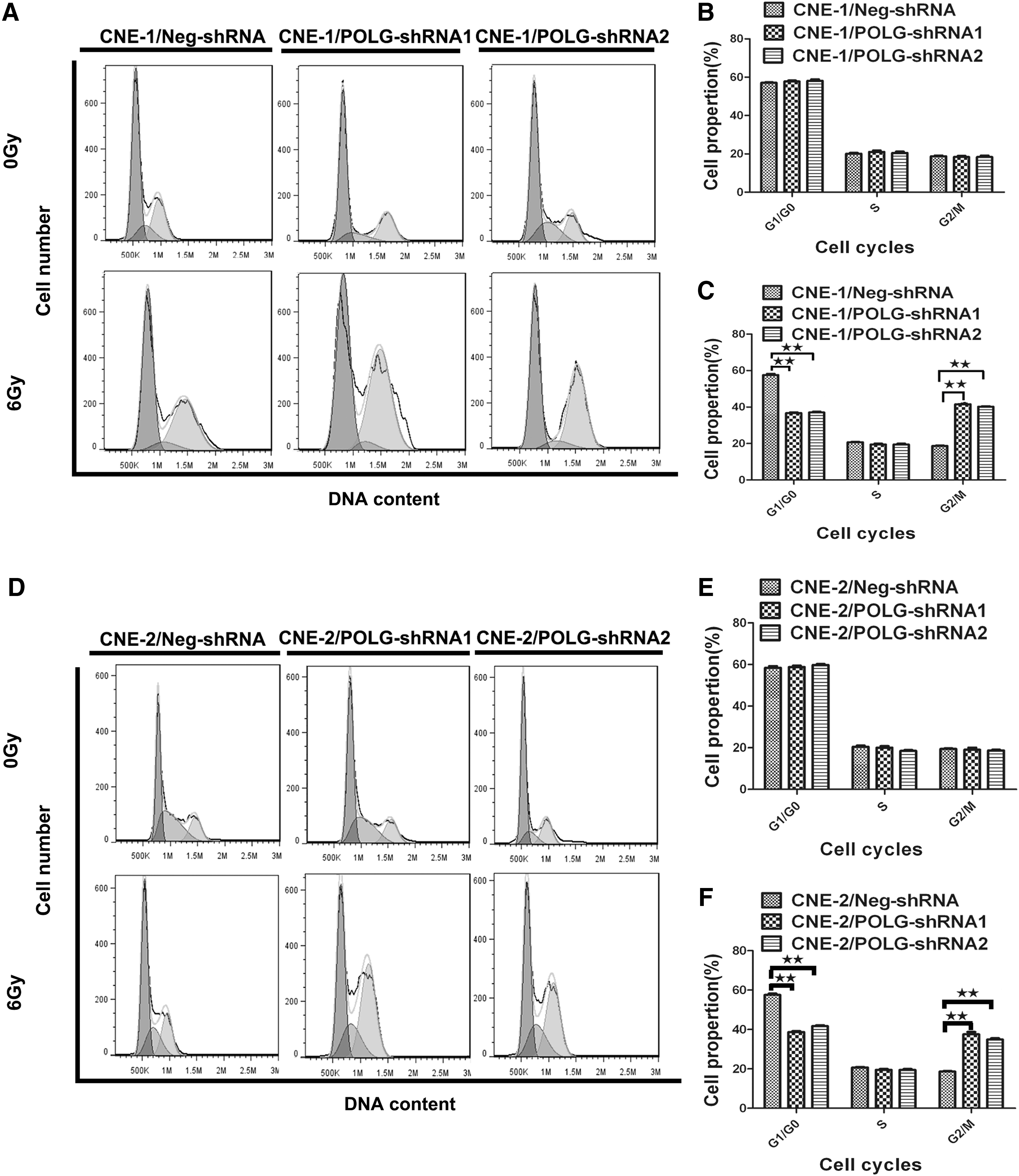
The effect of cell cycle response to POLG knockdown in CNE-1 and CNE-2 cells.

The effect of apoptosis response to POLG knockdown in CNE-1 and CNE-2 cells.
POLG knockdown modified the expression of epithelial–mesenchymal transition and cell apoptosis-related proteins
To further investigate the underlying mechanisms of cell apoptosis and radiosensitivity induced by POLG, several epithelial–mesenchymal transition (EMT) and cell apoptosis-related proteins were detected by Western blot analysis as shown in Figure 5. The expression levels of cleaved caspase-3, cleaved caspase-9, and cleaved PARP, apoptosis markers, were dramatically upregulated in POLG-shRNA cells compared with Neg-shRNA cells after IR exposure (Fig. 5D, H), whereas in the absence of IR, the difference of caspase-3, cleaved caspase-9, and cleaved PARP was not significant (Fig. 5C, G). This result is consistent with that of flow cytometry analysis. POLG-shRNA cells also exhibited a significant increase of E-cadherin and decrease of N-cadherin to Neg-shRNA cells (p < 0.001; Fig. 5C, D, G, H), indicating that the POLG gene may be associated with EMT.
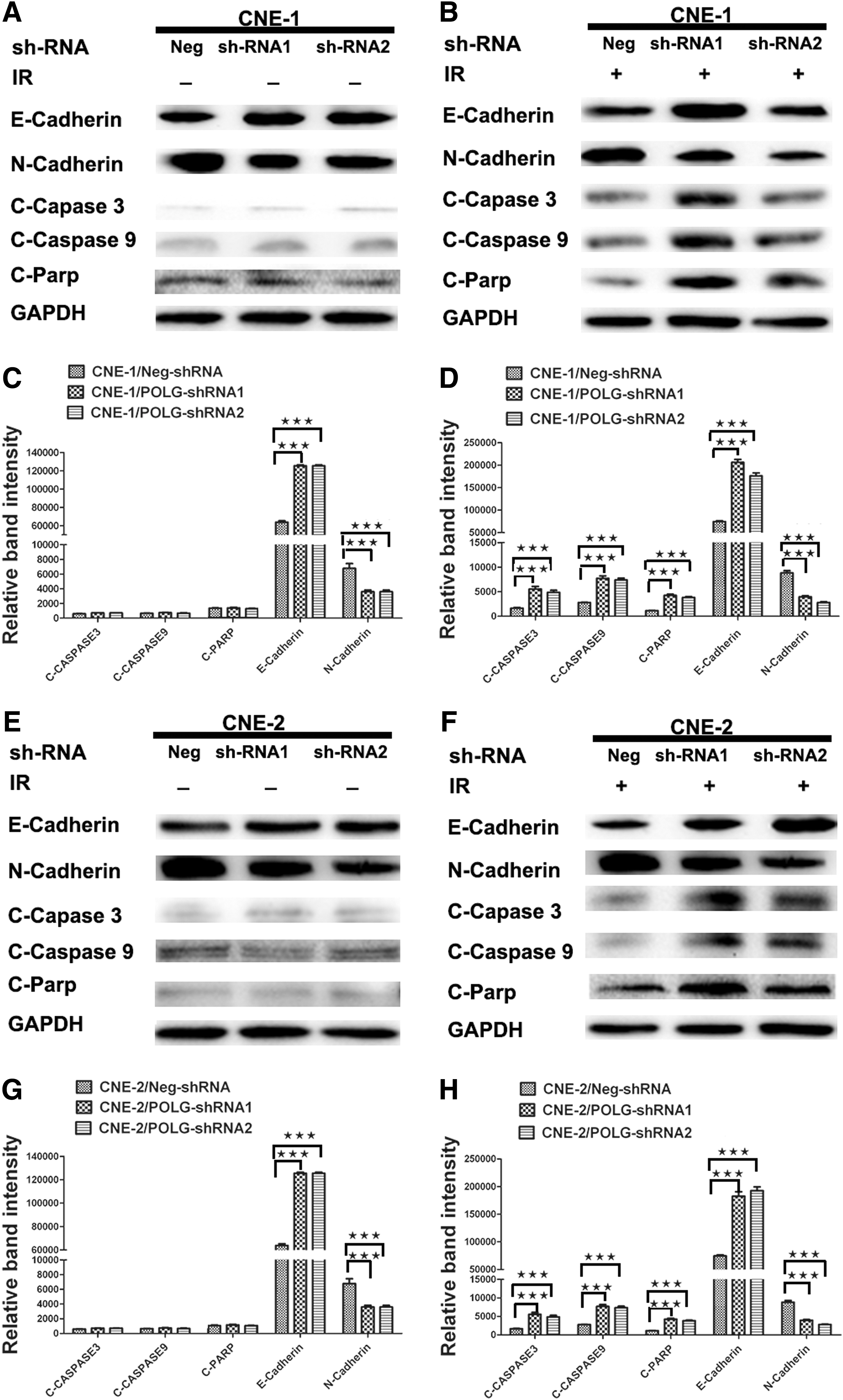
POLG modulates CNE-1 and CNE-2 radioresistance through apoptosis and EMT signaling pathways.
Discussion
Radiotherapy is the preliminary treatment for NPC, but radioresistance often remains an obstacle in NPC clinical management. The development of radioresistance is a complicated process, involving DNA repair genes, cell cycle regulatory and apoptosis genes, and microRNA. Cancer stem cells are considered to be closely related to NPCs with regard to radiation resistance. Results from several other investigations have confirmed that BMI-1, SIXI, Akt1, and miR-195 play key roles in the development of radioresistance in other human cancers. 7 –12 In NPCs, Huang et al. 13 found that Annexin A1 was involved in the p53-mediated radiation response of NPC cells. Zhang et al. 14 found that Twist1 played a significant role in the radioresistance of NPCs by promoting the accumulation of DNA damage repair and inhibition of NPC cell apoptosis. Finally, the Liu et al. 15 group found that miR-324-3p could regulate the radioresistance of NPC cells by targeting WNT2B. These previous reports collectively show the potential role of those genes as biomarkers of radioresistance in NPC cells. 16 –22 This study's findings confirm that POLG knockdown increases the radiosensitivity of CNE-1 cells. This study's data reveal that POLG is involved in processes including cell cycle regulation, cell apoptosis, cell migration, and cell invasion.
The POLG gene encodes the catalytic subunit of DNA polymerase-γ, a protein essential for mtDNA replication and repair. Da Pozzo et al. 23 have reported a link between POLG mutations and a spectrum of clinical phenotypes, resulting in autosomal recessive or dominant mitochondrial diseases. Interestingly, a report by Kasahara et al. 24 suggested that POLG1 variants were significantly more prevalent in patients with bipolar disorder than in healthy controls. This study's data expand the POLG repertoire to reveal that POLG can adjust the radioresistance and radiosensitivity of NPC cells in vitro.
IR is able to incite DNA damage and apoptosis through the generation of reactive oxygen species by radiolytic hydrolysis. 25 Apoptosis plays a key role in the cellular death pathway when exposed to IR, and previous studies have proposed that apoptosis is an important influence on the effectiveness of radiotherapy. 26,27 In this study, POLG knockdown was able to increase radiation-induced apoptosis of CNE-1 and CNE-2 cells. These results suggest that the downregulation of POLG improves NPC radiosensitivity by promoting apoptosis.
It is well known that radiosensitivity is different across cell cycle stages, with cells becoming more sensitive during the G2/M stage, and even more so in the S stage. 27 –29 Cell cycle results of this study show that a high percentage of POLG-shRNA cells were arrested in the G2/M phase after IR. These discoveries denoted that stable downregulation of POLG by shRNA was able to facilitate the arrest of NPC cells with DNA damage in the G2/M phase, thus increasing the radiosensitivity of NPC cells by impacting their cell cycle distribution.
To further elucidate the mechanism of these phenomena, several EMT and cell apoptosis-related proteins such as caspase-3, caspase-9, N-cadherin, and E-cadherin were detected by Western blot. Results reveal that the mechanisms by which cells produce radiation resistance may be related to the EMT process, likely due to cell-acquired stem cell-related properties through EMT, thereby producing radiation resistance. 30 –32 The results show that after IR, cleaved caspase-3 and caspase-9 expression was upregulated in POLG-shRNA cells, whereas N-cadherin expression was downregulated in POLG-shRNA cells.
In conclusion, the results of this study indicated that POLG may make a crucial role in the radiosensitivity of NPCs, and that downregulation of POLG may decrease the radioresistance of CNE-1 and CNE-2 cells by decreasing colony growth, increasing apoptosis, decreasing EMT, and altering cell cycle progression. Therefore, POLG may be a viable candidate for a novel biomarker in the prediction of the radiosensitivity of NPC. Consequently, predicting a patients' response to radiotherapy may allow clinicians to map optimal courses of treatment for patients in an individualized manner in the future. This study marks the first to elucidate a relationship between POLG gene and radiosensitivity of NPC cells in vitro, although this remains to be substantiated in vivo. In the meantime, the intricacies of the molecular mechanisms underlying the role of POLG in NPC radiosensitivity remain unknown and require further investigation in future researches.
Footnotes
Acknowledgment
This study was supported by Fujian Natural Fund Project (Grant No. 2017J01186).
Disclosure Statement
No competing financial interests exist.