
Abstract
Purpose:
The cross-reacting material 197 (CRM197) is a mutation of the diphtheria toxin. The protein of CRM197 was used successfully for the therapy of various tumors in the recent studies. In this study, the recombinant adenoviruses containing the CRM197gene(AdCRM197) were used to enhance the cellar toxicity of gemcitabine in human glioma U87, U251, and H4 cells.
Procedures:
MTT assay and flow cytometric analysis were performed to test the apoptosis of the U87, U251 and H4 cells with the combined treatment of AdCRM197 plus gemcitabine. Western blotting analyses were carried out to detect the cell apoptosis of the mitochondrial pathway. And the xenograft nude mice were used to observe the enhanced antitumor effect of AdCRM197 in vivo.
Results:
AdCRM197 sensitizes human glioma cells to gemcitabine in vitro by the mitochondrial pathway. Tumor volume was inhibited and survival time was prolonged in the U251 or U87 xenografted nude mice with gemcitabine plus AdCRM197. The enhanced antitumor effect of AdCRM197 was also detected by the immunohistochemical analyses and TUNEL staining.
Conclusion:
The authors found that AdCRM197 sensitized the human glioma to gemcitabine not only in vitro but also in vivo. They provide the first evidence that adenovirus-mediated CRM197 may be a potential chemosensitizing agent for the treatment of cancer. The diphtheria toxin is of great toxicity that even one molecule of diphtheria toxin is enough to kill one cell. However, because of the high toxicity, the diphtheria toxin would kill the packing cells when it is being packaged into the recombinant viruses. Therefore, the diphtheria toxin is hard to be used in the gene therapy for virus vectors. The cross-reacting material 197 (CRM197) is a mutation of the diphtheria toxin. Unlike DTA, CRM197 exhibit a weak toxicity. The week toxicity of CRM197 is a good feature for the virus packaging. In the present study, we used a recombinant adenovirus which carried a CRM197 gene (AdCRM197) to enhance the cellar toxicity of gemcitabine in human glioma cells.
Introduction
Gliomas are the very common brain tumors in adult in the United States. 1 They are classified into astrocytic, oligodendroglial, and mixed tumors, according to the morphologic characteristics. 2,3 The most common and most malignant type of glioma are the high-grade gliomas (grades III and IV), which are the fourth deadly tumors of mankind in adults, killing ∼140,000 people in the planet each year. 2 Traditional treatments of the high-grade gliomas are based on surgical resection, radiotherapy, and chemotherapy. Complete surgical resection of the high-grade gliomas is unrealistic due to tiny invisible tumors in the surrounding healthy brain tissues. The radiotherapy on high-grade gliomas is also limited by the side effect of destroying the surrounding normal tissues. The major challenges of chemotherapy are drug resistance and their systemic toxicity. Accordingly, a new strategy should be carried out to improve the antitumor effect of traditional chemotherapy. Chemotherapy is a main treatment in clinical cancer therapy, and adenovirus-mediated gene therapy is a novel treatment and may be a kind of assisted treatment in clinical use. Based on this consideration, the adenovirus-mediated cross-reacting material 197 (CRM197) was used to sensitize human glioma cells to gemcitabine in this report.
The corynebacteriophages, which are the phages infecting the Corynebacterium diphtheriae, encode the diphtheria toxin (DT) gene. 4 DT is composed of two subunits: subunit A and subunit B. The subunit B recognizes the receptors of the target cells; the subunit A is a poisonous part that could kill the cells. The subunit A of diphtheria toxin (DTA) is an attractive and widely used suicide gene in cancer gene therapy. 5 It has been demonstrated that a single molecule of DTA was enough to kill one cell. 6
CRM197 is a mutation of DT. CRM197 is different from DT in a single missense mutation at position 52, substituting glutamic acid for glycine. 7,8 This mutation results in an enzymatically inactive product. 9 As CRM197 possesses the same immunological characteristics with DT, it is often used as a vaccine against DT. In addition, CRM197 can induce T cell-dependent responses, so it is widely used as an immunological adjuvant to enhance the protective effects of the vaccines. 10 –13
CRM197 is often thought to be a nontoxic variant of DT. However, recent studies found that CRM197 is not a nontoxic agent; it showed a mild cellular toxicity. 14 The toxicity of CRM197 is about 10 6 times less compared with DT of wild type. 15 The protein of CRM197 exhibits an experimental antitumor effect on several types of tumors, including breast cancer, oral cancer, ovarian cancer, T cell acute lymphoblastic leukemia, and human U251 glioma cells. 16 –23 The protein of CRM197 even has been used in the treatment of ovarian cancer in human phase I study. 24
Although CRM197 was used successfully for the therapy of various tumors in recent studies, most of the studies used the protein of CRM197. Gene therapy is a new approach for cancer therapy. As an efficient transfer system, adenoviral vector system is an excellent delivery vehicle for cancer gene therapy. In this study, the authors used an adenovirus vector that carried the CRM197 gene to sensitize the glioma cells to gemcitabine. This study provides the first report that adenovirus-mediated CRM197 could be used as a novel chemosensitizing agent in the therapy of human glioma.
Materials and Methods
Cell culture
The human embryonic kidney 293 cells, human glioma cells lines U87, U251, and H4, and human normal astrocyte cell line HA1800 were grown in DMEM, which contains 10% Fetal Bovine Serum (FBS). U251, U87, H4, and HA1800 cells were divided into six groups. Control group (1) nontreated cells; control group (2) AdEmpty-treated cells (10 multiplicity of infection [MOI] of AdEmpty treated the cells for 2 h); AdCRM197 group—AdCRM197-treated cells (10 MOI of AdCRM197 treated the cells for 2 h); gemcitabine group—the cells with a treatment of 0.1 μg/mL gemcitabine; combination group (1) AdEmpty-treated cells with a treatment of 0.1 μg/mL gemcitabine; combination group (2) AdCRM197-treated cells with a treatment of 0.1 μg/mL gemcitabine. The following experiments were carried out 48 h after the above treatments.
Virus production
The plasmid pCite-CRM197 was a kind gift from Dr. Manuel Caruso. The authors amplified the fragment A of the CRM197 gene with the following pairs of primers: forward primer: 5′-GGAGGTACCATGGGCGCTGATGATGTTGTTG-3′(Kpn I); and reverse primer: 5′-CTTCTCGAGTTATGATCGCCTGACACGATTTC-3′(Xho I). The polymerase chain reaction product was inserted into pShuttle-CMV, and pShuttle-CMV-CRM197 was verified by nucleotide sequencing. The linearized (PmeI-digested) pShuttle-CMV-CRM197 plasmid and the plasmid pAdEasy-1 were cotransfected into the Escherichia coli BJ5183 cells. Then, the recombinant adenoviral plasmid Ad-CRM197 was selected by kanamycin and was transfected to E. coli DH5a to produced large amounts of the plasmid. Ad-CRM197 plasmid was digested by Pac I. Then, the linearized plasmid (Pac I digested plasmid) was transferred to AD-293 cells using liposomes to form the adenoviral CRM197. After amplification in the HEK293 cells, lysates of the cells containing the adenoviral CRM197 (AdCRM197) were purified by CsCl gradient ultracentrifugation. The empty adenovirus, which was used as a control, was generated by the same method.
Western blot analysis
The protocol of Western blotting analysis was described previously. 25 The expression COXIV is used as a loading control for the mitochondrial fraction and the expression of β-actin is used as a loading control for the cytosolic fraction. The enhanced chemiluminescence was used to detect the reactivity. The antibody of caspase-3, poly ADP ribose polymerase (PARP), and cleaved caspase-9 was form Cell Signaling Technology, Danvers, MA. The antibody of Bax was from Santa Cruz, CA. The anti-Smac was form Sigma; and antibody of cytochrome c and COXIV was form Molecular Probes, Grand Island, NY.
MTT assay
The authors used MTT assay to measure the proliferation of cells. The cell viability was calculated with the average of five repeated experiments. The proliferation of the untreated cells was defined as 100% cell viability.
Flow cytometric analysis
FITC labeled Annexin V and PI nuclei stain (V-FITC/PI) staining was performed following the instructions of the Annexin V-FITC/PI staining kit (KeyGen BioTECH). Apoptosis rate of the cells was evaluated by the proportion of Annexin V-stained cells and not PI-stained cells.
The treatments of the xenograft mice
The treatments of xenograft mice were performed as described previously with some modifications. 25 Briefly, each female nude mouse was injected with U87 or U251 cells (5 × 10 6 cells) in the right flank by subcutaneous injection. Seven days after inoculation with the tumor cells, the mice were randomly divided into six groups, and each group consisted of five mice. Then the following treatments were performed: (1) Control group 1: mice injected intratumorally with 100 μL Phosphate buffered saline (PBS); (2) Control group 2: mice injected intratumorally with 5 × 10 8 plaque-forming unit (PFU) Adempty twice a week; (3) Gemcitabine group: mice with an intraperitoneal treatment of 100 mg/kg gemcitabine every week for a month; (4) AdCRM197 group: mice injected intratumorally with 5 × 10 8 PFU AdCRM197 twice a week; (5) Gemcitabine plus the empty viruses (EV) group: (2) and (3) combined; and (6) Gemcitabine plus AdCRM197 group: (3) and (4) combined; tumor size = length × width 2 × 0.52. 26 At last, tumor tissues of the mice were collected for the following histopathological experiments. This work was approved by the Animal Care Committee of the institute (ID:SYXK-15-196).
Examination of the tissues
The authors soaked the tissues into 4% formaldehyde. Then, they cut the fixed tissues into 4 μm paraffin sections. Immunohistochemistry of Ki67 was performed with the specimens of the tumors (anti-Ki67 antibody was obtained from Abcam; Cambridge, MA). They counted the number of Ki67-positive cells of 20 randomly chosen fields, and recorded the percent of Ki67-positive cells of the specimens. The authors used the In Situ Cell Death Detection Kit (Roche) for TUNEL staining. The TUNEL-positive cells of 20 randomly chosen fields were calculated by a blind manner. The percentage of TUNEL-positive cells of the specimens was recorded as the apoptotic index.
Side effects observation
To observe the possible side effects in mice treated with the AdCRM197, the authors recorded the weight change, the appetite, and their behavior as described previously. 25 And in the end, tissues of the heart, the liver, the lungs, the spleen, and the kidneys were made into 4 μm paraffin sections. With hematoxylin and eosin staining of the paraffin sections, the pictures were taken under the microscope at 400 × magnification.
Data analysis
All the values in the pictures are represented by mean–standard deviation. The authors used SPSS 17.0 software to analyze the statistics by one-way analysis of variance (ANOVA), and analyze the differences of the values of the two groups by Tukey–Kramer multiple comparison test. The authors used the log-rank test to analyze the statistical significance of the survival curves. p-Value less than 0.05 means significant differences.
Results
AdCRM197 sensitizes human glioma cells to gemcitabine in vitro
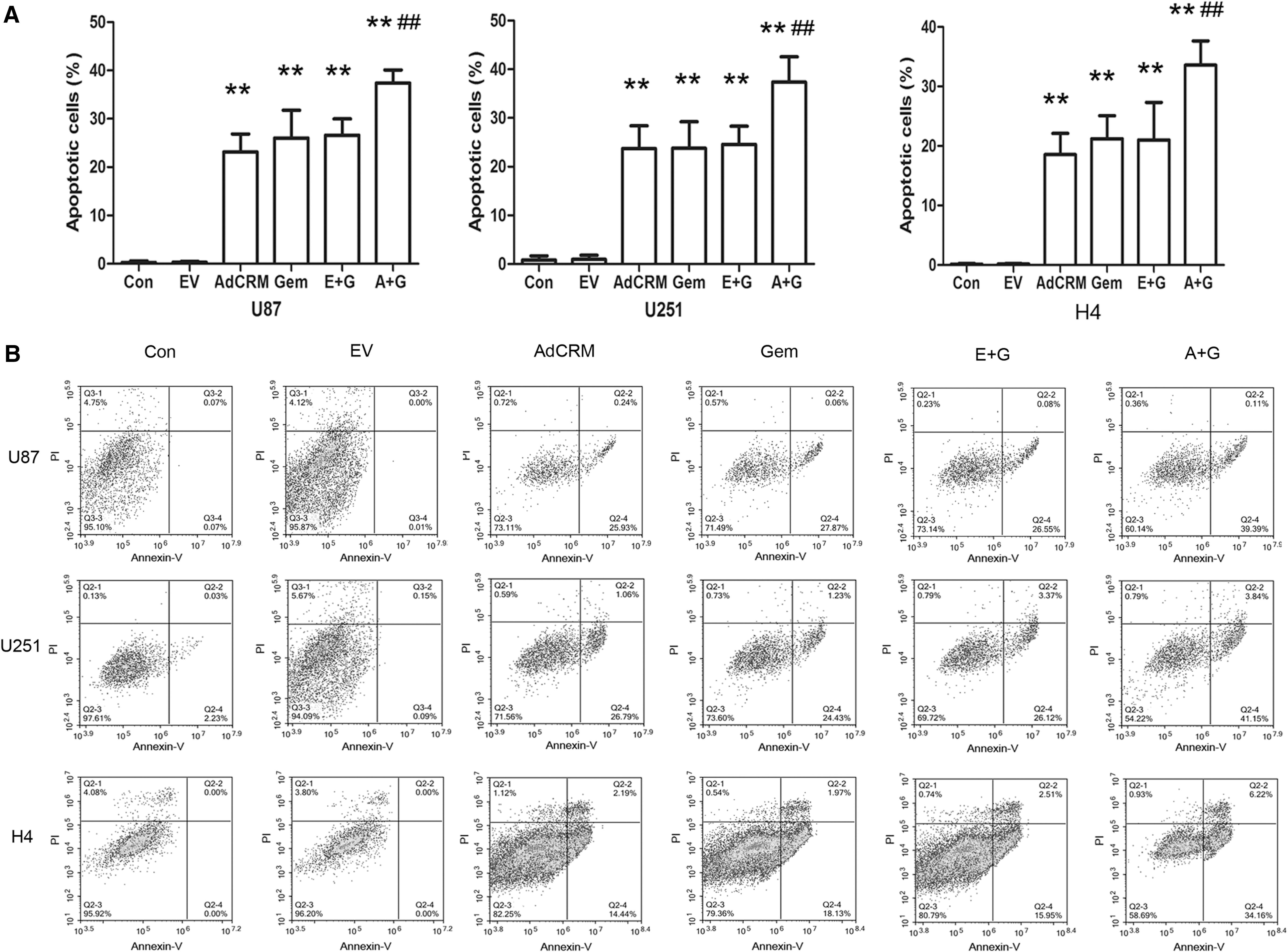
MTT assay was performed to evaluate the viability of the cells. Compared with the cells with gemcitabine or Adempty treatment, the viability of the cells with the combined treatment of AdCRM197 plus gemcitabine was much lower (p < 0.01). Also, in comparison with the U87, U251, or H4 cells, HA1800 cells were relatively insensitive to AdCRM197 or gemcitabine (p < 0.05). The toxicity of AdCRM197 to human normal astrocyte cells was much lower in comparison with that of human glioma cells (Fig. 1A). Hoechst staining showed the state of the nucleus of the cells. Compared with the cells that were treated with gemcitabine or the EV, more cracked nuclei or condensed nuclei of U87 cells appeared when they were treated with gemcitabine plus AdCRM197 (Fig. 1B, 400 × magnification). Annexin V-FITC/PI staining was used to evaluate cell apoptosis (Fig. 2). The apoptotic rate of cells with the combined treatment of AdCRM197 plus gemcitabine was much higher, compared with that of cells treated with gemcitabine (p < 0.01). The results suggest that the AdCRM197 can enhance the cytotoxicity of gemcitabine in the treatment of human glioma U87, U251, or H4 cells.

The cellar toxicity of gemcitabine was enhanced by AdCRM197. The cells untreated (Con), with the treatment of EV, with the treatment of AdCRM197 (AdCRM), with the treatment of gemcitabine (Gem), with the treatment of EV plus gemcitabine (E+G), or with the treatment of AdCRM197 plus gemcitabine (A+G).
AdCRM197 enhances the cytotoxicity of gemcitabine by the mitochondrial pathway
The caspase expression of U87 cells was estimated by Western blotting analysis. In comparison with the cells treated with gemcitabine, the amount of activated caspase-9 and activated caspase-3 was much higher in the U87 cells with the combined treatment of AdCRM197 plus gemcitabine. Moreover, more cleaved PARP was also found in the cells that were treated with both gemcitabine and AdCRM197. The caspases and cleaved PARP expression of HA1800 cells showed the slight toxicity of gemcitabine and AdCRM197 (Fig. 3A).
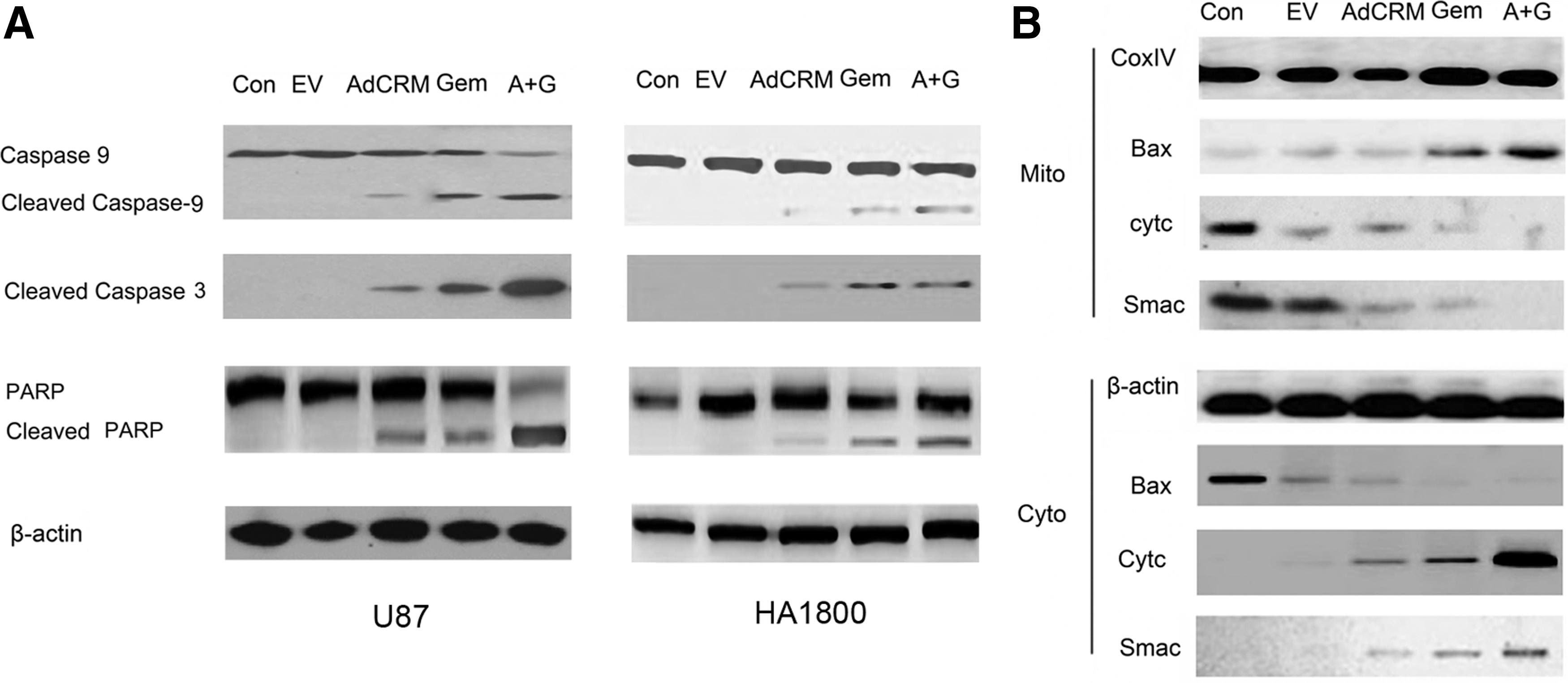
To test if the mitochondria pathway plays an important role in the enhanced cytotoxicity of gemcitabine, the expression of several important proteins was analyzed by Western blotting. The release of the cytochrome C from mitochondria in U87 cells is shown in Figure 3. In comparison with the U87 cells treated with gemcitabine, the cytochrome C and Smac in the mitochondria decreased greatly when the cells were treated with gemcitabine plus AdCRM197. Also, more quantities of Bax, a BCL2 family protein, translocated from the cytosol to the mitochondria (Fig. 3B).
The antitumor effect of gemcitabine in vivo was enhanced by AdCRM197
In comparison with the gemcitabine group, the tumor volume was inhibited remarkably in the gemcitabine plus AdCRM197 group (p < 0.01) (Fig. 4). Synergistic indices of gemcitabine plus AdCRM197 were calculated according to a previous study. 27 Gemcitabine plus AdCRM197 showed an additive effect on d 20, and showed a synergistic effect on d 25–35, in the U87 tumor-bearing mice. Gemcitabine plus AdCRM197 showed a synergistic effect in the U251 tumor-bearing mice on d 20–35 (Supplementary Data A).
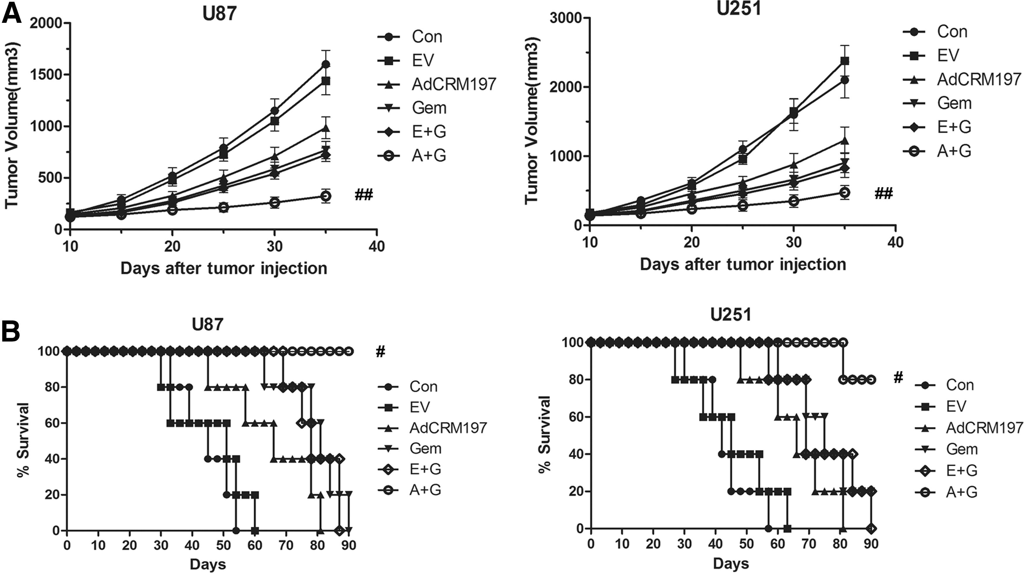
Tumor volume
Also, both in U87 tumor-bearing mice and U251 tumor-bearing mice, the mean survival time was prolonged obviously the in gemcitabine plus AdCRM197 group, compared with that in the gemcitabine group (p < 0.05) (Fig. 5). These results indicated that the tumor volume of the nude mice was inhibited and the time of survival was prolonged by AdCRM197 in the combined treatment.
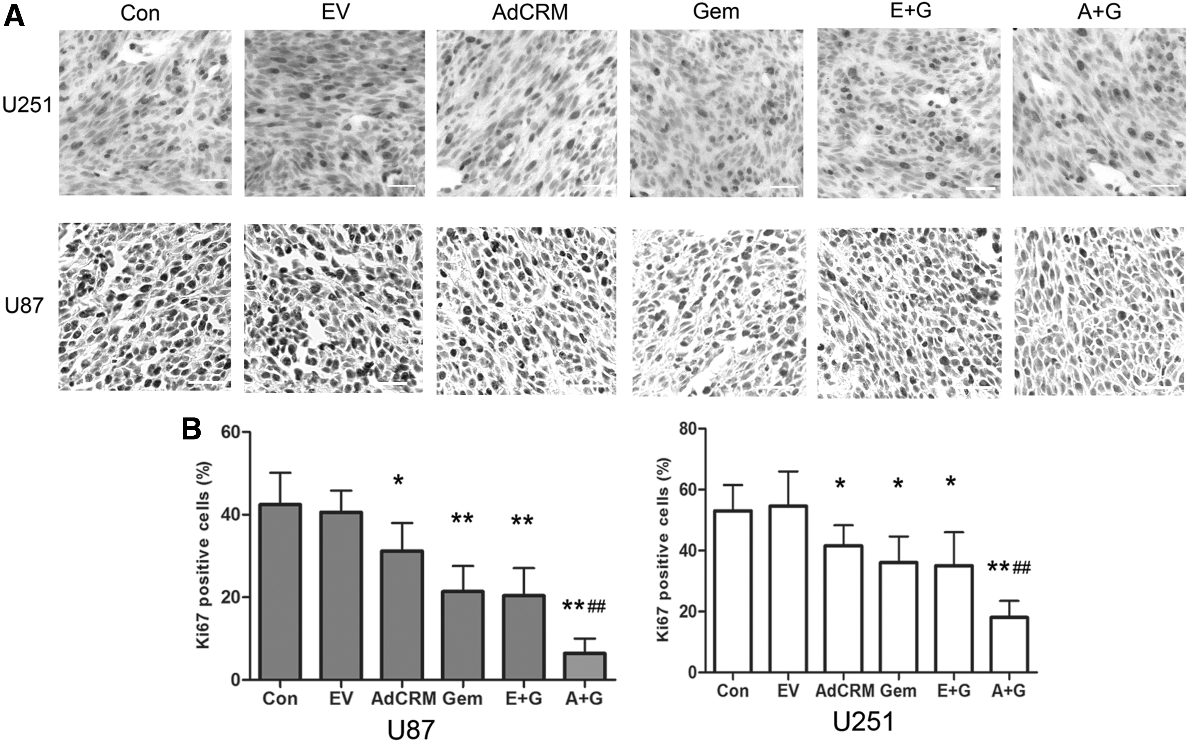
Immunohistochemistry of Ki67 of xenografted tumors. Representative fields of U251 and U87 xenografted tumors are shown
The percentage of Ki67-positive cells decreased in the gemcitabine plus AdCRM197 group, compared with that in the gemcitabine group (p < 0.01). More apoptotic cells could be detected in the gemcitabine plus AdCRM197-treated mice, compared with that in the gemcitabine-treated mice (p < 0.01) (Fig. 6, 200 × magnification).

TUNEL staining of the xenografted tumors. Representative fields of the TUNEL staining of the xenografted tumors
No obvious side effect was found in the treatment of AdCRM197
In comparison with the PBS-treated mice, mice that were treated with AdCRM197 or Adempty did not lose weight and showed normal appetite. AdCRM197- or AdEmpty-treated mice acted as the PBS-treated mice. The mice treated with gemcitabine or with gemcitabine plus AdCRM197 or Adempty showed decreased appetite and weight loss (Weight of the mice were shown in Supplementary Data B). Also, no ulcers were found in any of the mice with the various treatments. The sections of the vital organs of AdCRM197-treated nude mice, such as the liver, the heart, the spleen, the kidney, and the lungs, were examined by the microscope (Supplementary Data C). No obvious change in the sections was found under the microscope. These results indicated that AdCRM197 may be relatively safe to mice.
Discussion
CRM197 showed a weak EF2-ADP-ribosyl activity in recent researches. 15 Also, a study suggested that CRM197 could inhibit the protein synthesis of the cells. 14 CRM197 could remarkably suppress the tumor growth in the xenografted mice. 28,29 Furthermore, a treatment with the protein of CRM197 in the xenografted tumors reduced angiogenesis and inhibited the invasion and migration of adrenocortical carcinoma cells. 21
Previous studies have shown that CRM197 protein inhibits the cell proliferation by inhibition of heparin-binding epidermal growth factor-like growth factor (HB-EGF), a member of the EGF family. 30 As a specific inhibitor of HB-EGF, the protein of CRM197 could inhibit the mitogenic activity of the cells, for its ability of inhibiting epidermal growth factor receptor (EGFR) binding. 30,31 The protein of CRM197 was also demonstrated to be a possible chemosensitizing agent for ovarian cancer for its ability of inhibiting HB-EGF. 17 –19 It has been reported that CRM197 sensitizes ovarian cancer cells to paclitaxel through the balance of antiapoptotic and proapoptotic pathways. 17 The combined treatment of paclitaxel and CRM197 could inhibit ERK and Akt, and activate p38 and JNK, leading to the enhanced antitumor effect. 17 Moreover, CRM197 was also found to inhibit MDR1 in paclitaxel-resistant human ovarian cancer cells. 32 CRM197 can induce the T cell-dependent responses, and was found to be an immune modulator in cancer therapy. There are two ongoing clinical trials in cancer with CRM197 (NCT02310464 and NCT02795988) and one clinical trial has been recently completed (NCT01852591). These trials are using CRM197 as an immune modulator in various cancer types. In a clinical study, CRM197 was injected subcutaneously into 25 outpatients with various advanced tumors. The treatment of CRM197 increased the number of circulating neutrophils and the serum level of tumor necrosis factor (TNF)-α, caused tolerable toxicities, and exerted an antitumor effect. 33
Cells undergo apoptosis by two major pathways: the intrinsic pathway and the extrinsic pathway. The extrinsic pathway of apoptosis is mediated by the death receptor and the intrinsic pathway is mediated by the mitochondria. So the intrinsic pathway is also called “the mitochondrial pathway.” The mitochondrial outer membrane permeabilization (MOMP) plays a key role in the apoptosis of the intrinsic pathway. 34 –37 In this study, AdCRM197 showed a weak cellular toxicity that caused MOMP, and released the cytochrome c and the Smac from the mitochondria. These results indicated that AdCRM197 could strengthen the antitumor effect of gemcitabine in glioma by the mitochondrial pathway.
As an attractive novel option of cancer therapy, gene therapy is developing very fast in recent years. A plasmid encoding CRM197 gene was used for pancreatic cancer gene therapy under the control of the heat shock protein promoter. 38 Despite the main safety concern of the immune response, as an efficient transfer system, the adenovirus vectors are good options in gene therapy. 39 –41 The adenoviral vectors have been successfully applied for several years. 42 –44 In this study, the authors used the adenovirus-mediated CRM197 to sensitize the human glioma cells to gemcitabine. They found that AdCRM197 sensitized the human glioma to gemcitabine not only in vitro but also in vivo. These results indicated that AdCRM197 would be a potential chemosensitizing agent for cancer therapy.
Footnotes
Acknowledgments
This work was supported by the National Natural Science Foundation of China (Grant No. 81471848, 81870940), the Education Department of Sichuan Province Natural Science Research Program (Grant No. 15ZB0250, 18ZA0148), Program of Chengdu Medical College (Grant No. CYCG15–02), Initial Project for Post-Graduates of Hubei University of Medicine (Grant No. 2015QDJZR08), the Scientific Research and Technological Development Project of Shiyan Science and Technology Bureau (Grant No. 16K70), and the Science and Technology Program of Guangzhou (Grant No. 201607010365). Research Found of Department of Education of Hubei province (Grant No. B2018117).
Disclosure Statement
No competing financial interests exist.
Supplementary Material
Supplementary Data A
Supplementary Data B
Supplementary Data C
References
Supplementary Material
Please find the following supplemental material available below.
For Open Access articles published under a Creative Commons License, all supplemental material carries the same license as the article it is associated with.
For non-Open Access articles published, all supplemental material carries a non-exclusive license, and permission requests for re-use of supplemental material or any part of supplemental material shall be sent directly to the copyright owner as specified in the copyright notice associated with the article.