
Abstract
Cancer poses a major health problem, not only due to cancer-related deaths but also because of treatment toxicities. This review discusses early diagnosis and strategies to overcome treatment difficulties, to facilitate recovery, and prevent deaths. Generally, noninvasive techniques such as computed tomography (CT), magnetic resonance imaging (MRI), single photon emission computed tomography (SPECT) and positron emission computed tomography (PET), and their hybrid systems, including SPECT/CT, PET/CT, and PET/MRI, are used in diagnosis of cancer. Cancer treatment in clinics still comprises conventional methods such as chemotherapy, radiotherapy, and surgery. However, these techniques and methods are often inadequate. Therefore, new approaches, including the formulation of actively and/or passively targeted nanosized drug delivery systems and combined treatment protocols, are being investigated. In this article, conventional cancer imaging and treatment are reviewed. In addition, the formulation of nanosized systems and their use in cancer treatment are discussed and combined diagnostic and therapeutic (theranostic) approach are proposed as additional cancer therapies.
Introduction
Cancer is a disease characterized by abnormal and uncontrollable cell proliferation, with the potential to metastasize to other parts of the body, including the brain, liver, and bones, through blood or lymph circulation. It is a major health problem and a leading cause of death worldwide. 1 According to the Global Burden of Cancer Study (2012), 14.1 million new cancer cases and 8.2 million cancer deaths are reported by the International Agency for Research on Cancer annually. 2,3 The aim of this article is to review conventional cancer imaging techniques and treatments and targeted specific nanocarriers for accurate diagnosis and/or effective therapy as a theranostic approach.
For this purpose, first, conventional cancer imaging techniques, their advantages and disadvantages, and their comparisons with each other are mentioned. In the part of conventional cancer treatment, current cancer treatment protocols, difficulties of treatment, and new approaches (combination therapy and nanosized systems) are explained to solve these problems. Following, definition, advantages and targeting mechanisms of nanosized systems are summarized. In addition, the theranostic approach, which gains importance day by day in effective cancer treatment and imaging, takes place. Finally, some studies are given clarifying future aspects in cancer imaging, treatment, and the theranostic approach.
Conventional Cancer Diagnosis and Imaging
To decrease and potentially eliminate cancer, early diagnosis is critical. Moreover, cancer diagnosis and imaging play a large role in prognosis, staging, treatment planning, and the evaluation of treatment response. 4 Cancer can be diagnosed using invasive and/or noninvasive techniques. Generally, cancers are detected with noninvasive imaging techniques such as magnetic resonance imaging (MRI), X-ray-based systems (computed tomography [CT], digital radiography [DR], and fluoroscopy), nuclear imaging (single photon emission computed tomography [SPECT] or positron emission computed tomography [PET]), and ultrasound systems and their hybrid techniques, including PET/CT, SPECT/CT, and PET/MRI. 5 Each of these techniques has properties that make them uniquely suited to some diagnostic scenarios and unsuitable to others. The advantages and disadvantages of each technique are summarized in Table 1. 5 –7
CT, computed tomography; MRI, magnetic resonance imaging; SPECT, single photon emission computed tomography; PET, positron emission computed tomography.
Nuclear magnetic resonance imaging is known as MRI and it uses radiofrequency (RF) energy. It does not utilize ionizing radiation, which is important for the patients who need to limit their exposure to radiation. 5 Under normal circumstances, the cells in the body are insensitive to energy in the RF range of the electromagnetic spectrum; however, in a strong magnetic field, protons become ready to be stimulated and spin state of protons interact with RF energy. Generally, hydrogen atoms in water molecules are used as the proton source in the human body. By removing RF, energized protons return to their original spin and simultaneously emit a signal. These signals are detected by the MRI detector and an image is created. 8 –10 MRI consists of various imaging techniques such as diffusion weighted (DW), T1 weighted, and T2 weighted. 11 Most tumor tissues have a lower apparent diffusion constant than healthy tissues; therefore, DW imaging is often used in cancer imaging. DW imaging is helpful in detecting and evaluating pancreatic and prostate cancer following the detection of changes in these tissues. MRI can be combined with mammography to increase its sensitivity in the diagnosis of breast cancer. 12 It is also essential in the assessment of cancer treatment such as the evaluation of breast cancer response to chemoembolization. 13 To obtain T1-weighted and T2-weighted images, paramagnetic gadolinium (Gd) chelates and iron oxide are used as contrast agents, respectively. Gd can be encapsulated in nanosized drug delivery systems that can be actively and passively targeted to tumor tissues. In addition, superparamagnetic iron oxide nanoparticles can be modified with various tumor receptor-specific ligands for targeting. In a previous study, a higher diagnostic accuracy was obtained in prostate carcinoma patients when DW and T2-weighted MRI was combined. 14
X-ray imaging systems, including DR, CT, and fluoroscopy, are faster, cheaper, and easier to use than other imaging techniques. Chest radiography (chest X-ray) is the main screening technique for chest abnormalities, including lung cancer. While CT is more sensitive, X-ray is inexpensive, easy to use, and the radiation dose is low. 15 Virtual bronchoscopy, another radiological technique, is useful in confirming tumor localization. 16 Mammography is a DR technique that is used in the imaging of breast cancer. Digital mammography can be combined with tomosynthesis to produce three-dimensional images in contrast to the two-dimensional images produced by standard mammography. As tomosynthesis is a more sensitive imaging method, more breast lesions can be detected and false negatives are minimized. Furthermore, iodinated contrast agents can be used for imaging of angiogenic areas in breast lesions. Tomosynthesis can be combined with contrast-enhancement mammography to monitor the distribution of contrast agents in the breast tissue and obtain information about the separation of benign-malign lesions. 4,17 CT, another X-ray-based imaging system, is a tomographic imaging modality that has the highest resolution of the imaging techniques. 5 Three-dimensional and cross-sectional images can be obtained by CT. In colon cancer specifically, information can be obtained about staging, metastases, and planning of treatment. 10,18
Noninvasive nuclear imaging modalities are frequently and successfully used to obtain accurate images after administration of radiopharmaceuticals to patients. While conventional gamma cameras create two-dimensional images, three-dimensional SPECT images can be obtained by rotating cameras around the patient. The radiopharmaceuticals used for nuclear imaging purposes depend on their emission types and properties. Radiopharmaceuticals labeled with 99mTc, 111 In, and 123I radionuclides decay gamma rays. PET radiopharmaceuticals labeled with 11C, 15O, 18F, 13N, 82Rb, 64Cu, and 68Ga radionuclides decay positron emission, which causes the release of two 511 keV gamma rays. 5,19 Among nuclear imaging techniques, PET is more sensitive than SPECT. 5 The gold standard in cancer diagnosis, the PET radiopharmaceutical 18F-Fluorodeoxyglucose (FDG), is a glucose analog that accumulates in higher quantities in tumor tissue depending on the overexpression of tumor cell membrane transporters. 5 Furthermore, detection of bone metastases can be carried out using 99mTc-MDP, 99mTc-MIBI, and 99mTc(V)-DMSA radiopharmaceuticals. Neuroendocrine tumors can be detected and diagnosed with 111 In-labeled octreotide, 68Ga-DOTA GOC, and 123I labeled meta-iodobenzylguanidine. 4,20,21 While prostate cancer can be diagnosed with 111 In-labeled capromab pendetide, which recognizes prostate membrane-specific antigen (PMSA), angiogenesis or response to antiangiogenic therapy in breast cancer can be diagnosed and evaluated with 99mTc-RGD (Arg-Gly-asp) or 18F- RGD. 4,22,23
In ultrasound (US) imaging, high-frequency (3–7.5 MHz) sound waves are applied to the body and reflected sound waves are detected by a transducer to create images. US is used to diagnose various types of cancer, including transrectal and prostate cancers. Endoscopic US is useful for detecting gastrointestinal tumors. 4,24 Microbubbles, including free gas such as perfluorocarbon, administrated intravenously to patients, can be used as contrast agent in US imaging. Passive or active targeting of microbubbles to tissues is currently being investigated. The sensitivity of US imaging is increased through the use of these microbubbles, specifically in metastatic areas. 25 Another example is performed in a preclinical study. The imaging efficiency of microbubbles targeted to the angiogenic factors integrin and vascular epithelial growth factor receptor 2 (VEGFR2) in xenograft animal models has been evaluated. 26,27 Therapeutic agents can be encapsulated in the oily layer of the microbubbles to be used in targeted cancer imaging and/or therapy. 25
Among noninvasive imaging methods, while MRI and CT have the ability to obtain better anatomic images, molecular changes in cancer-like receptors or antigens can be specifically imaged with functional hybrid imaging techniques such as PET/CT. In addition, PET/CT has the ability to obtain images at the molecular level due to the use of F, C, O, and N having roles in biological pathways such as glycolysis compared to SPECT/CT or planar gamma scintigraphy. Therefore, PET/CT can differentiate tumors from other lesions such as inflammation or necrosis following radiotherapy.
Biopsy is an invasive diagnostic technique that is performed by removing tissue samples from the tumor and investigating these samples pathologically. In clinical practice, a lesion will usually be biopsied if there is a high uptake of 18F-FDG. Imaging methods such as CT or MRI can be used to guide the clinician during a biopsy. In addition, biopsy is relatively easy to perform and is useful in discriminating between malignant and benign lesions. 5
Conventional Cancer Treatment
Current cancer treatment methods can be classified into three groups, including 28
• Pharmaceutical treatment (chemotherapy, hormone therapy, and immunotherapy)
• Radiation treatment (radiotherapy [external and internal], photodynamic therapy, high-intensity focused ultrasound [HIFU], laser ablation treatment, and RF ablation)
• Physical intervention (resection of tumors with surgical methods and cryotherapy)
In chemotherapy, chemical medicines having different mechanisms of action, are used. Alkylating agents (nitrogen mustards, cyclophosphamide, platin derivatives, etc.), antimetabolite analogs (methotrexate, pemetrexed, fludarabine, gemcitabine, etc.), and/or natural or seminatural products (vinca alkaloids and taxanes) can be used alone or in combination with each other during conventional cancer treatment. 28
Some types of cancer, including breast, prostate, and endometrium cancer, are hormone dependent or hormone sensitive. Nuclear receptors have an important role in the tumorigenesis, owing to having high expression rate like expression of estrogen receptor-α in breast cancer or androgen receptor in prostate cancer. Therefore, in the treatment of these types of cancers, hormone therapy is used to change and regulate the level of total hormones in the body. 29 In hormone receptor-positive breast cancer, the levels of hormones (estrogen and/or progesterone) in the body can be reduced or the hormone receptor can be blocked with tamoxifen. 30 In clinical studies, abiraterone acetate, an androgen biosynthesis inhibitory molecule, has been used in the treatment of prostate cancer. 31
The purpose of immunotherapy in cancer patients is the activation of immunity using monoclonal antibodies (mAb), vaccines, or cytokines. 32,33 Prophylactic treatment is provided using vaccines to produce antibodies against oncogenic infection such as human papilloma virus, hepatitis B virus, and Helicobacter pylori. Cytokines can have immune-modulating properties (e.g., interleukin-2) or cytotoxic effects (e.g., tumor necrosis factor-α). 34 mAb can directly inhibit receptors (e.g., epidermal growth factor receptor) or ligands of receptors (e.g., vascular endothelial growth factor [VEGF]). These receptors and ligands play essential roles in the development of tumors and angiogenesis. 35,36
Radiotherapy, a treatment method using high energy ionizing radiation, involves external or internal radiotherapy. External radiotherapy (or external beam radiation therapy) is the most common type of radiation therapy. In this type of therapy, charged proton particles or high-intensity X-ray beams generated from a linear accelerator, are applied from outside the body to cancerous tissue in the body. Internal radiotherapy is also called brachytherapy, radionuclide therapy, or close therapy, and involves radioactive sources that are placed near or inside the tumor tissues. 37 Radiolabeled antibodies such as 90 Y-ibritumomab tiuxetan or 90 Y-J591 (anti-PMSA) can be used for targeting of cellular antigens in radionuclide therapy. 38,39
Photodynamic therapy relies on a photosensitizing agent, light, and oxygen to exert a toxic effect on tumor cells. A photochemical reaction occurs through the interaction of red light or infrared radiation with a photosensitizing agent and oxygen, which forms cytotoxic reactive oxygen radicals. 40,41 The wavelength of light (red light or infrared radiation) must be below 800 nm for the formation of singlet oxygen ( 1 O2). 40 Park et al. 42 reported the use of photodynamic therapy in the treatment of non-small cell lung cancer. They formulated hyaluronic acid-coated nanoparticles, added Gd ions for magnetic resonance contrast, and encapsulated Chlorin e6 as the near-infrared (NIR) photosensitizer agent. After the application of the nanoparticles, the tumor was irradiated with light and an enhanced therapeutic effect was observed as a result of the formation of reactive oxygen radicals. 42
During treatment with HIFU, high-energy sound waves with frequencies between 0.8 and 3.5 MHz are applied to the skin, causing the temperature to increase in the tumor, ultimately destroying the cancer cells. 43,44 HIFU is used in the treatment of both benign and malignant tumors of the prostate, liver, bone, breast, kidney, pancreas, and soft tissue. In addition, it is a noninvasive alternative method to surgery. As there is no upper limit of tissue tolerance, HIFU can be applied more than once, which gives it an advantage over ionizing radiation. 43
Laser ablation treatment is another noninvasive method; it is also referred to as photo-thermal therapy, laser interstitial therapy, and laser interstitial photocoagulation. 45 In this therapy, focused electromagnetic radiation beams generated from lasers are used. The beam denatures the proteins because of an increase in temperature within the cells. Laser ablation can be performed with US or MRI to determine the exact location of the tumor. 46 MRI-guided laser ablation treatment has been applied to prostate cancer patients in a phase-1 study. 47
A high-frequency electric current that has a frequency of 10 kHz-900 MHz is used in RF ablation. This electric energy converts to heat, which causes the formation of coagulation necrosis in 4–6 min at 50°C–52°C. 48 A study to evaluate the use of gold nanoparticles during RF ablation of tumor cells found that, while gold nanoparticles were not cytotoxic in vitro hepatocellular carcinoma cells, they facilitated the destruction of 80% of cells upon exposure of RF. In addition, according to the results of in vivo studies in rats, the temperature significantly increased in the group injected with gold nanoparticles compared to that in the control group. 49
In cryotherapy, the cytotoxic effect of cold is used to kill cancer tissue. Generally, two types of low-temperature probes are used: liquid nitrogen probes and nitrous oxide-driven cryoprobes. Because of the adherence of these probes to tissue, ice crystals occur in the intracellular and extracellular spaces, which cause damage to the organelles and lead to cellular dehydration. 50 –52 Clarke et al. evaluated the effect of a combination of chemotherapy and cryotherapy in prostate cancer cell line (PC-3) by using 5-fluorouracil as the chemotherapeutic agent. Their results indicated that, when cells were exposed to temperatures between −5°C and −20°C, cell viability was significantly lower after combination treatment than after treatment with chemotherapy or cryotherapy alone. In addition, all cells exposed to temperatures between −40°C and −80°C were killed during combination treatment. 53
Chemotherapy is the most common cancer therapy. However, the properties of chemotherapeutic drugs make them difficult to use. These properties include the following: 54
• Antineoplastic drugs have a narrow therapeutic index.
• Serious toxic systemic effects occur slightly above the therapeutic dose, including myelotoxicity, cardiomyopathy, nephrotoxicity, and immunosuppression.
• When applied singly, they cannot overcome drug resistance mechanisms that are pre-existing in tumor tissue or developed during treatment.
• Their cytotoxic effects are not limited to cancer cells. They also affect normal cells that proliferate rapidly such as cells lining the gastrointestinal tract (GIT), hair follicles, and germ cells, which lead to mucositis in the GIT and alopecia. In addition, some drugs can damage spermatogenesis or oogenesis causing infertility, amenorrhea, early menopause, or impotence.
• Most chemotherapeutics cannot be used during the first trimester of pregnancy because of their teratogenic effects.
• Chemotherapy has a mutagenic effect and can cause secondary malignant neoplasms to develop.
• Some anticancer drugs are cycle specific, therefore only affecting cells that are in a specific cell cycle such as S phase.
• Tumor cells that proliferate rapidly are more sensitive to chemotherapy than tumor cells that proliferate slowly (e.g., cancer “stem cells”).
• Patient-related factors should be considered during chemotherapy. Age, sex, nutrition, immunity, and comorbidities of the patients play an important role in the response to treatment.
As a result of the difficulties associated with conventional chemotherapeutic drugs, novel approaches to cancer therapy are being investigated in various scientific disciplines, including pharmaceutical sciences, polymer chemistry, medicine, molecular biology, tumor biology, and molecular imaging. These novel approaches involve passive/active targeted drug delivery systems and combined treatment protocols.
Combination therapy (also polytherapy and multiple therapy) is the use of more than one drug or method in treating patients for a single disease. When treating cancer, the combined use of chemotherapy and radiotherapy is known as combined therapy. 55 Numerous pathophysiological processes are involved in complex diseases such as cancer and cardiovascular and infectious diseases, and many related therapeutic targets exist. Knowledge of the processes and targets has made it possible to determine which drug combinations are most suitable in the treatment of these complex diseases. 56 Bactrim, which consists of trimethoprim and sulfamethoxazole, was the first instance of a combination of two drugs approved by the Food and Drug Administration in 1974. 57 Following Bactrim, combined drug therapy became increasingly important, especially in treating diseases like HIV-AIDS, tuberculosis, malaria, diabetes, and hypertension. The first antiviral combination, Combivir (Lamivudine+Zidovudine), used to treat HIV infection was approved in 1997. 58
Currently, combination therapy in cancer treatment consists of surgical methods and radiotherapy with drug therapy. However, surgical methods and radiotherapy are limited to local cancers and cannot be applied in metastatic cases. On the other hand, in the treatment of metastatic cancer cases, cytotoxic cancer drugs can kill the cancer cells, but they have less chance of cure than localized disease. This is because the metastasis can spread out to different places of the body. 59,60 In the literature, anticancer drugs that target different pathophysiological processes have been applied to cancer cell lines in vitro and animal models in vivo. The results of these experiments have shown that cancer cell death is increased as a result of the synergistic effect of drugs. Furthermore, the toxic effects during the treatment decreased, leading to an increase in life span in the animal models. 61,62
There are many advantages of combined therapy: (1) the toxic effects observed in monotherapy are decreased by reducing the dosage of individual therapy, (2) targeting of different action mechanisms minimizes the development of drug resistance, and (3) the clinical response increases with the synergistic drug effects. Using several drugs at optimal doses minimizes intolerable side-effects. In addition, combined chemotherapy is useful in patients with advanced cancer because radiotherapy and surgery can usually not be used in these patients. 63 –65 Platinum-based combined cancer therapy consists of carboplatin or cisplatin and a second cytotoxic agent. This treatment is the backbone for cytotoxic combined chemotherapy. 66 According to the results of a study on patients with advanced non-small cell lung cancer, life expectancy was found to be 8 months when patients were treated with platinum-based chemotherapy. 67 Combination of gemcitabine+ docetaxel/pemetrexed/paclitaxel/vinorelbine or paclitaxel+vinorelbine has been found to be useful in patients who are unable to use the platinum derivatives because of their toxic effects. 66 Examples from the literature of nanosized co-delivery systems for the treatment of a variety of cancers are summarized in Table 2.
PLGA, poly (lactic-co-glycolic acid).
Nanosized System Approach in Cancer Treatment
Advantages
Nanotechnology is the design, simulation, and fabrication of materials, systems, and devices, having sizes between 1 and 100 nm. It is used currently in various areas from textile research to organ transplantation, including drug development. Nanotechnology is remarkable in the delivery of drugs and nanosized systems are one of the most commonly studied drug delivery systems because of various advantages including the following: 88,89
• They are biocompatible, nontoxic, and nonimmunogenic.
• They improve parenteral administration through their ability to enhance drug solubility.
• They improve the drug bioavailability as a result of changes in solubility.
• They are able to cross blood–brain barrier easily.
• Drugs with different physicochemical properties, such as hydrophobic or hydrophilic drugs, can be delivered using these systems.
• Drugs are released at a fixed rate depending on zero-order release kinetics in the disease area.
• They have the ability to carry multiple therapeutic agents, which helps to overcome multidrug resistance.
• Sensitive molecules like polypeptide are protected from degradation during blood circulation.
• They can be targeted actively and/or passively to disease areas within the body using surface modification. Targeting results in higher concentration of drug in the disease areas, thereby decreasing toxic reactions in healthy tissues.
• Their surface can be coated with a polymer like polyethyleneglycol (PEG), which helps the system to escape immune cells. They can be formulated at nanometer particle size, which reduces renal clearance and enhances the biological half-life of the drugs. The size and surface properties of nanoparticles include passive targeting mechanisms.
• They can be modified with multiple ligands specific to disease area for active targeting.
Targeting
Targeting of nanosized drug delivery systems is essential for the diagnosis and treatment of complex diseases such as cancer, because of the difficulties associated with conventional cancer treatment. Through targeting of nanocarriers, drug concentrations can be increased in the disease area and decreased in other parts of the body, therefore reducing the risk of side-effects in healthy tissues. Ultimately, better therapeutic effects can be achieved with standard or even decreased drug concentrations. Moreover, targeted therapeutic agents can access hard-to-reach areas, such as intracellular spaces, bacteria, viruses, and organs, including the brain. Ideally, the aim of cancer treatment is to reach deep within tumor tissues to selectively kill tumor cells, thereby preventing the adverse and toxic reactions in healthy cells. 90 Nanosized drug delivery systems can be specifically formulated to target tumor tissues. Two main types of targeting strategies exist: passive and active targeting.
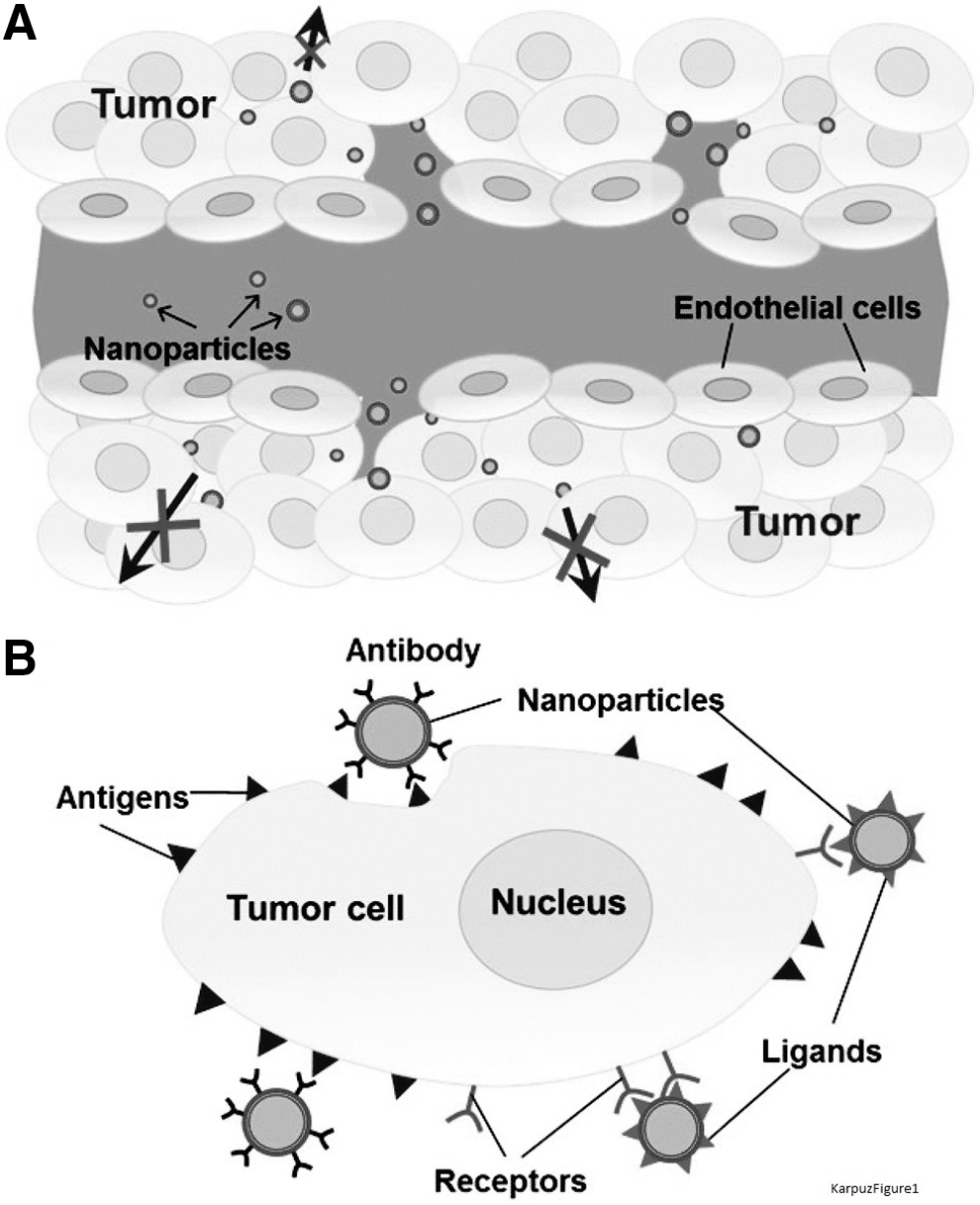
For passive targeting, particle sizes can be decreased and surface characteristics can be modified to escape the reticuloendothelial system (RES). Compared to nanosized systems, the accumulation of small drug molecules in the tumor area is limited due to faster blood elimination, shorter circulation half-life (t1/2), higher volume of distribution (Vd), and lower area under the curve (AUC). 91 Particle sizes in the nanometer range allow nonspecific accumulation of these systems in the tumor area, an effect that is referred to as enhanced permeability and retention (EPR) of tumor tissue. Mainly, dysfunctional lymphatic drainage and leaky vasculature in the tumor area cause EPR. Normally healthy vascular endothelial cells are lined up tightly, but rapid tumor proliferation and growth lead to the formation of a leaky endothelial cell structure (Fig. 1). 92
Consequently, in many tumor types, the blood vessel wall becomes more permeable than in healthy tissues. The EPR effect provides advantages in treating cancer with drugs that can be delivered with nanosized systems. Particles with a molecular weight higher than 50 kDa and between 100 and 600 nm in size have been reported to be the most suitable for nanosized drug delivery systems. 93,94 To achieve better therapeutic outcomes with this type of targeting strategies, drug delivery systems should remain in the blood, circulating for an extended period of time. Normally, opsonin proteins in the circulation bind to nanoparticles to facilitate recognition by RES cells (e.g., macrophages) for rapid removal from the bloodstream. In addition, particles with hydrophobic surfaces are eliminated from the blood circulation primarily by the liver as well as the spleen and lungs. Therefore, to extend the half-life of nanoparticles, particle surface characteristics can be modified with polymer coatings to evade the RES. Various natural or synthetic biodegradable polymers can be used to coat nanoparticles, including poly (D-lactic acid) (PDLA), poly (vinyl alcohol), poly (glycolic acid), poly (acrylic acid), and the most frequently used PEG. 95 By coating the particle with PEG, the surface takes on hydrophilic properties, which allow it to remain in the blood circulation. Some PEGylated liposome formulations used in the cancer treatment are available commercially, e.g., doxorubicin encapsulated Doxil® and Lipo-dox® approved for Kaposi sarcoma, ovarian cancer, and breast cancer; daunorubicin encapsulated DaunoXome® approved for the treatment of blood tumors. 96
Active targeting can include ligand- or receptor-mediated targeting and physical targeting. Ligand mediated targeting involves linking a ligand, which recognizes receptors on the target side, directly to drugs or drug delivery systems. These receptors are expressed or overexpressed in the endothelial cell membranes or disease sites such as tumors, whereas they are less expressed or not expressed in healthy cells. A variety of molecules can act as ligands, including mAb, that bind specifically to surface antigens or tumor cell receptors, integrins, and other peptides that bind to overexpressed integrin receptors, nucleic acid ligands such as aptamers and folates that bind to overexpressed folate receptors on tumor cells because of enhanced DNA proliferation and transferrin uptake. 93 Conjugation of NL-1 gmAb to Adriamycin®, which is specific to many types of cancer, is an example of an active drug targeting ligand conjugate. With these conjugates, growth inhibition of cancer cells, specifically, can be achieved. 97
Angiogenesis, the formation of new blood vessels, is essential for growth and metastasis of tumor cells, which makes it a potential target for therapy. Normally, there is a balance between angiogenesis inhibitor molecules and stimulating agents in healthy tissues. However, when the tumor tissue requires nutrients and oxygen, angiogenesis can be stimulated with the upregulation of angiogenesis factor and the downregulation of inhibitor molecules. Consequently, inhibition of angiogenesis presents an essential target in cancer treatment. Inhibition of vascular endothelial growth factor (VEGF) proteins, their receptors (VEGFR), and the intracellular signaling pathways of VEGFR by mAb, such as bevacizumab, or tyrosine kinase inhibitors like, sorafenib, is foremost in this type of targeting for cancer treatment. 98
Another active targeting method is physical targeting, which includes pH-sensitive systems, thermo-sensitive systems, magnetic targeting, ultrasound targeting, light-sensitive systems, and enzyme-sensitive systems. 93 The umbrella term for these systems is stimulus-sensitive carriers, since they release drugs in the disease area as a result of external stimulation or presence of certain enzymes, such as matrix metalloproteinases, phospholipase-A2, and alkaline phosphatase, in the tumor microenvironment. 99,100 Enzyme-sensitive nanocarrier systems are disrupted by enzymes to allow drugs to be released in the tumor area. Thermo-sensitive liposomes comprise thermo-sensitive lipids (e.g., 1, 2-dipalimitoyl-sn-glycero-3-phosphocholine) that can be used depending on their phase transition region. The tumor area is usually heated to 42°C using external energy like microwaves, US, or RF and thus thermo-sensitive lipids control the drug release. 101 ThermoDox® liposomes, a commercially available formulation containing doxorubicin, are used in the treatment of hepatocellular carcinoma. 96 Other polymers, like poly (N-isopropylacrylamide), can be used for the development of thermo-sensitive micelles or nanoparticle formulations. 102 A schematic representation of a drug-loaded, multifunctional, stimulus-sensitive nanoparticulate pharmaceutical drug delivery system is given in Figure 2. 103
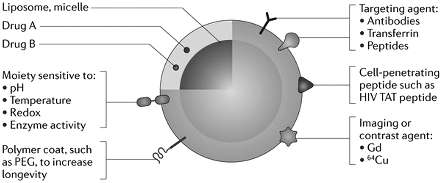
Schematic representation of a drug-loaded, multifunctional, stimulus-sensitive nanoparticle pharmaceutical drug delivery system. Reprinted by permission from Ref. 104.
Normal physiological pH is in between 7.35 and 7.45; however, in tumor interstitial fluid, the pH value decreases to acidic levels because of anaerobic glycolysis by cancer cells. Therefore, in the formulation of pH-sensitive systems, changes in local pH values can be an advantage. To this end, ionic polymers, including poly-acrylic acid, poly-methacrylic acid, 104,105 or phosphatidylethanolamine, 106 can be used. Drug delivery systems that are prepared using these types of polymers or lipids are stable at physiological pH. These systems degrade under acidic conditions to release the drugs at the tumor area. US targeting mechanisms that make use of low-frequency US benefit from EPR effect that increases permeability of blood capillaries. 93 Furthermore, low-frequency US can aid the release of drugs from the liposomes as a result of enhanced lipid permeability in the tumor area. 107
Theranostic Approach and Advantages
Theranostic is the combination of therapeutics and diagnostics that specifically relates to targeted drug delivery systems. It includes both a therapeutic agents/radionuclide and a diagnostic agents/radionuclide in the same system. These systems make it possible to obtain the diagnostic, imaging, and therapeutic information required to treat diseases. 108 A schematic representation of nanosized theranostic (nanotheranostic) systems is given in Figure 3. 109

Schematic representation of nanosized combined diagnostic and therapeutic (theranostic) systems. Reprinted by permission from Ref. 110.
The advantages of these systems are listed below
110
–112
: • Nanotheranostics have multifunctional properties, including the capacity to contain multiple drugs or radiocontrast agents, making them more effective in tumor diagnosis, imaging, and therapy than conventional drugs and/or radiocontrast agents. • They are useful for rapid diagnosis and treatment of malignant diseases. • Treatment of fatal diseases such as cancer is more straightforward with imaging of treatment efficiency and better information about disease prognosis can be obtained. • They have the ability to monitor the therapeutic agents and treatment efficiency. • More accurate images can be obtained by the use of different imaging methods. • They have the ability of designing new imaging-guided therapeutics. • By using theranostic systems, the pharmacokinetics of therapeutic agents and in vivo targeting can be monitored. • Undesirable differences in biodistribution of therapeutic and diagnostic agents can be overcome. • Improved information about cellular phenotype and tumor heterogeneities can be obtained. • Patient compliance is improved over conventional systems because diagnostic and therapeutic agents are provided using a single delivery system. Therefore, patients' quality of life can be enhanced.
Nanosized System Applications for Cancer Treatment and/or Imaging
Given the importance of nanosized drug delivery systems in treating cancer, numerous studies have been performed to improve the diagnostic signal and therapeutic effect of these systems.
Therapeutic nanocarriers
In this study, the authors review three studies on combination therapeutics in cancer treatment.
To overcome multidrug resistance in cancer treatment, Assanhou et al. 113 prepared a cationic liposome formulation, which contains paclitaxel as a chemotherapeutic agent and lonidamine as a chemosensitizer agent. The formulation was functionalized with D-α-tocopheryl poly ethylene glycol 1000 succinat to increase the accumulation of paclitaxel and inhibit P-glycoprotein efflux pump. This system targeted hyaluronic acid to CD44 receptors, which are overexpressed in multidrug-resistant MCF-7 breast cancer cells. According to the results of the in vitro MTT assay, this system was more cytotoxic than nontargeted, nonfunctionalized systems with liposomes containing only paclitaxel. Moreover, the IC50 value of this formulation was found to be lower than those of other formulations. Pharmacokinetic studies demonstrated higher AUC, longer mean residence time in the blood, longer elimination half-life, and slower clearance from the blood than those of other formulations. Functionalized, targeted liposomes containing paclitaxel and lonidamine showed the highest rate of accumulation in the tumor tissue. Ultimately, administration of this formulation led to a substantial decrease in tumor volume. 113
Shim et al. 114 prepared a co-delivery liposome formulation with doxorubicin and omacetaxine to obtain a synergistic effect on cervical carcinoma HeLa cells. Cell viability was the lowest and antiapoptotic cell populations were the highest after administration of the co-delivery formulation compared to all other formulations in vitro. In mice, tumor inhibition rate was higher with the application of co-delivery liposomes than with liposomes containing only omacetaxine or doxorubicin. 114
Zhang et al. 115 formulated co-delivery micelles with chemotherapeutic doxorubicin and tyrosine kinase inhibitory dasatinib. The efficacy of this micelle formulation was tested in breast cancer (4T1.2), human prostate cancer (PC3), and human colonic carcinoma cell lines (HCT-116). The co-delivery micelle formulation was found to be the most cytotoxic in vitro as a result of the synergistic effect between doxorubicin and dasatinib. In breast cancer mouse models treated with the co-delivery micelle formulation, tumor volume was the lowest, the percentage of tumor growth inhibition was the highest, and life expectancy was the longest compared to the groups treated with doxorubicin or dasatinib solutions or micelles containing only doxorubicin or dasatanib. 115
Nanocarriers for imaging
To obtain better images in cancer diagnosis, formulations that can be used in hybrid imaging techniques are essential. Examples of suitable formulations are reviewed below.
In a study by Lee at al., 116 iron oxide nanoparticles coated with poly (aspartic acid) and radiolabeled with 64Cu were synthesized. This system was targeted with the RGD peptide to integrin αvβ3, an important receptor in tumor angiogenesis. The imaging efficiency of this system was tested using micro-PET in U87MG human glioblastoma cell-bearing mice. Tumor uptake of integrin αvβ3-targeted iron oxide nanoparticles was found to be significantly higher compared with nontargeted systems. Moreover, in MRI, the highest T2-weighted signals were obtained in mice administered the active targeted iron oxide nanoparticles. 116
Another nanosized, multifunctional imaging system for cancer diagnosis was formulated by Silindir et al. 117 Nanosized, multifunctional immunoliposome formulations for SPECT/CT imaging were actively targeted with the mAb 2C5 to human breast adenocarcinoma (MCF7) and mouse lymphoma (EL4) cells. While iopromide contrast agent was encapsulated in the water phase of liposomes for CT imaging, 99mTc was radiolabeled on the bilayer of formulations for SPECT imaging. Liposomes modified with 99mTc-labeled tumor-specific antibody demonstrated a 3- to 8-fold increase in cell surface association in vitro. This enhanced binding of mAb 2C5-modified liposomes to cancer cells was confirmed by fluorescence microscopy. 117
Sun et. al. 118 prepared a dual imaging system comprising superparamagnetic poly (lactic-co-glycolic acid) (PLGA) microcapsules, which were formulated by integrating iron oxide nanoparticles to the microcapsules. The efficiency of the formulation in MRI and US dual imaging was evaluated along with potential synergistic effect of HIFU in VX2 rabbit tumor cells in vitro and animal models in vivo. Iron oxide-containing microcapsule formulations can be used as a dual imaging agent in MRI and US. To obtain information about potential synergistic effects between HIFU and iron oxide/PLGA microcapsules, the coagulative necrosis volume was measured in VX2-bearing rabbits and the results indicated that the coagulative necrosis volume was the highest in the group treated with a combination of iron oxide/microcapsules and HIFU. 118
Theranostic nanocarriers
Theranostics offer a new and attractive approach in obtaining an enhanced therapeutic effect and improved imaging signals in cancer treatment and imaging. Below, the authors review three recent studies that were performed to evaluate the efficacy of theranostics.
In a study by Xiao et al., 119 a liposome formulation containing tyrosine kinase inhibitor sorafenib as a therapeutic agent for the treatment of hepatocellular carcinoma and Gd as an imaging agent for MRI was evaluated. Sorafenib and Gd co-loaded liposomes demonstrated a lower cytotoxicity in vitro than sorafenib solution due to the slow release of sorafenib from the liposomes in the human hepatocellular liver carcinoma cell line (HepG2). A higher signal intensity was obtained from the heart, liver, tumor, and muscles of H22 tumor-bearing mice administered theranostic liposomes than mice administered Magnevist®. The antitumor activity of theranostic liposomes was more effective against tumor weight and tumor volume in vivo. 119
In another study, Li et al. 120 prepared a multifunctional doxorubicin liposome formulation modified with Gd for MRI, infrared dye (IRDye) as an NIR fluorescent tracer, 99mTc for SPECT imaging, and 64Cu for PET imaging. A xenograft tumor model was established in nude rats by using squamous cell carcinoma of the head and neck (SCCHN) cells. It was observed that Gd liposomes were distributed in the tumor tissue of SCCHN xenograft nude rats according to T1-weighted images. Higher fluorescent intensity, according to intratumoral retention, was observed with the liposomal formulation than with Free IRDye. In addition, when the tumor tissue and other organs were dissected to obtain NIR images, liposomes were found in 15% of the total tumor volume. It was reported that this amount may be sufficient after one more injection and accumulation of liposomes in other organs was insignificant. According to the results, stable labeling with 99mTc and 64Cu radionuclides, 99mTc-Gd-liposomes, and 64Cu-Gd-liposomes indicated a sufficient uptake in the tumor tissue based on micro-PET and gamma camera planner scanner images. 120
Wang et al. 121 prepared a micelle formulation that contains paclitaxel as a therapeutic agent and superparamagnetic iron oxide nanocrystals as an MRI agent in the hydrophobic core. This formulation was modified with folic acid for active targeting to ovarian cancer cells (SKOV-3). The actively targeted formulation showed a higher cellular uptake and distribution than nontargeted formulations in vitro. Furthermore, actively targeted formulations resulted in notably changed T2 values, and thus were more effective as MRI contrast agents than nontargeted formulations. A higher therapeutic efficacy was observed with the targeted formulation since a similar antitumor effect was observed with the paclitaxel at the 0.2 μM micelle concentration as a result of enhancing intracellular accumulation of micelle. 121
Conclusion
It is widely known that cancer is an important health concern owing to its high incidence of death, the reduction in quality of life, and the toxicity of treatment. Numerous methods are available for imaging and treatment of cancer. Each imaging and diagnostic method has some advantages and disadvantages. Current imaging methods are not always effective in early diagnosis at a cellular or molecular level. It is also essential to differentiate the cancer from other lesions by proper imaging modalities based on being a sensitive, specific method. The key point is to select a proper imaging method and modality depending on the type and origin of the disease to obtain accurate images and diagnosis correctly. Moreover, cancer treatment methods should also be selected according to the type and stage of the cancer (e.g., malign, benign, metastatic) and the most curative treatment type (e.g., chemotherapy, radiotherapy, or combination therapy) should be performed. Conventional treatment methods are not effective in all cases and may cause serious side-effects. To prevent and reduce the incidence of death, improved diagnostic methods and more effective and less toxic treatments are needed. Nanosized drug delivery systems could play a significant role in diagnosing and treating cancer based on their surface modification capacity, ability to carry multiple imaging or therapeutic agents, and the possibility to combine imaging and/or treatment approaches. In addition, these systems can be targeted to the disease area. Therefore, treatment can be administered earlier and more effectively using lower drug concentration, thereby decreasing the risk of side-effects in other parts of the body. Using combined treatment systems, chemotherapeutic agents can affect different stages of cancer to achieve more effective therapy. Moreover, treatment and imaging can be carried out simultaneously by using theranostic nanocarriers. Through the use of nanosized, targeted drug delivery systems and theranostics, personalized medicine could become more common in the future.
Footnotes
Disclosure Statement
No competing financial interests exist.