
Abstract
Cancer Biotherapy and Radiopharmaceuticals
officially retracts the paper entitled, “MiR-17 Regulates Prostate Cancer Cell Proliferation and Apoptosis Through Inhibiting JAK-STAT3 Signaling Pathway,” by Hong Dai, Chunmei Wang, Zhihai Yu, Donglin He, Kun Yu, Yin Liu, and Sheng Wang (Cancer Biother Radiopharm 2018;33(3):103–109. doi: 10.1089/cbr.2017.2386) due to the authors' admission that the paper was submitted by a paper mill. A message from the corresponding author reads as follows:
“I'm very sorry to tell you that some of the experiments in this paper were delegated to an outsourced laboratories inc. because the experimental conditions are limited in our affiliation. The terrible thing is that the laboratories inc. has been found out experimental misconduct. Unfortunately, our paper is involved in. We have to apply for withdrawal of this paper, which is also under investigation of our affiliation.
[sic]”
The Editor and Publisher of Cancer Biotherapy and Radiopharmaceuticals are committed to preserving the scientific literature and the community it serves and does not tolerate any violations of scientific misconduct.
Introduction
Prostate cancer (PCa) is the most common malignant tumor in male urinary system. It accounts for the sixth in male malignancy incidence and second in the male malignancy mortality. 1 It is estimated that the incidence of PCa is about 60.3/100,000, while the mortality rate is 26.6/100,000 in China. 2 Older men are the high risk population of PCa, with more than 90% patients aged between 60 and 80 years old. 3
Janus kinase (JAK)-signal transducer and activator of transcription (STAT) signaling pathway widely exists in multiple tissues and cells and plays a critical role in a variety of biological processes, such as cell proliferation, cell cycle, apoptosis, migration, and invasion. JAK-STAT signaling pathway abnormality is closely associated with the tumorigenesis. STAT3 is an important transcription factor in JAK-STAT signaling pathway, including STAT1, STAT2, STAT3, STAT4, STAT5a, STAT5b, and STAT6. As the most important member, STAT3 plays a key role in JAK-STAT signaling pathway to promote target gene transcription and expression, facilitate cell proliferation, and antagonize cell apoptosis, thus to act as an oncogene during tumorigenesis. JAK-STAT3 signaling pathway activation resulting from STAT overexpression is related to the occurrence, progression, drug resistance, and poor prognosis in various cancers. 4 –7 It was shown that STAT3 expression was significantly increased in PCa tumor tissue, which affected tumor progression and distant metastasis. 8,9
MiRNA is a type of endogenous single-stranded noncoding RNA with a length of 20–24 nt. It plays a degrading or inhibiting role on mRNA by binding to the 3′-UTR of target genes, thus negatively regulating gene expression at post-transcriptional level. 10 Numerous studies revealed that miRNA abnormal expression and function play a crucial role in multiple tumor pathogenesis. 11 –13 MiR-17 was found to be significantly reduced in prostate cancer tissue and cells, suggesting that it might be a tumor suppressor in prostate cancer tumorigenesis. 14,15 Bioinformatics analysis showed the complementary binding site between miR-17 and STAT3. This study aimed to investigate the role of miR-17 in regulating JAK-STAT signaling pathway, as well as affecting prostate cancer cell proliferation and apoptosis.
Materials and Methods
Main reagents and materials
Human PCa LNCaP and normal prostate epithelial RWPE-1 cells were purchased from Shanghai Zhong Qiao Xin Zhou Biotechnology Co., Ltd. Dulbecco's modified Eagle's medium (DMEM) and Keratinocyte-SFM medium were purchased from Gibco. Fetal bovine serum (FBS) was acquired from Gemini Bio Products. Total RNA extraction reagent TRNzol Universal was bought from Tiangen (Beijing, China). Lipofectamine 2000 was derived from Invitrogen. QuantiTect SYBR Green RT-PCR Kit was obtained from Qiagen (Germany). MiR-17 mimic and mimic NC were purchased from RiboBio (Guangzhou, China). Si-NC and si-STAT3 were bought from GE Dharmacon. Mouse anti human STAT3 and Bcl-2 antibodies were purchased from Abcam. Rabbit anti human p-STAT3 antibody was bought from CST. HRP-labeled secondary antibody was purchased from Jackson ImmunoResearch. Dual-Luciferase reporter gene vector pGL3 and Dual-Luciferase® Reporter Assay System were provided by Promega. Click-iT EdU Flow Cytometry Assay Kit was derived from Molecular Probes.
Cell culture
LNCaP cells were cultured in DMEM containing 10% FBS and maintained at 37°C and 5% CO2. RWPE-1 cells were cultured in Keratinocyte-SFM medium containing 5 ng/mL EGF and 0.05 mg/mL Bovine Pituitary Extract and maintained at 37°C and 5% CO2. Cells in logarithmic phase were used for experiments.
Dual-luciferase reporter gene assay
The full length of STAT3 gene 3′-UTR segment was cloned into pGL3 to construct pGL3-STAT3-3′-UTR-wt (or pGL3-STAT3-3′-UTR-mut). Next, it was co-transfected into HEK293T cells using Lipofectamine 2000 together with miR-17 mimic (or mimic NC). The luciferase activity was detected according to the Dual-Luciferase Reporter Assay System manual after being cultured for 48 h.
Cell transfection and grouping
LNCaP cells were cultured in vitro and divided into four groups, including mimic NC, miR-17 mimic, si-NC, and si-STAT3 groups. The cells were collected after 72 h to test the related index. siRNA sequences were designed as follows: si-STAT3, forward, 5′-CAUCUGCCUAGAUCGGCUA-3′, reverse, 5′-UAGCCGAUCUAGGCAGAUG-3′; si-NC, forward, 5′-UUCUCCGAACGUGUCACGU-3′, reverse, 5′-ACGUGACACGUUCGGAGAA-3′.
qRT-polymerase chain reaction
Total RNA was extracted using TRNzol Universal Kit, which was for polymerase chain reaction (PCR) using QuantiTect SYBR Green RT-PCR Kit. The reaction system contained 10 μL 2 × QuantiTect SYBR Green RT-PCR Master Mix, 1.0 μL primers at 0.5 μm/L, 2 μg template RNA, 0.5 μL QuantiTect RT Mix, and ddH2O. The PCR was composed of 95°C predenaturation for 15 min, followed by 40 cycles of 94°C for 15 s, 60°C for 30 s, and 72°C for 30 s. Real-time PCR was performed on Bio-Rad CFX96 to test the relative expression.
Western blot
Total protein was extracted using RIPA lysis buffer. A total of 40 μg protein was separated on 8%–10% SDS-PAGE and transferred to a membrane. Next, the membrane was blocked with 5% skim milk and incubated with primary antibody at 4°C overnight (STAT3, p-STAT3, Bcl-2, and β-actin at 1:4000, 1:1000, 1:3000, and 1:20,000, respectively). Then the membrane was incubated with HRP-labeled secondary antibody (1:30,000) after being washed by PBST for three times. At last, the protein expression was detected by ECL chemiluminescence.
Flow cytometry
The cells were digested by trypsin and resuspended in 500 μL binding buffer. Next, the cells were incubated with 5 μL Annexin V-FITC and 5 μL PI. At last, cell apoptosis was measured by Beckman Coulter FC 500 MCL/MPL flow cytometry.
EdU staining
Ten micromolar EdU solution was added into the cultured cells for 2 h. After incubation for 48 h, the cells were digested and collected. After being washed by PBS, fixed, and penetrated, the cells were incubated with reaction liquid containing Alexa Fluor 488 at room temperature under dark for 30 min. Then the cells were washed and tested by Beckman Coulter FC 500 MCL/MPL flow cytometry.
Statistical analysis
All data analyses were performed on SPSS 18.0 software. The measurement data are depicted as mean ± standard deviation (SD) and compared by t-test. p < 0.05 was considered as statistical significance.
Results
miR-17 targeted regulated STAT3 expression
Bioinformatics analysis (
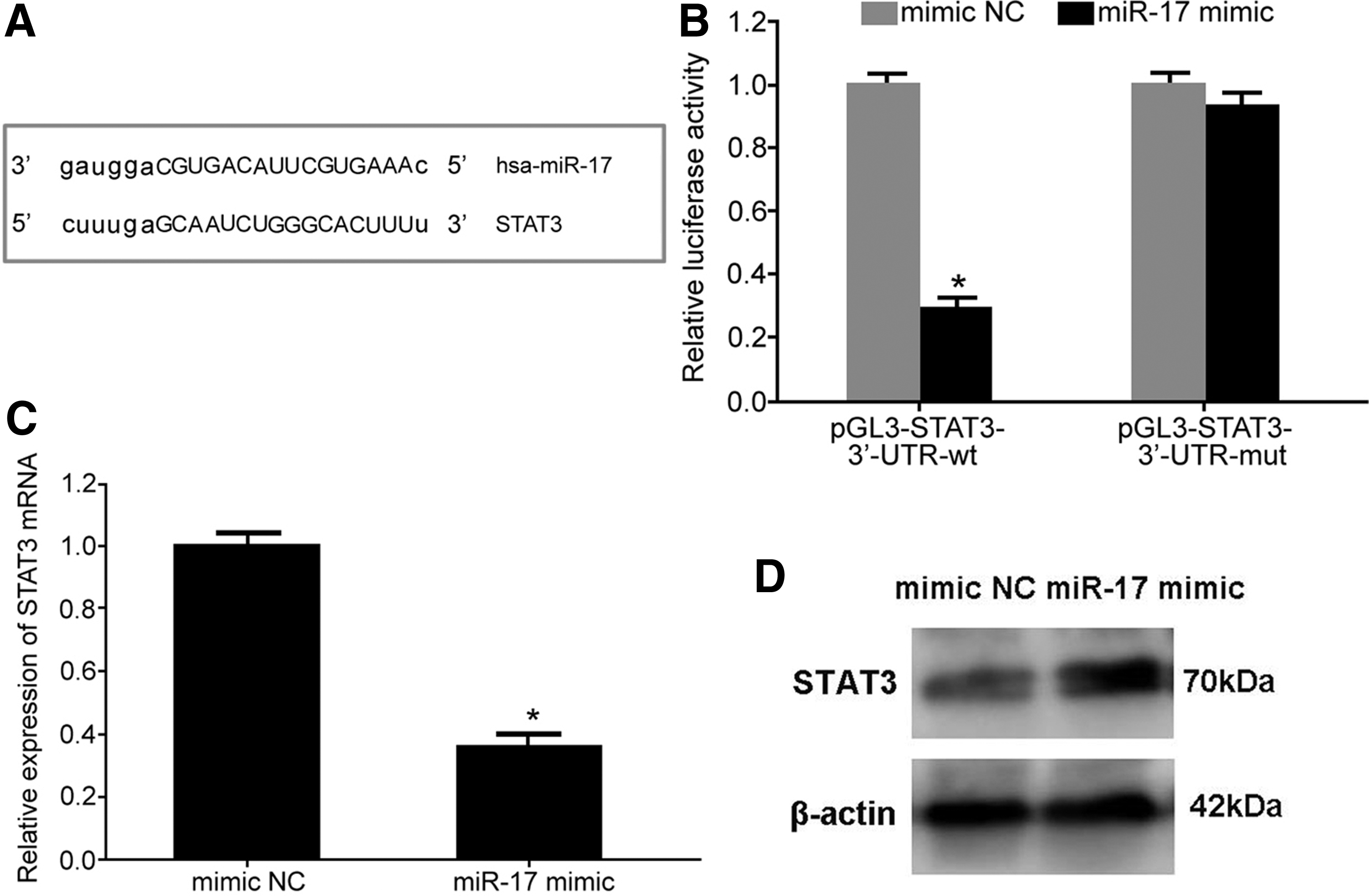
MiR-17 targeted regulated STAT3 expression.
Downregulated MiR-17 and increased STAT3 expression in LNCaP cells
To assess the expression profile of miR-17 and STAT3 in prostate cancer cell line, qRT-PCR was performed and showed that miR-17 expression was significantly reduced, while STAT3 mRNA was obviously elevated in LNCaP cells compared with RWPE-1 cells (Fig. 2A), consistent with the target relationship between miR-17 and STAT3. Western blot revealed that STAT3 protein expression was markedly enhanced in LNCaP cells compared with RWPE-1 cells (Fig. 2B). Flow cytometry demonstrated that cell proliferation was increased, while cell apoptosis was attenuated in LNCaP cells compared with RWPE-1 cells (Fig. 2C, D).
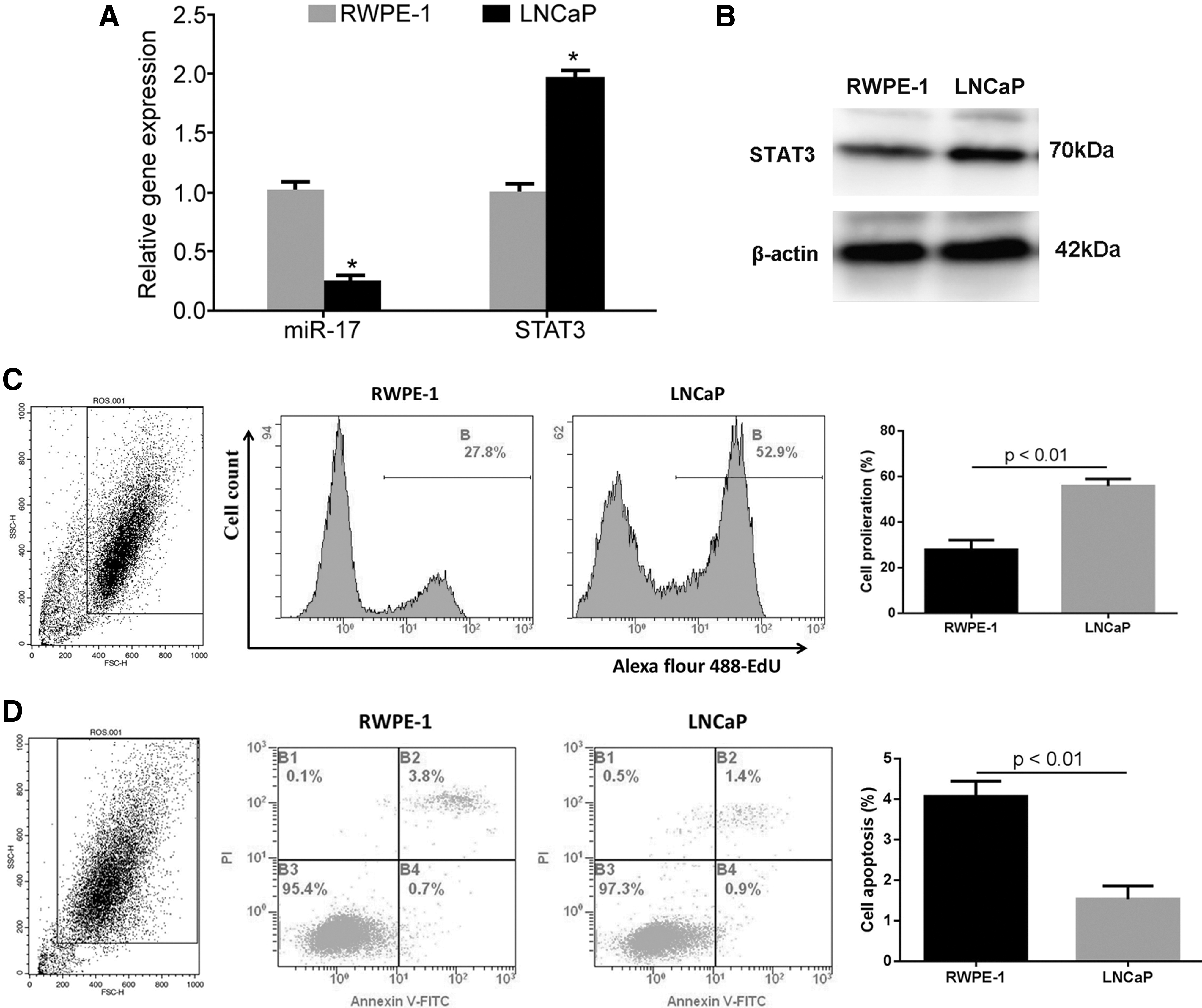
MiR-17 downregulated, while STAT3 increased in LNCaP cells.
MiR-17 overexpression suppressed cell proliferation and promoted cell apoptosis
To investigate the role of miR-17 in the biological behaviors of LNCaP cells, MiR-17 mimic was transfected into the cells. Their results showed that miR-17 mimic transfection markedly downregulated the expression of STAT3, p-STAT3, and Bcl-2 (Fig. 3A, B), attenuated cell proliferation (Fig. 3C), and enhanced cell apoptosis (Fig. 3D) in LNCaP cells.
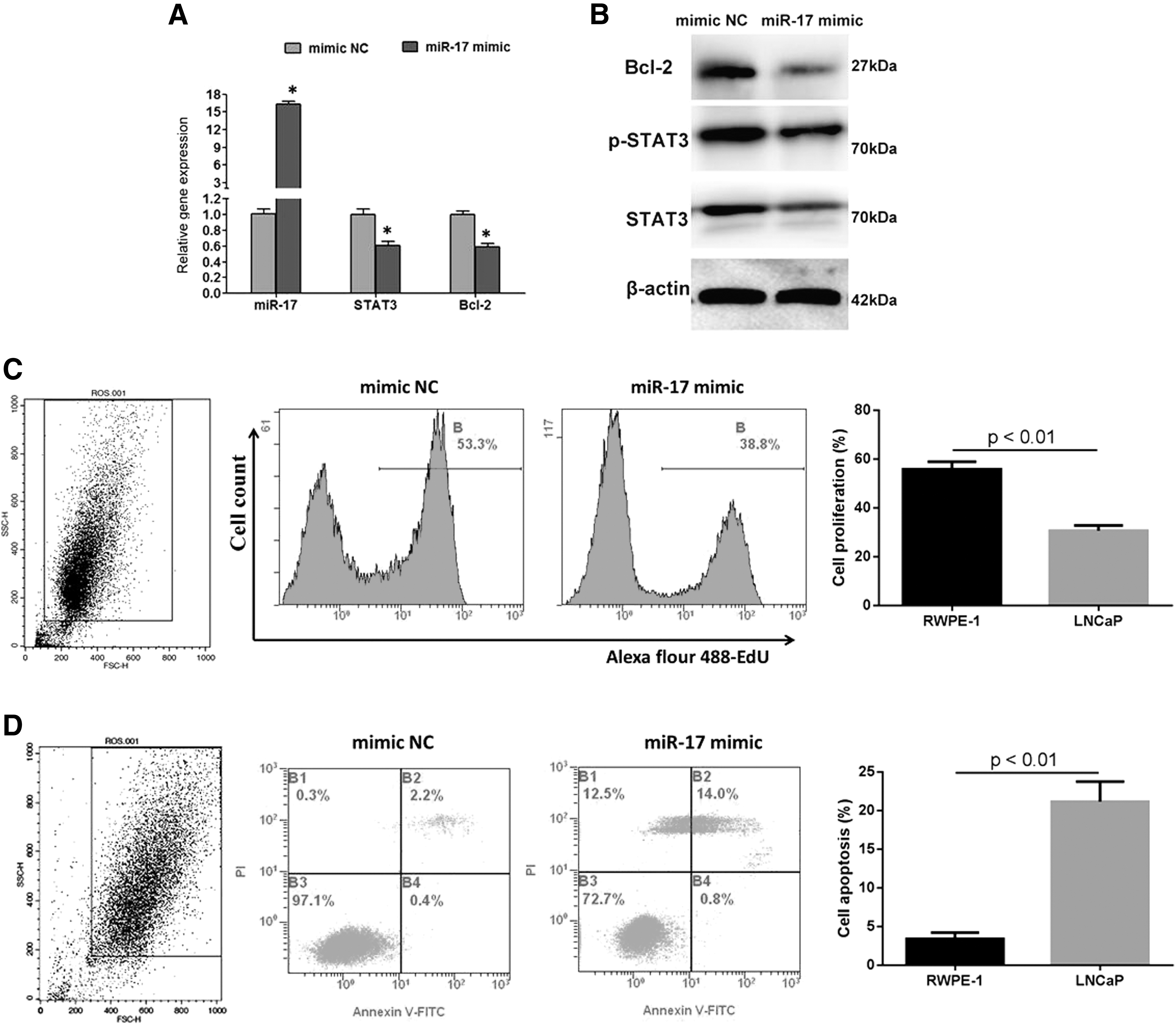
MiR-17 overexpression suppressed cell proliferation and promoted cell apoptosis in LNCaP cells.
STAT3 siRNA restrained cell proliferation and facilitated cell apoptosis
Si-STAT3 transfection significantly reduced the expression of STAT3, p-STAT3, and Bcl-2 (Fig. 4A, B), inhibited cell proliferation (Fig. 4C), and elevated cell apoptosis (Fig. 4D) in LNCaP cells.

STAT3 siRNA restrained cell proliferation and facilitated cell apoptosis in LNCaP cells.
Discussion
JAK-STAT plays a crucial role in extracellular factor response, intracellular signal transmission, cell proliferation, apoptosis, differentiation, and angiogenesis. 16 JAK-STAT3 signaling pathway can respond to multiple extracellular growth factor and mitogen stimulus. JAK-STAT activation makes the cell membrane receptor dimerization, leading to phosphorylation of JAK, which further phosphorylates receptor tyrosine kinases. It promotes STAT3 substitute to the tyrosine phosphorylation site of receptor complex through SH2 structural domain. Meanwhile, JAK activates adjacent STAT3 protein, leading to STAT3 dissociation from receptor complex, dimer formation, and nuclear translocation. It thus facilitates gene transcription and expression, which were related to cell proliferation, cycle, apoptosis, migration, and invasion. 17 STAT3 is an important transcription factor in JAK-STAT signaling pathway, including STAT1, STAT2, STAT3, STAT4, STAT5a, STAT5b, and STAT6, among which STAT3 is the most important one related to tumorigenesis. 17 STAT3 plays the key role in JAK-STAT signaling pathway to promote target gene transcription and expression, facilitate cell proliferation, and antagonize cell apoptosis, thus to act as an oncogene during tumorigenesis.
Numerous studies revealed that miRNA abnormal expression and function play a crucial role in multiple tumor pathogenesis. 11 –13 MiR-17 was found to be significantly reduced in prostate cancer tissue and cells, suggesting that it might be a tumor suppressor in prostate cancer tumorigenesis. 14,15 As a type of transcription factor, STAT3 regulates multiple target gene transcription and expression, such as Cyclin D1, VEGF, and Survivin, thus to accelerate cell proliferation and antagonize apoptosis. Bcl-2 is a kind of proto-oncogene that regulates mitochondrial dependent apoptosis signaling pathway. 18 Bcl-2 plays an anti-apoptotic role by restraining mitochondrial cytochrome C (Cyt C) release, reducing ROS production and lipid peroxide formation, inhibiting calcium ion transmembrane fluxes, and blocking apoptosis signaling transduction. 19 Several studies demonstrated that Bcl-2 is an important target gene of JAK-STAT3 signaling pathway. 20,21 JAK-STAT3 signaling activation resulting from STAT3 overexpression is associated with the tumorigenesis, progression, drug resistance, and poor prognosis of colorectal cancer, 5 lung cancer, 4 and breast cancer. 7 It was shown that STAT3 expression was significantly increased in PCa tumor tissue, which affected tumor progression and distant metastasis. 8,9 Considering the opposite expression profile of miR-17 and STAT3 in PCa tumor tissues, whether there was a relationship between them remains unclear. Using bioinformatics analysis, the authors showed the complementary binding site between miR-17 and STAT3. This study aimed to investigate the role of miR-17 in regulating JAK-STAT signaling pathway and affecting prostate cancer cell proliferation and apoptosis.
Dual luciferase assay revealed that miR-17 mimic transfection significantly declined the relative luciferase activity of HEK293 cells, indicating the regulatory relationship between miR-17 and STAT3 mRNA. MiR-17 mimic transfection obviously declined the STAT3 expression in LNCaP cells, further confirming the targeted regulatory relationship between miR-17 and STAT3. Flow cytometry demonstrated that cell proliferation was increased, while cell apoptosis was attenuated in LNCaP cells compared with RWPE-1 cells. Moreover, miR-17 expression was significantly reduced, while STAT3 level was obviously elevated in LNCaP cells compared with RWPE-1 cells. It indicated that downregulation of miR-17 plays a role in increasing the expression of STAT3, p-STAT3, and Bcl-2, facilitating LNCaP cell proliferation and antagonizing apoptosis. Ottman found that miR-17 expression was markedly reduced in PCa cell line DU145, M12, PC3, and LAPC4 compared with human benign prostatic hyperplastic cells BPH-1. The level of miR-17 expression also apparently decreased in PCa tissue compared with adjacent normal control. 15 Gong et al. reported that miR-17 level was significantly downregulated in PCa cells LNCaP, C4-2B, and PC3 compared with normal prostate epithelial cells RWPE1 and PrEC. 14 Yu et al. suggested that miR-17 level was reduced in PCa cells compared with normal prostate epithelial cells. 22 Xu et al. exhibited that miR-17 was declined in PCa cells DU145 and PC3 compared with PrEC cells. 23 Zhang et al. discovered that miR-17 was obviously downregulated in PCa tissue compared with normal prostate epithelium, and it decreased following pathological upgrading and tumor size enlargement. 24 In this study, miR-17 expression was markedly decreased in PCa cells, suggesting that miR-17 may be involved in PCa tumorigenesis, which was consistent with previous studies conducted by Gong et al., 14 Yu et al., 22 Xu et al., 23 and Zhang et al. 24 Abdulghani et al. showed that STAT3 expression was significantly upregulated in PCa tissue. 25 This study indicated that STAT3 level was obviously higher in PCa cells than that in normal epithelial cells, suggesting that STAT3 might be an oncogene in PCa, which was similar with Abdulghani et al. 25 Further investigation demonstrated that MiR-17 mimic transfection markedly downregulated the expression of STAT3, p-STAT3, and Bcl-2, attenuated cell proliferation, and enhanced cell apoptosis in LNCaP cells. STAT3 siRNA also induced similar effect as miR-17, revealing that miR-17 regulates PCa cell proliferation and apoptosis by targeting STAT3. Ottman proposed that upregulation of miR-17 suppressed PCa cell proliferation, restrained tumorigenesis in animal model, attenuated EMT process and invasion, and enhanced sensitivity to docetaxel. 15 Gong et al. found that miR-17 overexpression inhibited PCa LNCaP cell proliferation through targeting p300/CBP-associated factor (PCAF) expression. 14 Yu et al. demonstrated that Phenethyl isothiocyanate (PEITC) treatment attenuated PCa cell proliferation through upregulating miR-17 to target suppressed PCAF expression. 22 Zhang et al. found that miR-17 overexpression obviously restrained PCa M12 cell proliferation and alleviated cell migration and invasion. 24 Xu et al. revealed that miR-17 weakened PCa DU145 and PC3 cell proliferation by targeting MnSOD, GPX2, and TrxR2 expressions. 23 Abdulghani et al. showed that STAT3 overexpression apparently enhanced DU145 cell motility, migration, and metastasis, while inhibition of STAT3 activation attenuated PCa cell motility and migration. 25 In this study, upregulation of miR-17 alleviated the malignancy of PCa cells, which was similar with Zhang et al. 24 and Xu et al. 23 Downregulation of STAT3 also reduced PCa cell malignancy, which was in accordance with Abdulghani et al. 25 This study revealed the role of miR-17 reduction in regulating STAT3 and Bcl-2 expression and promoting PCa pathogenesis. Except Bcl-2, Survivin and Cyclin D1 are also the target genes of STAT3. This study only showed that miR-17 plays a role in regulating STAT3 and Bcl-2 and affecting PCa cell proliferation and apoptosis; other potential mechanisms still need further investigations.
Conclusion
MiR-17 overexpression inhibited LNCaP cell proliferation and induced cell apoptosis by downregulating the expression of STAT3, p-STAT3, and Bcl-2.
Footnotes
Acknowledgment
This work was supported by Wanzhou Sanxia Central Hospital of Chongqing (No.201303005).
Disclosure Statement
No competing financial interests exist.