
Abstract
Hepatocellular carcinoma (HCC) is one common malignancy. The authors previously demonstrated that miR-570 regulates the development of HCC. This study detected the effect of miR-570 on cell apoptosis, angiogenesis, T cell activation, and proliferation in a tumorigenicity assay in nude mice. miR-570 mimics and negative control (NC) were transfected into SMMC7721 cells, and then, the cells were subcutaneously injected in the right flank in nude mice. Six weeks later, the dissected tumors and peripheral blood were collected. Tumor weight and volume were measured, and expression of miR-570 and apoptosis-related gene Bax/Bcl-2 was detected by quantitative real-time polymerase chain reaction. Hematoxylin and eosin, immunohistochemistry of CD31 and vascular endothelial growth factor (VEGF), TUNEL assay, and flow cytometry detection of CD4 and CD8 in peripheral blood were performed. miR-570 mimics suppressed tumor growth compared with the NC, with decreases in tumor weight and tumor volume. Very few CD31 and VEGF were found in tumor sections in miR-570 mimics group. Bax level was significantly increased, while Bcl-2 level was significantly downregulated. Significant lower ratio of CD3+CD4+ T cells and higher ratio of CD8+IFN-γ+ T cells were found in peripheral blood and tumor tissues in miR-570 mimics than NC. Collectively, miR-570 plays an important role in the proliferation, angiogenesis, and immune escape of HCC, which might be potential diagnostic and predictive biomarkers.
Introduction
Hepatocellular carcinoma (HCC) is the most common type of liver cancer, which accounts for 70%–90% of primary liver cancer worldwide. 1 During 2012, in the world, there were ∼782,500 new cases of liver cancer and 745,500 deaths. 1 There is a trend of increasing year by year. New cases and deaths of HCC in China account for about 50% worldwide. 1 In China, the incidence and mortality of HCC are higher than other malignancies. HCC has become the second leading cause resulting in cancer death, only less than gastric cancer in the country and lung cancer in the city.
The treatment for HCC depends largely on the age of the patients, the liver function, the size of tumor, and the presence of metastasis and vascular invasion. 2 At present, the treatment strategies of HCC include surgery such as hepatectomy, palliative surgery, and liver transplantation, and nonsurgical treatment such as local ablation therapy, arterial embolization, chemotherapy, radiotherapy, and molecular target and biological therapies. 3,4 Among them, the surgical resection is still the most effective way for improving the long-term survival. 5 For HCC, there were no effective adjuvant therapies. After resection of HCC for a certain period of time, hepatoma cells remain in dormant state with an estimated doubling time up to 10–20 months. However, the 5-year recurrence and metastasis rates of HCC are still as high as 50%–70%. The postoperative recurrence of HCC has become the major cause resulting in HCC patient deaths. Although the pathology negative margins were evaluated after surgical resection, a small residual HCC tissue might exist. The residual HCC cell in the dormant state without obvious proliferation, thus escape from the immune surveillance, not be recognition and killing. 6 –9 Those cells have the potential of proliferation, which can activate and proliferate to cause HCC recurrence and metastasis. 10 –13 Therefore, the postoperative dormancy is the key to the outcome and prognosis of HCC.
The mechanism of HCC dormancy is not clear, which may involve angiogenesis and immune response. 7,14,15 A variety of vascular growth factors can promote tumor angiogenesis, and the tumor itself can induce the secretion of these vascular growth factors by a variety of ways. Among them, vascular endothelial growth factor (VEGF) plays an important role in the growth of tumor blood vessels. 6,10 VEGF mainly regulates the growth and survival of endothelial cells. 16 The secretion of VEGF not only promotes tumor angiogenesis and accelerates the tumor development but also induces the dormant tumor cells to rejuvenate the cell cycle progression, leading to tumor recurrence and metastasis. In addition, the immune system has no ability to kill and eliminate the intolerable tumor cells, and specific immunophenotypes were exhibited. 7,8
The authors previously demonstrated that miR-570 targets on B7-H1 (also known as PD-L1 or CD274) involving in HCC. 17 B7-H1 inhibits activation and proliferation of T cells, suppressing immune response and affecting the immune escape of cancer cells. 18,19 This study detected the roles of miR-570 in HCC cell apoptosis, angiogenesis, T cell activation, and proliferation in a tumorigenicity assay in nude mice.
Materials and Methods
Cell culture and transfection
Human HCC line SMMC7721 cells were purchased from ATCC (Rockville, MD) and were cultured in RPMI 1640 medium with 10% fetal bovine serum at 37°C, 5% CO2. The experiments were performed, while cells reached 70%–80% confluency.
The miR-570 mimics and miR-negative control (NC) were synthesized by GenePharm, China. 17 Before transfection, 2 × 10⁵/well cells were seeded in a 6-well plate. After 24 h, cells were transfected with miR-570 mimics (50 nM) or miR-NC (50 nM) using FuGENE® HD transfection reagent (Promega, Madison, WI) according to the manufacturer's instruction. The transfected cells were cultured in an antibiotic-free medium for 48 h, and then 100–300 μg/mL G418 (Geneticin; Life Technologies, Inc., Rockville, MD) was used for selection about 20 d.
Tumorigenicity assay in nude mice
The tumorigenicity assay was performed as described previously. 17 Six-week-old female BALB/c athymic nude mice (n = 12) were obtained from Vitalriver Laboratory Animals (Beijing, China), and randomly divided into two groups, including miR-570 stable transfection cell group and NC transfection cell group. The stable transfection cells in exponentially growing were collected and resuspended in a serum-free medium. For subcutaneous injection, cells were resuspended in 2.0 × 10⁶ cells/0.1 mL phosphate buffered saline, 0.1 mL cells were injected in the right flank. After tumor formed, the volume of tumors (V) was measured by caliper daily. The formula for volume calculation was V = (L × W2)/2, where L was the length of tumor and W was the width. Once the tumor reached an average 30–69 mm3, the mice were inoculation of miR-570 and NC for 42 d. The mice were randomly divided into two groups (n = 6) for inoculation of miR-570 and NC for 42 d after tumors reached. After 6 weeks, the mice were euthanized, and the dissected tumors and peripheral blood were collected. Tumors were weighted and calculated volumes. The expression levels of miR-570, CD31, and VEGF, change in apoptosis (TUNEL, Bax/Bcl-2), and levels of CD4 and CD8 in peripheral blood were performed. The animal experimental protocols were approved by the Laboratory Animal Center of the First Affiliated Hospital of Sun Yat-Sen University.
Quantitative real-time polymerase chain reaction
Total RNAs from cells were isolated using TRIZOL (Invitrogen, Inc., Carlsbad, CA). The concentration of RNAs was measured using GeneQuant II (Pharmacia, Uppsala, Sweden) at 260 nm. According to the manufacturer's manual (Invitrogen), reverse transcription reaction and complementary DNA synthesis were performed. Then, polymerase chain reaction (PCR) analysis was performed using the SYBR Premix Ex Taq GC Kit (Takara, Japan) on ABI 7500 System (ABI, Foster. City, CA). For PCR amplification, the stem-loop primers were synthesized by RiboBio, China. GAPDH and U6 were used as controls, and the final expression levels were normalized to GAPDH or U6 using the relative ΔΔCT method. 20 Three independent experiments were performed.
Hematoxylin and eosin staining, immunohistochemistry, and TUNEL assay
The dissected tumors were processed for immunohistochemistry (IHC) as standard protocols. Three-micrometer thick paraffin section using anti-CD31, anti-VEGF, anti-Bax, anti-Bcl-2, anti-CD4 and anti-CD8 antibody (Santa Cruz), and anti-rabbit poly-HRP-IgG. For histopathology, tissues were fixed in 4% neutral buffered formaldehyde and paraffin wax. Sections were cut into 4 mm thickness and stained with hematoxylin and eosin (H&E) for histologic assessment. For TUNEL assay, a fluorometric TUNEL Detection Kit (Beyotime, China) was used to detect apoptotic DNA strand breaks. The tumors were cut into 10 mm thick sections in a freezing cryostat at 200 µC. Sections were fixed with 4% neutral buffered formaldehyde for 20 min, permeated with proteinase K for 20 min, and incubated with the labeling reaction mixture for 2 h. Then, nuclei were stained with DAPI and images captured by fluorescence microscope (Olympus, Japan).
Flow cytometry analysis of CD4 and CD8
Peripheral blood specimens were collected from mice. Specimens were filtered using a 40 μm nylon mesh filter. Total T cells were stained with primary antibodies against CD4-FITC and CD8-FITC. Cells were acquired in flow cytometry (BD, Germany).
Statistical analyses
All data were shown as mean ± standard deviation from three independent experiments.
Statistical analyses were performed using the SPSS version 18.0 (SPSS, Inc., Chicago, IL) with analysis of variance and Tukey's t-test. p-Value <0.05 was considered to be statistically significant.
Results
miR-570 inhibits the tumorigenicity in vivo
The authors previously demonstrated that miR-570 is downregulated in Bel-7404, Huh-7, and HepG2 cells, and miR-570 mimics significantly induced cell apoptosis and attenuated the proliferation of HepG2 cells. 17 To further detect the role of miR-570, miR-570 mimics-transfected SMMC7721 cells were injected into nude mice. After 6 weeks, the tumor size in the group with miR-570 mimics was smaller than the NC (Fig. 1A), with decreases in tumor weight (Fig. 1B, p < 0.01) and tumor volume (Fig. 1C, p < 0.05). In the dissected tumors, miR-570 was upregulated in the group with miR-570 mimics (Fig. 1D, p < 0.01).

miR-570 suppresses tumorigenicity in vivo. Exponentially growing SMMC7721 cells collected from miR-570 stable transfections were harvested and resuspended in serum-free medium. Six-week-old female mice were subcutaneously injected in the right flank with SMMC7721 cells. Tumor volume and weight were measured, and expression of miR-570 was confirmed by qRT-PCR.
miR-570 promotes cell apoptosis and inhibits the proliferation of endothelial cells
The cell apoptosis and proliferation in tumor sections were detected (Fig. 2). H&E assays showed a significant decrease in cell density in tumor section (Fig. 2A). TUNEL assays showed significantly increased apoptotic cells in tumor section (Fig. 2B). These results further supported that miR-570 promotes cell apoptosis and inhibited cell proliferation. To detect whether miR-570 suppresses the proliferation of endothelial cells, IHC of CD31 and VEGF was performed (Fig. 2C, D). Very few CD31 and VEGF were found in tumor sections. Thus, miR-570 also inhibited the proliferation of endothelial cells and might contribute to decrease in angiogenesis in nude mice.
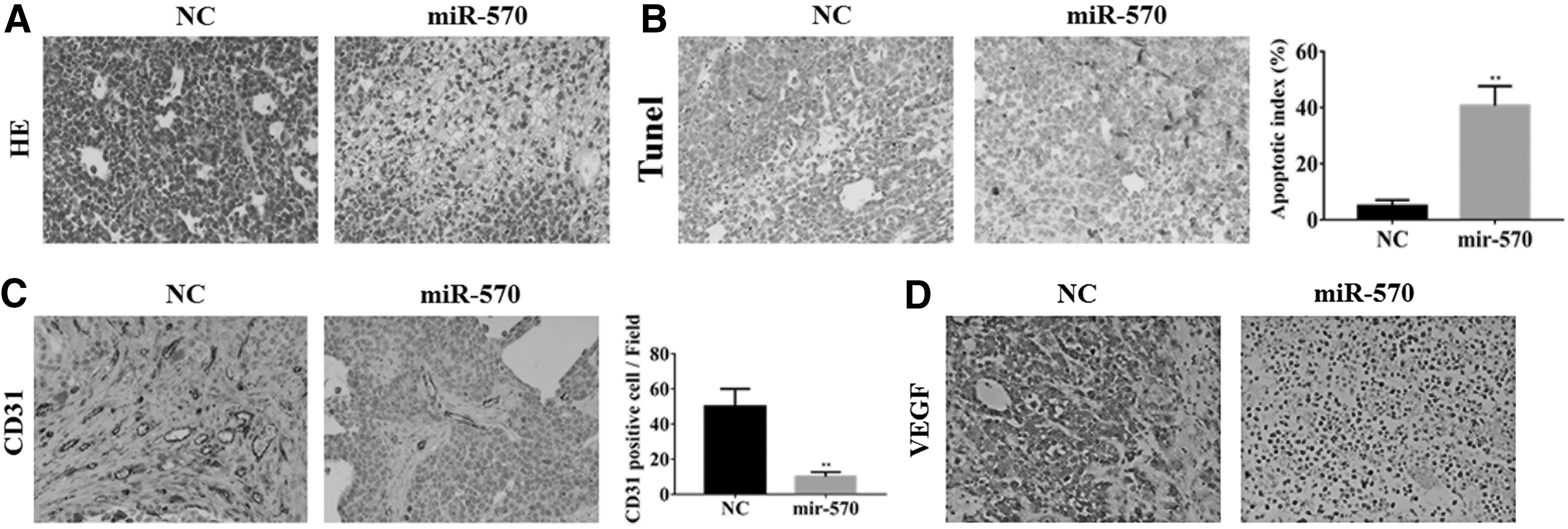
Cell apoptosis and proliferation in tumor sections.
miR-570 promotes expression of Bax, while it inhibits the Bcl-2 expression
To further analyze the potential mechanism of cell apoptosis induced by miR-570, the authors detected the levels of proapoptotic gene Bax and the antiapoptotic gene Bcl-2 in tumor sections (Fig. 3). IHC of Bax and Bcl-2 showed that Bax level was significantly increased (Fig. 3A) and Bcl-2 level was significantly downregulated (Fig. 3B). The significant increase and decrease in levels of Bax and Bcl-2 were confirmed by quantitative real-time PCR, respectively (Fig. 3C, p < 0.01).
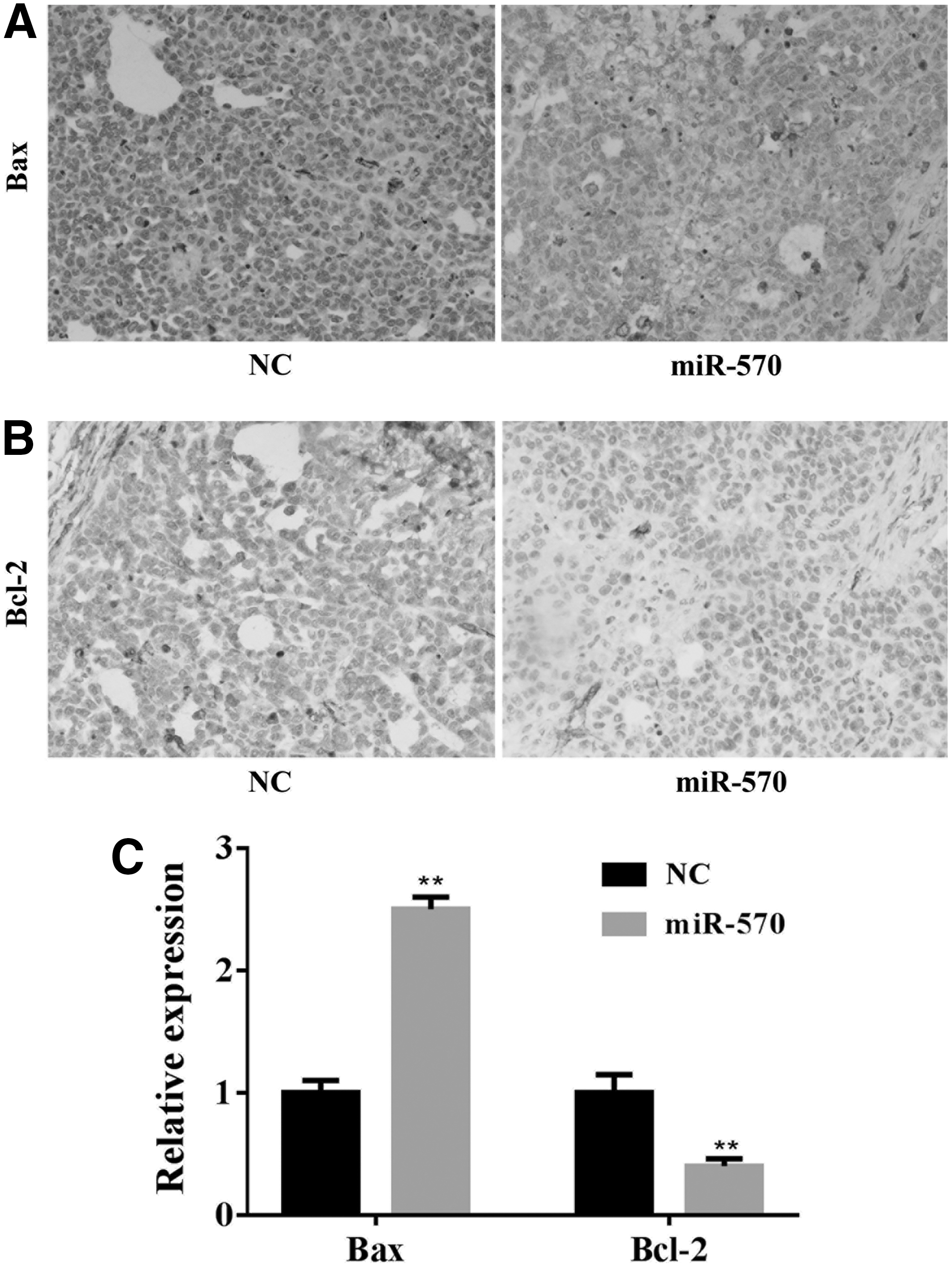
Levels of Bax and Bcl-2 in tumor sections.
miR-570 enhances CD3+CD8+IFN-γ+ T cells, while it decreases CD3+CD4+ T cells
To assess the influence of miR-570 on T cells, the authors measured the CD3+CD4+ and CD3+CD8+IFN-γ+ T cells in nude mice subcutaneously injected with miR-570 mimics or NC-transfected SMMC7721 cells by flow cytometry. Significant lower ratio of CD3+CD4+ T cells (Fig. 4A) and higher ratio of CD8+IFN-γ+ T cells (Fig. 4B) were found in peripheral blood in miR-570 mimics than NC (Fig. 4C). Besides, the expression of CD4 and CD8 was analyzed. The CD8 level was increased by miR-570 and the CD4 level was decreased by miR-570 in tumor tissues (Fig. 4D). Whether this expansion is related with a clinical benefit in patients remains to be elucidated.

Flow cytometry analysis of CD3+CD4+ and CD8+IFN-γ+ T cells.
Discussion
This study was designed to investigate the role of miR-570 in cell proliferation, apoptosis, and immune escape in HCC tissues in nude mice subcutaneously injected with miR-570 mimics or NC-transfected SMMC7721 cells. Future study needs to be designed to evaluate the clinical significance of miR-570 in HCC diagnosis, prognosis, and treatment.
MicroRNAs (miRNAs) are endogenous, with ∼22 nucleotides in mammals. miRNAs were served as negative regulators in gene expression. 21 It induced the target messenger RNA (mRNA) degradation through pairing with the 3′-untranslated regions of target mRNAs, or inhibited the translation of the target mRNAs through integrating into RNA-inducing silencing complexes, 22 and thus acted as tumor suppressors or oncogenes involving in various cancers, including HCC. 5,23,24 It was found that the dysregulation of miRNAs was different between noninvasive and muscle-invasive cancers 25,26 ; they are potential biomarkers and treatment targets for early diagnosis and treatment of HCC. 27
It was demonstrated that miR-570 inhibited the proliferation of gastric cancer and colorectal cancer cells. 28 Recently, miR-570 has been shown as one of the most important miRNAs in the miRNA-gene networks in alcoholic liver disease. 29 As is known, alcoholic liver disease has the ability to develop to HCC. In their previous studies, the authors found that in human HCC cell lines Bel-7404, Huh-7, and HepG2, the expression level of miR-570 was significantly lower than in nonmalignant cell lines L-02 and HL-7702. 6 Epithelial–mesenchymal transition is believed to promote the HCC cell migration, which ultimately facilitates tumor metastasis. 30 miR-570 suppresses cell proliferation and migration in HCC cells. 18 In functional studies, using MTT assay, invasion assay, and flow cytometry assay, the data illustrated that transfection of miR-570 significantly inhibited proliferation, migration, and invasion of HCC cells, and induced the cell early/total apoptosis. There was no significant difference between NC cell group and control HepG2 cell group. Reintroduction of miR-570 significantly suppressed the capability of migration and invasion in the three human HCC cell lines. 17 In the dissected tumors, miR-570 was upregulated by miR-570 mimic transfection; the levels of proapoptotic gene Bax and the antiapoptotic gene Bcl-2 in tumor sections were significantly increased and downregulated, respectively. These results suggested that miR-570 promoted cell apoptosis. CD31 and VEGF are well-studied markers of endothelial cells. 16 Very few CD31 and VEGF were found in tumor sections. Thus, miR-570 not only inhibited the proliferation of HCC cells but also inhibited the proliferation of endothelial cells, and might contribute to decrease in angiogenesis in nude mice.
The mechanism of HCC dormancy may involve both angiogenesis and immune response. 7,14,15 The authors previously found that B7-H1 was directly targeted by miR-570. Tregs play a critical role in tumor dormancy or recurrence. 8 MiR-570 significantly decreased ratio of CD4+ T cells and increased ratio of CD8+ T cells, indicating that miR-570 promotes immune function and might inhibit the immune escape of HCC cells.
Taken together, miR-570 plays a critical role in the proliferation, angiogenesis, and immune escape of HCC cells, which might be potential diagnostic and predictive biomarkers. The molecular mechanisms by which miR-570 affects HCC and modulates the immune escape of HCC cells would be studied in the future.
Footnotes
Acknowledgments
This study was supported by the National Natural Science Foundation of China (Grant No. 81373582) and the Administration of Traditional Chinese Medicine of Guangdong, China (Grant Nos. 20161002 and 20171001).
Disclosure Statement
No competing financial interests exist.