
Abstract
Background:
To study the distribution and imaging of 99mTc-nGO-PEG-FA in human pancreatic cancer Patu8988 tumor-bearing nude mice, and to explore its usefulness as an imaging reagent for pancreatic cancer.
Materials and Methods:
Natural graphite powder was used as raw material to prepare the nanosized graphene oxide (nGO) by using the modified Hummers method, and then was covalently modified by polyethylene glycol (PEG) on the surface of nGO. The nGO was further optimized by in vitro cell experiment, and then conjugated with the targeting molecule folic acid (FA) to form nGO-PEG-FA system. The nGO-PEG-FA was finally labeled by radioactive nuclide 99mTc by direct labeling method to form the 99mTc-nGO-PEG-FA molecular imaging probe. Nude mice bearing patu8988 pancreatic cancer xenografts were intravenous injection (I.V.) injected with 99mTc-nGO-PEG-FA, and the distribution of 99mTc-nGO-PEG-FA in nude mice at different time course was investigated by determination of tissue uptake of radioactivity (%ID/g), as well as the single photon emission computed tomography (SPECT) imaging at different time course.
Results:
The labeling rate of nGO-PEG-FA with 99mTc was (90.08 ± 2.34)%, and the highest binding rate of 99mTc-nGO-PEG-FA with Patu8988 cells was (3.15 ± 0.31)%. The radioactive uptake in tumor reached (5.11 ± 1.23)%ID/g at 6 h after I.V. injection of 99mTc-nGO-PEG-FA in nude mice. Meanwhile, the radioactive uptake in liver, spleen, and lung was also high and reached (10.33 ± 1.22)%ID/g, (5.86 ± 0.59)%ID/g, and (3.55 ± 0.93)%ID/g, respectively, whereas less radioactivity uptake was observed in the heart (1.12 ± 0.33)%ID/g and blood (2.76 ± 0.39)%ID/g, respectively. The tumors can be clearly imaged at 4.0–6.0 h after 99mTc-nGO-PEG-FA injection.
Conclusions:
99mTc-nGO-PEG-FA can efficiently target pancreatic cancer, which may be developed as an imaging agent for pancreatic cancer.
Introduction
P
Graphene is a new, two-dimensional material composed of single layer of carbon atoms, and is composed of sp2-hybridized carbon atoms arranged in a hexagonal honeycomb lattice. Because nanographene has a large specific surface area, it can be used as a variety of biomolecule carrier for biological detection, drug and gene delivery. 7 –10 Furthermore, it was found that nanographene that has been modified by biocompatible polyethylene glycol (PEG) can be accumulated in tumor area of tumor-bearing animal model without the help of targeted antibody by tumor tissue permeability enhancement and retention effect (enhanced permeability and retention effect [EPR] effect). 11
As a common target molecule, folic acid (FA) is composed of glutamic acid, p-aminobenzoic acid, and pteridine nucleus. FA is a necessary vitamin for cells, especially for proliferative cells. It has been found that free FA and FA conjugates can enter cells through the folate receptor-mediated endocytosis. Folate receptor is overexpressed in the surface of most malignant tumor cells, such as the pancreas, breast, ovary, kidney, lung, brain, bone marrow, and other tumor cells, 12 –15 whereas there is less expression on the surface of normal cells. Because FA has a small molecular weight and is not immunogenic, it is an ideal carrier for tumor-targeted therapy. Therefore, the application of FA complex in tumor targeting imaging and treatment has become of the great interest in the area. 16 –19
In this experiment, nanosized graphene oxide (nGO) was prepared by modified Hummers method 20 and its surface was covalently modified with PEG, and then ligated to molecular folate (FA) to form the nGO-PEG-FA system, and finally, nGO-PEG-FA was labeled with radionuclide 99mTc by direct labeling method 21 . The in vivo tumor targeting and biological properties of 99mTc-nGO-PEG-FA were studied by radionuclide imaging of the tumor-bearing animal model and its usefulness as a molecular probe for tumor-targeted imaging was further explored. As far as the authors know, this is the first report in the area and similar studies have not been reported so far.
Materials and Methods
Materials and reagents
Analytical purity of natural graphite powder, stannous chloride (SnCl2·2H2O, analytical grade), and ascorbic acid (Vc, analytical grade) 1-(3-Dimethylaminopropyl)-3-ethylcarbodiimide Hydrochloride (EDC·HCl), N-hydroxysuccinimide (NHS-HCl), sodium chloride, polyethylene glycol (PEG), sulfuric acid, hydrogen peroxide (H2O2), and sodium hydroxide (NaOH) were purchased from Sinopharm Chemical Reagent Co., Ltd. FA was purchased from Haoran Biological Co., Ltd. Sodium nitrate was purchased from China Pharmaceutical Group Shanghai Chemical Reagent Co., Ltd. Potassium permanganate (analytical grade) was purchased from Shanghai Meixi Chemical Co., Ltd. Na99TcmO4 eluent was purchased from Shanghai Xinke Pharmaceutical Co., Ltd. Roswell Park Memorial Institute (RPMI) 1640, fetal bovine serum (FBS), and trypsin were obtained from Gibco.
Radionuclide calibrator CRC-15R (US Capintec company), SN-695B type radioimmunoassay (RIA)-γ meter (Shanghai Rihuan Photoelectronic Instrument Co., Ltd., Shanghai Institute of Applied Physics), BS224S electronic balance (Germany Sartorius Co., Ltd.), CR3 type Low temperature centrifuge and carbon dioxide (CO2) incubator (Jouan, France), inverted microscope (Olympus, Japan). Millipore ultrafiltration centrifuge tube (Shanghai Kenqiang Instrument Co., Ltd.). High-speed refrigerated centrifuge (Hitachi, Japan), ultrasonic cleaning instrument (SK8200H; Shanghai Branch Guided Ultrasound Equipment Co., Ltd.), and HWS12 constant temperature water bath (Shanghai Heng Technology Co., Ltd.).
Preparation of nGO
One gram of natural graphite powder and 60 g of sodium chloride were weighed and mixed in a mortar, and thoroughly ground. After 30 min, the mixture was dissolved with the appropriate amount of deionized water and filtered to get the expanded graphene powder. The filtered material was dried and then put into a 250 mL single-necked flask. A total of 0.5 g of sodium nitrate and 23 mL of concentrated sulfuric acid with a concentration of 98% were added into the flask and the mixture was stirred for 30 min. Three grams of potassium permanganate was then gradually added into the flask and continuously stirred for another 8 h at 10°C condition. The reaction mixture was stirred at 35–40°C for 30 min and then at 65–80°C for 45 min. After that, 46 mL of deionized water was added to the flask and continuously stirred at 98–105°C for 30 min. Finally, 12 mL of 3% H2O2 was added into flask and the reaction was terminated after 30 min of stirring. The reaction solution was sonicated in an ultrasonic machine for 2 h and then washed with 5% of hydrochloric acid several times by centrifugation, and then washed with deionized water to get a pH value of 7.0 for supernatant after centrifugation. The sample was then freeze-dried to obtain a single layer of oxide graphene (nGO). The structure is shown in Figure 1.
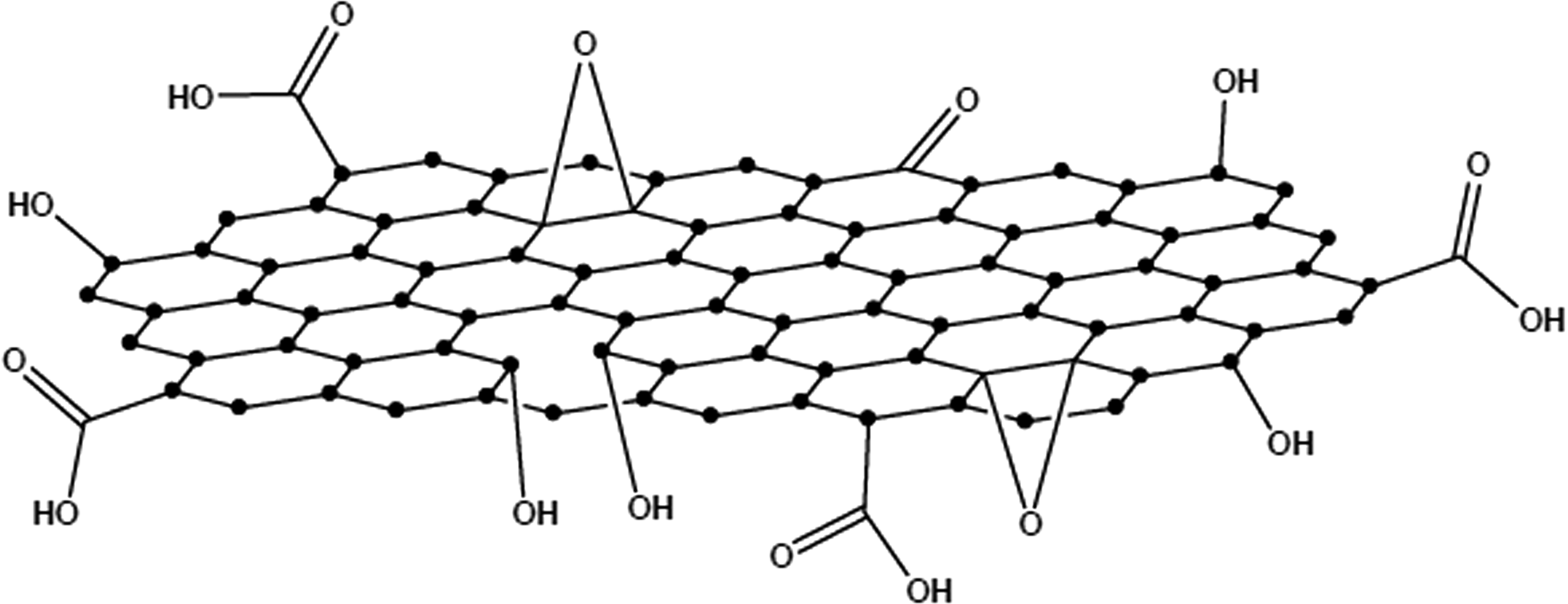
Molecular structures of nanographene oxide.
Preparation of nGO-PEG
Fifty milligrams of prepared graphene oxide was weighed and dispersed in water at a concentration of 1 mg/mL. Seven hundred fifty milligrams of NHS was added to the solution and stirred for 30 min to activate the carboxyl group. Two hundred fifty milligrams of EDC was then added to the solution and the pH value of the solution was adjusted to 5.76 with 1 mg/mL of NaOH. At this time point, 520 mg PEG was added to the solution and stirred at room temperature for 24 h. The solution was washed with deionized water several times with centrifugation and then freeze-dried for 24 h to get nGO-PEG. The structure is shown in Figure 2.

Molecular structures of nGO-PEG. nGO, nanosized graphene oxide; PEG, polyethylene glycol.
Preparation of nGO-PEG-FA
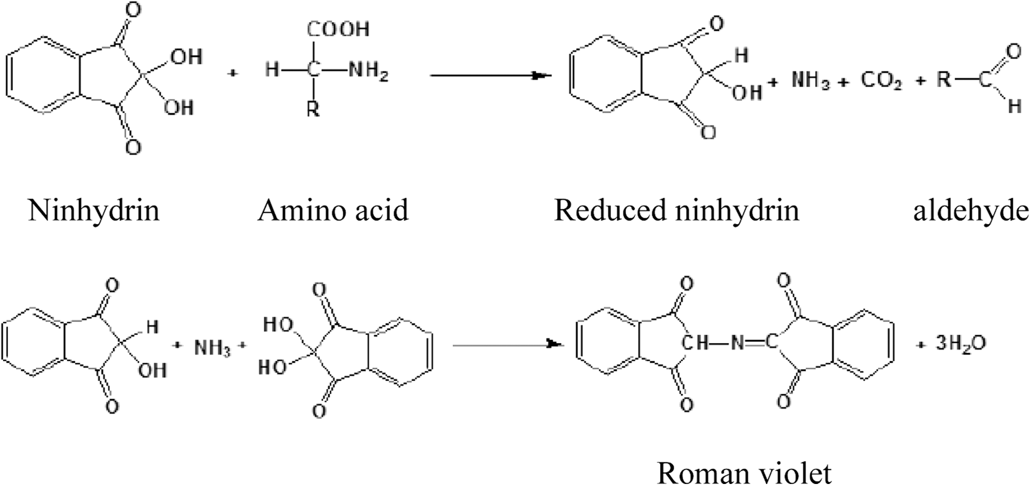
One milligram of FA was weighed and fully dissolved in 1 mL of dimethylformamide (DMF). Five milligrams of NHS was added into the solution and stirred for 30 min to activate the carboxyl group. Fifteen milligrams of EDC and 1 mL of nGO-PEG (1 mg/mL) were then sequentially added into the solution and stirred in the dark overnight. After that, a small amount of DMF was added to the reaction solution and centrifuged one to two times in an Amicon centrifugal filtration apparatus (molecule weight cut-off [MWCO] = 100 kDa) to remove the unreacted FA and then washed several times with deionized water to remove NHS and EDC presented in the reaction solution. Finally, the material was suspended into saline to get nGO-PEG-FA. The structure of nGO-PEG-FA is shown in Figures 3 and 4. To specifically modify the small molecule nanographene oxide with the target molecule FA, the surface amino group concentration of the nanoparticles was measured. The ninhydrin colorimetric method was used to determine the amino group concentration on the nanomaterial surface. Ninhydrin is a reagent that can be used to detect ammonia or primary and secondary amines, especially amino acids. These free amines and ninhydrin hydrates, when heated under weak acid conditions, can produce a dark blue or purple material called Roman violet. The substance has a maximum absorption peak at a wavelength of 570 nm. Since the size of the absorption peak is proportional to the concentration of the amino group, this method can be used to measure the amino concentration of an unknown concentration sample. The reaction mechanism is as follows:
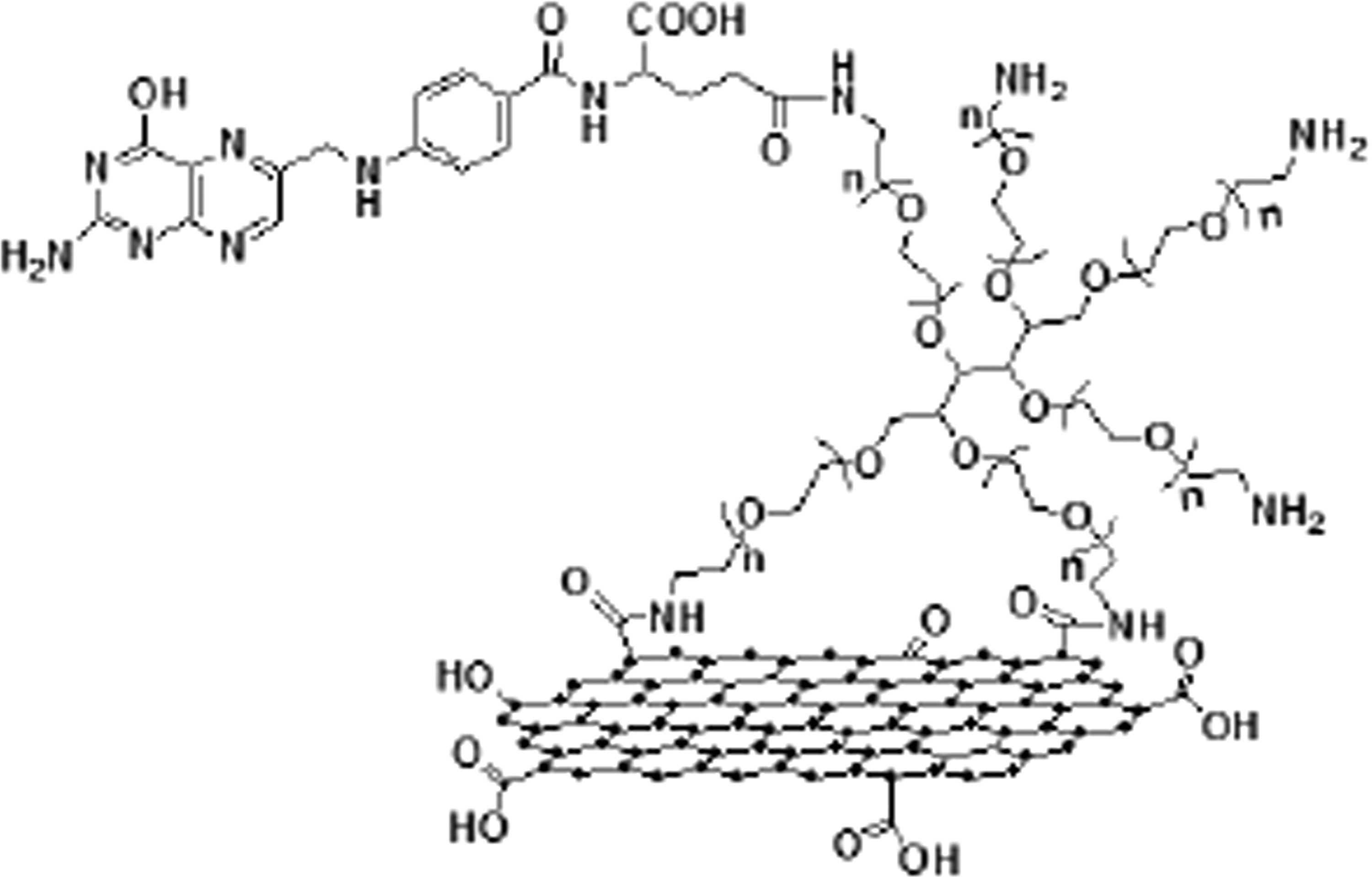
Molecular structures of nGO-PEG-FA. FA, folic acid.
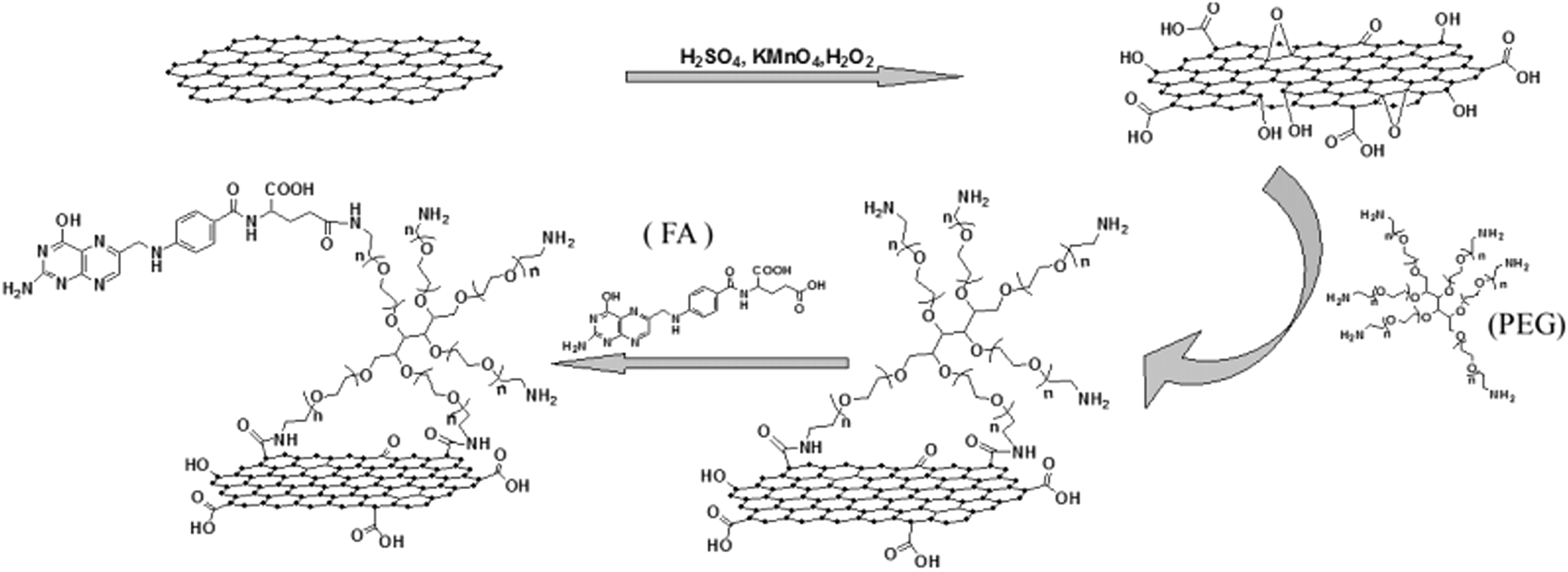
The procedure of nGO-PEG-FA.
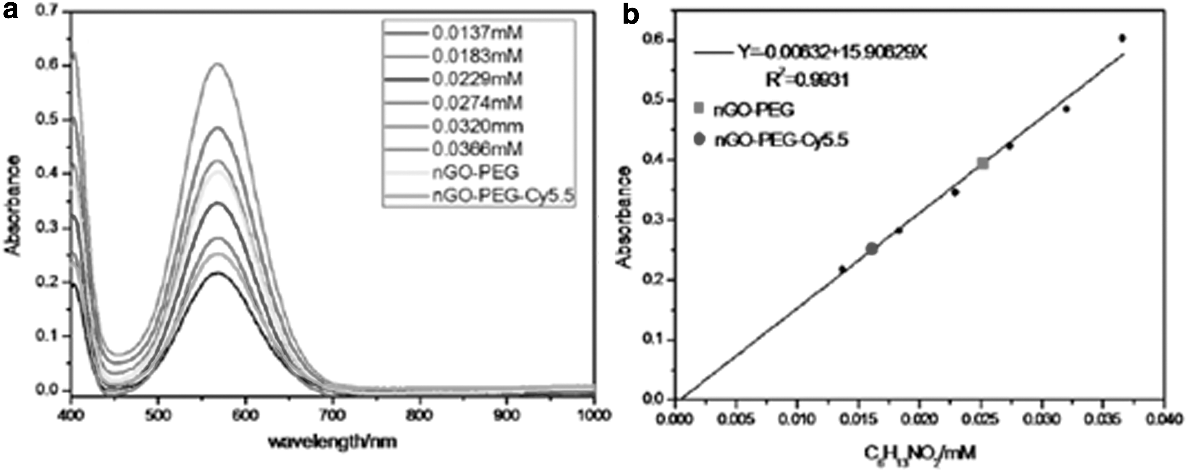
The ultraviolet (UV) absorption curves of L-Leucine standard solutions with different concentration gradients were plotted, and a standard curve was plotted using the relationship between the UV absorbance and the concentration of the solution substance at the wavelength λ = 570 nm. To determine the density of amino groups on the surface of the nGO-PEG nanomaterial, the solution after the reaction from the nanomaterials and ninhydrin was tested in a fluorescence cuvette for the UV absorption curve. According to its absorbance at wavelength λ = 570 nm (Fig. 5a), it is substituted into the standard linear relationship between absorbance and amino concentration (Fig. 5b) to calculate the amino concentration of the reaction, and the amino group is calculated together with the reaction volume. The value of nanomaterials divided by the ninhydrin reaction mass is the density of the amino groups on the surface of the nGO-PEG nanomaterial. According to the above calculation method, the surface amino group density of the nGO-PEG nanomaterial was approximately 2.51 × 10⁻⁵ mol/g, and the amino group capable of reacting with FA was ∼64.1%. FA molecules are labeled onto the nanomaterial surface by amide reaction. FA first activates its carboxyl group with NHS and EDC, and then forms an amide bond with the amino group on the surface of the nanomaterial, which is attached to the surface of the nanomaterial. Since the folate solution absorbs to varying degrees in the UV-visible (UV-Vis) region, the UV-Vis absorption curve can be used to characterize its functional success, as shown in Figure 6. According to the absorption curve of UV-Vis, it can be known that the characteristic absorption peak of pure folate molecules in water is at 280 and 365 nm. Since the unfunctionalized nGO-PEG nanomaterial has no characteristic UV absorption in solution, the nGO-PEG-FA nanomaterial with the folic modified by amide reaction exhibits two characteristic absorption peaks of folate in the UV-Vis absorption curve in solution, suggesting that FA was successfully conjugated on the nanoparticles.
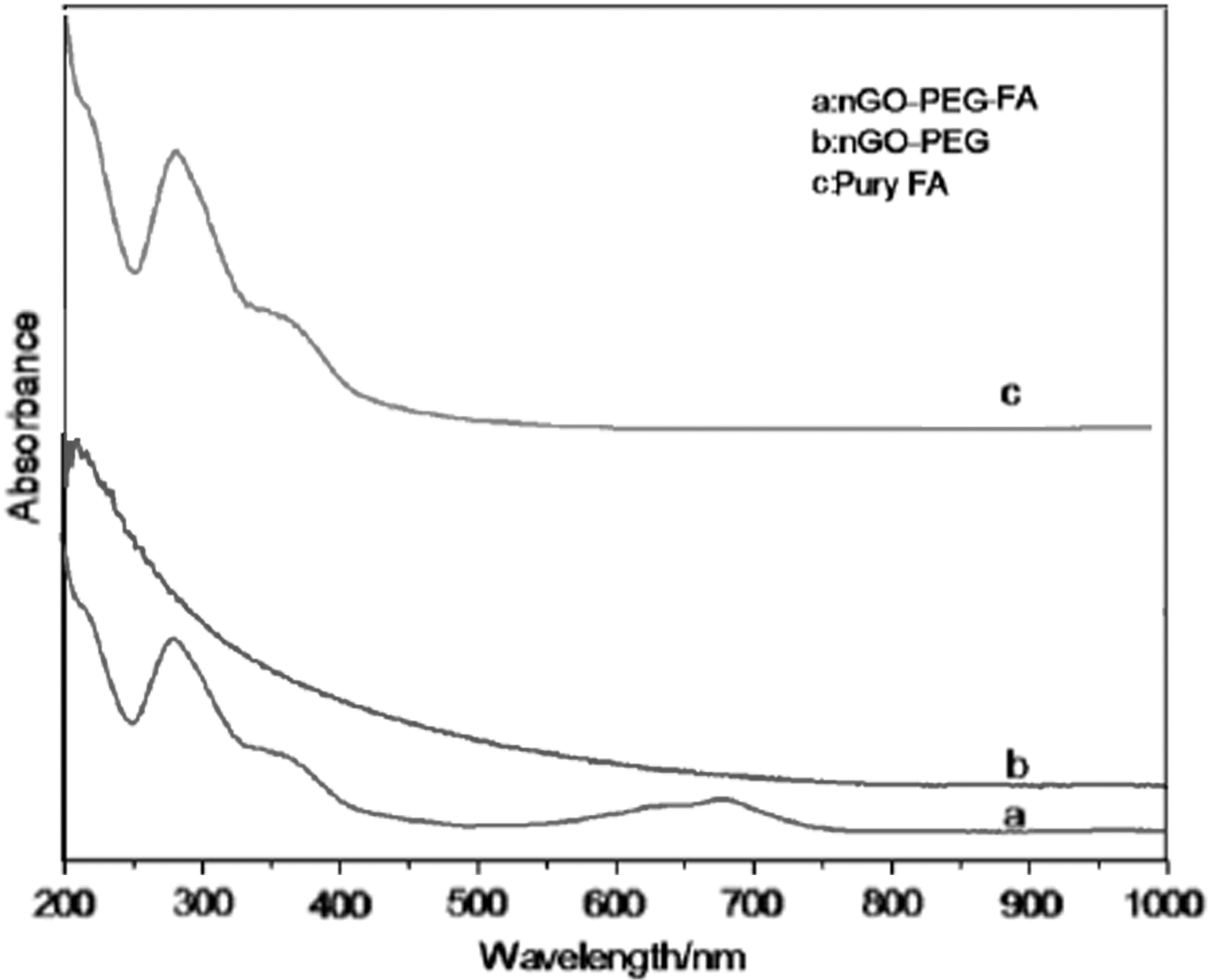
UV-visible absorption curves of nGO-PEG-FA, nGO-PEG, and pury FA in water.
Cell culture
Human pancreatic cancer Patu8988 cell line was provided by the Department of Nuclear Medicine, the First Affiliated Hospital of Suzhou University. Cells were maintained with the RPMI 1640 medium supplemented with 10% FBS, 100 μg/mL streptomycin, 100 μg/mL penicillin, and 1% L-glutamine in an incubator with 5% CO2 at 37°C. Cells were split every 48 h and the logarithmic growth phase cells were used for the experiments.
Cytotoxicity experiment
The cytotoxicity of prepared material to Patu8988 cell line was determined with the Cell Counting Kit-8 (CCK8) method. The cells with a density of 1 × 10⁵ cells/mL and RPMI medium containing 10% FBS were added to 96-well plate, respectively, and 50 μL per well. The plates were incubated in a 5% CO2 incubator at 37°C overnight. After the cells attached to the culture plate, phosphate buffered saline (PBS) solution containing different concentrations of nGO-PEG-FA (0, 5, 10, 20, 50, 100, and 200 μg/mL) was added to the cells and incubated with cells for 24 and 48 h, respectively. After incubation, CCK8 (10 μL/well) was added to each well and incubated for 1 h. The 96-well plate was read using a microplate reader at 450 nm. The proliferation rate of the cells was calculated according to the following formula: cell viability (%) = (average absorbance value of the experimental group – average absorbance value of the blank control)/(average absorbance value of the control group – average absorbance value of the blank control) × 100%. The experimental data were expressed as the mean ± standard deviation (SD) (n = 6).
Histopathological analysis of nGO-PEG-FA in nude mice
The nude mice bearing human Patu8988 xenografts injected with nGO-PEG-FA (300 μL, 1 mg/mL) were sacrificed, and the heart, liver, spleen, lung, kidney, stomach, muscle, lymph, and tumors were removed, and fixed with 0.5% formaldehyde. The tissue samples were then sliced for hematoxylin and eosin (H&E) staining.
Preparation of 99mTc labeled nGO-PEG-FA
Thirty microliters of Vc (10 mg/mL) and freshly prepared stannous chloride solution (10 mg/mL, made with 0.1 mol/L HCl solution) were added to 200 μL of 1 mg/mL nGO-PEG-FA. The pH of the solution was adjusted to about 6.0 with 20 μL of 0.5 mol/L NaOH, and then added to the saline solution containing Na99mTcO4. The mixture was then reacted in a 40°C water bath for 5–8 min. The labeled sample was then centrifuged with a refrigerated centrifuge for 15 min at 13,000 rpm/min, the supernatant was discarded, and the precipitate was ultrasonically dispersed with MilliQ water and centrifuged for another 15 min to further remove the residual Na99mTcO4 and possible 99mTc colloid. The precipitate was dispersed with MilliQ water and the radiochemical purity of sample was evaluated by paper chromatography. To evaluate the stability of 99mTc-nGO-PEG-FA in the cell culture medium, the RPMI 1640 medium with or without 10% FBS was added to a certain amount of purified 99mTc-nGO-PEG-FA, the samples were analyzed at different time point by paper chromatography, and the radiochemical purity of 99mTc-nGO-PEG-FA in RPMI 1640 with or without 10% FBS was calculated.
Determination of labeling rate
Xinhua No. 1 chromatographic paper was used as the stationary phase, and 3–5 μL solution of peaks from labeled 99mTc-nGO-PEG-FA purification was transferred to the origin of the chromatographic paper by capillary and developed in saline. The chromatographic paper was cut into 1-cm strips and the radioactivity of each strip was counted. The first peak was the labeled product and the second peak was the free 99mTcO4 −. Radiochemical purity (%) = radioactivity count of product peak/(radioactivity count of product peak + radioactivity count of free 99mTc O4 −.) × 100%.
The in vitro stability of 99mTc-nGO-PEG-FA
One hundred microliters of the labeled product was, respectively, added to human serum and mouse serum at a ratio of 1:20 and incubated at room temperature, and the radiochemical purity of the labeled product was examined at 2, 4, 6, and 24 h after incubation.
The binding assay of 99mTc-nGO-PEG-FA with Patu8988 cells
Effects of different cell densities on the binding of 99mTc-nGO-PEG-FA to Patu8988 cells
Patu8988 cells were routinely cultured in RPMI 116 medium containing 10% FBS at 37°C in a humidified atmosphere containing 5% CO2. The number of cells was adjusted to 2.0 × 10⁴, 4.0 × 10⁴, and 6.0 × 10⁴ cells/mL, and seeded into different 96-well plates and 200 μL per well. Each group had six replicates and 99mTcO4 − eluent was set up as a control group. The plate was incubated for 24 h at 37°C in a humidified atmosphere containing 5% CO2, and 10 μL of 99mTc-nGO-PEG-FA (370 kBq/mL) was then added to each well (the same volume and radioactive activity of 99mTcO4 − eluent were added to control group), and continually incubated for 60 min. The supernatant was aspirated and each well was washed twice with 200 μL normal saline, and the supernatant and saline were stored in RIA tube, labeled as the supernatant tube (F). Cells were trypsinized and were transferred to an RIA tube and labeled as a cell uptake tube (B). The radioactivity counts per minute from each tube were measured, and the cell binding rate of 99mTc-nGO-PEG-FA was calculated as follows: B/(B + F) × 100%, and the cell binding rate of the 99mTcO4 − eluent group was deducted. The optimal cell density was selected according to the cell binding rate of different plating densities.
Effects of different incubation times on the binding of 99mTc-nGO-PEG-FA to Patu8988 cells
Patu8988 cells were adjusted to a density of 4.0 × 10⁴ cells/mL and seeded into 96-well plates with 200 μL per well. The cell samples were divided into 60-, 120-, and 180-min incubation time groups, and six replicates for each group. 99mTcO4 − eluent was used as a control in each group. After 24 h of incubation in a CO2 incubator, 10 μL of 99mTc-nGO-PEG-FA or 99mTc O4 − eluent (370 kBq/mL) was added to cells and continuously incubated for different time courses. The cell binding rate of 99mTc-nGO-PEG-FA was measured, and the optimal incubation time was selected based on the cell binding rate for different incubation times.
Effect of different doses of 99mTc-nGO-PEG-FA on the binding rate of Patu8988 cells
The cells with a density of 4.0 × 10⁴ cells/mL were seeded into the 96-well plate and 200 μL per well. After the cells were incubated for 24 h in a 5% CO2 incubator, different radioactivity (370, 740, and 1480 kBq/mL) of 99mTc-nGO-PEG-FA and 10 μL 99mTcO4 − eluent were added to different groups of wells with a total of four groups, and six repeats for each group, and continuously to incubate for 120 min. The cell binding ratio of 99mTc-nGO-PEG-FA was determined. The optimized working dose of 99mTc-nGO-PEG-FA was determined according to the binding ratio at different incubation time.
The binding rate of 99mTc-nGO-PEG-FA, 99mTcO4, and 99mTc-nGO-PEG (no FA) with pancreatic cancer cell Patu8988
Patu8988 cells with a density of 4.0 × 10⁴ cells/mL were seeded into 96-well plates with 200 μL per well, and incubated for 24 h in a CO2 incubator. A total of 370 kBq/mL 99mTc-nGO-PEG-FA, 99mTc-nGO-PEG (no FA), and 99mTcO4 − eluent were added to different group of cells, respectively, and six replicates per group. Cells were continuously incubated for 120 min. The binding rates of 99mTc-nGO-PEG-FA, 99mTc-nGO-PEG (no FA), and 99mTcO4 − eluent with Patu8988 cells were determined.
Biodistribution and pharmacokinetic parameters of 99mTc-nGO-PEG-FA in nude mice
The study was approved by the institutional review board/local ethics committee, and the experiments followed the guidelines on experiments with animals. Six normal nude mice were assigned into two groups with three mice per group. Mice in each group were intravenously injected 0.1 mL of freshly labeled 99mTc-nGO-PEG-FA (9.25 MBq, i.e., 250 μCi), respectively. Their tail vein blood was collected at 2, 5, 10, and 30 min, as well as 1, 2, 4, 8, and 24 h postinjection, and used to measure the wet blood weight and count radioactivity using the γ-counter. The mean uptake of 99mTc-nGO-PEG-FA in blood of nude mice at different times was calculated according to the following formula: Uptake of 99mTc-nGO-PEG-FA in blood (%ID/g) = (the counts in blood/the total counts injected into the body)/the blood weight × 100.
99mTc-nGO-PEG-FA standards were prepared as follows, at the same time performing tail vein injection: a total of 0.1 mL 99mTc-nGO-PEG-FA was diluted by injecting water to 50 mL in a volumetric flask. After being mixed thoroughly, 200 μL of solution was transferred using a pipette into an RIA tube, counted simultaneously with the samples using the γ-counter, and used as the standard to calculate the uptake of 99mTc-nGO-PEG-FA in various organs (%ID/g). The pharmacokinetic parameters were calculated using the pharmacokinetics software DAS2.0. (Fig. 11).
Establishment of tumor model
Animal experiments were performed in accordance with the guidelines for animal care and use of the Commission. Patu8988 pancreatic cancer cell line was used to establish the tumor model in nude mice. Briefly, Patu8988 cells grown in the flask were digested with 0.05% trypsin-ethylene diamine tetraacetic acid (EDTA) solution and then pelleted by centrifugation. The cells were resuspended in PBS solution at a concentration of 1 × 10 7 cells/mL and 0.1 mL of cell suspension was injected into the armpit of right forelimb in each nude mouse. Two weeks after injection, the tumor grew to a size of 1.0 cm in diameter, which can be used for in vivo experiments.
In vivo biodistribution experiment of nude mice
Thirty-two nude mice were randomly divided into eight groups (n = 4) and intravenous injection (I.V.) injected by 0.1 mL of 99mTc-nGO-PEG-FA (∼4.81 MBq). Mice were sacrificed at 15 min, 30 min, 1 h, 2 h, 4 h, 6 h, 24 h, and 48 h after injection, respectively. The brain, heart, liver, spleen, lung, kidney, stomach, intestine, pancreas, muscle, thyroid, bone, tumor, lymph, and other tissues, as well as 100 μL carotid blood were collected, and their radioactive uptake (%ID/g) was calculated by measuring the weight of tissues and their radioactivity.
The single photon emission computed tomography (SPECT) imaging of 99mTc-nGO-PEG-FA in nude mice
The tumor bearing nude mice (n = 3) were I.V. injected with 0.1 mL 99mTc-nGO-PEG-FA (∼4.81 MBq). Single photon emission computed tomography (SPECT) imaging was performed at 30 min, 1 h, 2 h, 4 h, and 6 h after injection. The distribution of 99mTc-nGO-PEG-FA in tumor foci was observed by using GE Hawkeye dual probe SPECT instrument with low-energy, high-resolution collimator. The settings included the following: collection matrix (1024 × 1024), magnification (2), and the acquisition time (15 min).
Statistical analysis
The data are expressed as mean ± SD (
Results
Characterization of nGO-PEG
The size and morphology of graphene oxide were characterized by atomic force microscopy (AFM). The size of nGO was about 20–30 nm and the thickness was 1005 nm. The gravimetric method was characterized by modified Hummers oxidation method.
As shown in Figure 7, the size and morphology of graphene oxide were characterized by AFM. The size of nGO was about 20–30 nm and the thickness was 1005 nm.
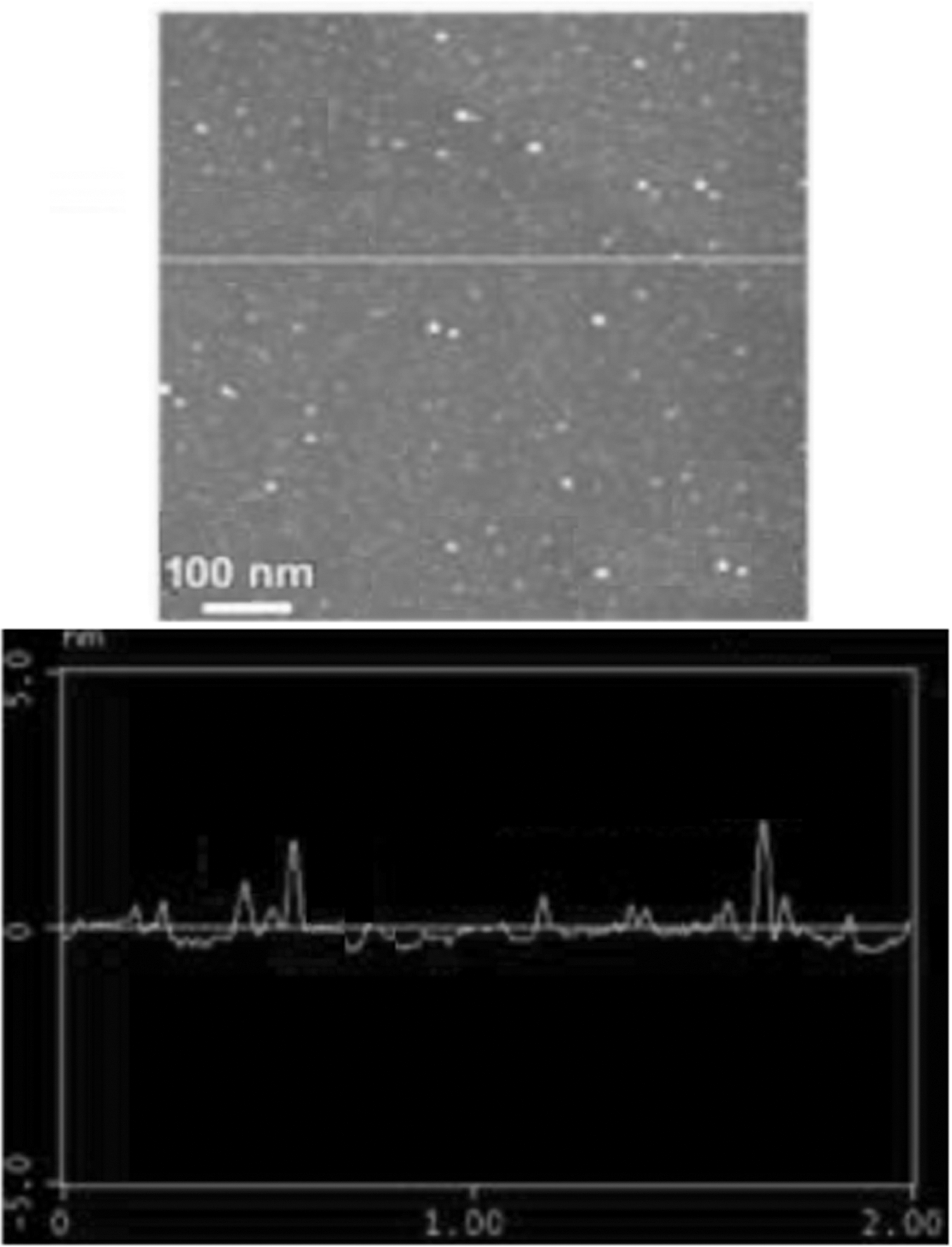
Atomic force microscopy of nGO.
In addition, the detection from Raman spectroscopy showed that two characteristic peaks were visible in the nGO-PEG (Fig. 8).

Raman spectrum of nGO-PEG.
Cytotoxicity of nGO-PEG-FA
In this study, the toxicity of nanomaterial nGO-PEG-FA was tested by CCK8 method. FA is a natural and nontoxic substance for cells, especially hyperplastic cells. Notably, the folate receptor is overexpressed in the surface of most malignant tumor cells, but not the normal cells. 22 These results indicated that the cell survival rate still reached over 80% when the cells were incubated with 200 μg/mL of nGO-PEG-FA for 48 h (Fig. 9), suggesting that nGO-PEG-FA had less toxicity to Patu8988 cells.
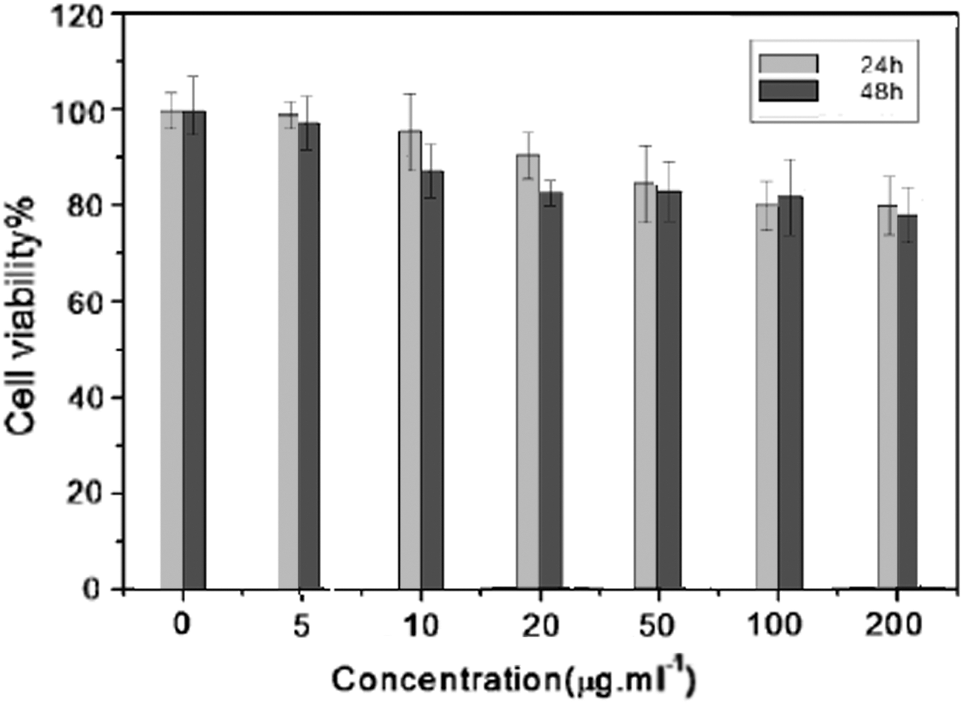
The cell viability of Patu8988 cells in the presence of nGO-PEG-FA. Patu8988 cells were incubated with different concentrations of nGO-PEG-FA at 37°C for either 24 h or 48 h, and the cell viability was determined with Cell Counting Kit-8 method.
Histopathological analysis of mice after the injection of nGO-PEG-FA
The H&E staining analysis showed that there was no significant change in the tissues of mice injected with nGO-PEG-FA nanocomposites, indicating that nGO-PEG-FA did not exhibit significant toxicity and had good biocompatibility in vivo (Fig. 10). This result demonstrated that nGO-PEG-FA can be applied for imaging experiments in living animals.
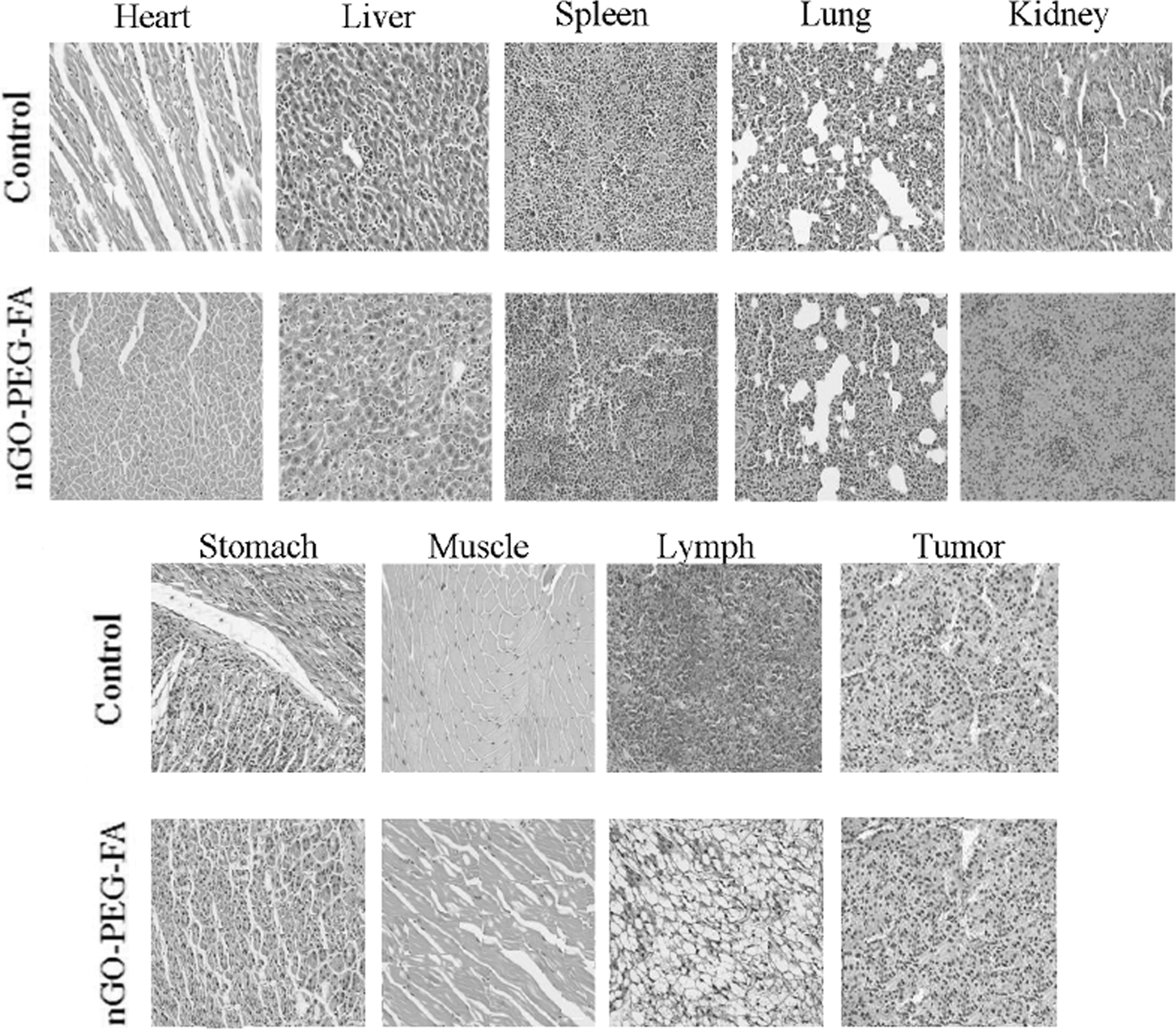
Hematoxylin and eosin staining of tissues of mice injected with nGO-PEG-FA.
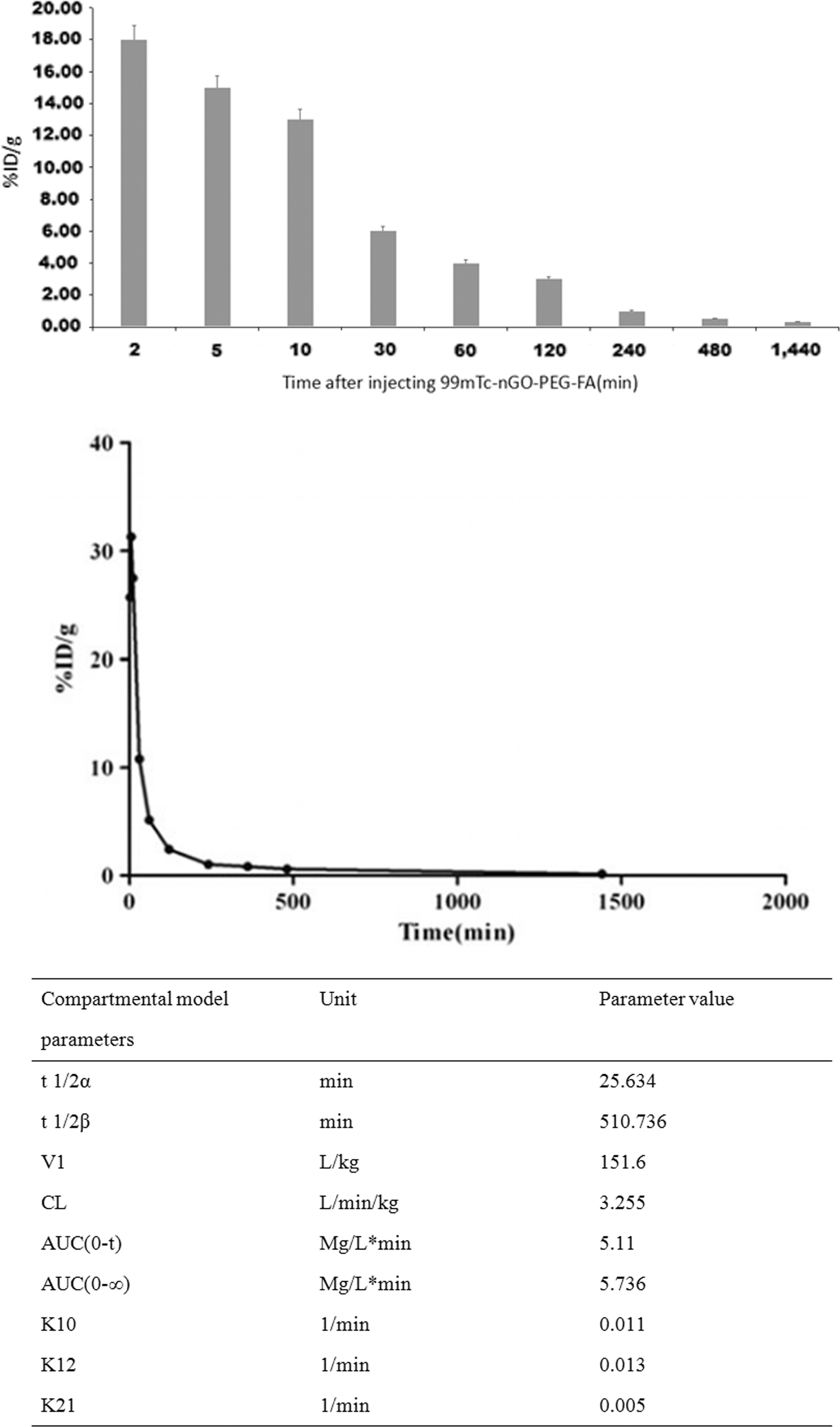
Metabolic and pharmacokinetic curves of 99mTc-nGO-PEG-FA in blood of nude mice.
Preparation of 99mTc-labeled nGO-PEG-FA
As described in Materials and Methods, the prepared nGO-PEG-FA nanomaterial was labeled with Na99mTcO4 to obtain the 99mTc-labeled nGO-PEG-FA. It was shown that the labeling ratio of the material was (90.08 ± 2.34)%, indicating that nGO-PEG-FA was labeled with Na99mTcO4 efficiently.
The stability of 99mTc-nGO-PEG-FA in vitro
The radiochemical purity of the labeled product in human and mouse serum after 2, 4, 6, and 24 h incubation is shown in Table 1. The radiochemical purity of 99mTc-nGO-PEG-FA in human serum decreased after 2 h of incubation, but was still >60% after 6 h of incubation. However, as shown in Table 1, the decrease of radiochemical purity in mouse serum is more obvious than that in human serum. The result is shown in Table 1.
Radiochemical Purity (%,
The in vitro binding assay of Patu8988 cells with 99mTc-nGO-PEG-FA
Effects of different cell densities on the binding of Patu8988 cells to 99mTc-nGO-PEG-FA
It was shown that the binding rates of 99mTc-nGO-PEG-FA to Patu8988 cells at cell densities of 4000, 8000, and 12,000 cells per well were (1.31 ± 0.22)%, (2.12 ± 0.34)%, and (1.97 ± 0.31)%, respectively, which was statistical significant (F = 19,355, p < 0.05). There was no significant difference in the binding rate of 99mTc-nGO-PEG-FA between the 8000 cells per well and 12,000 cells per well groups (p > 0.05), but the binding rate of 4000 cells per well group was significantly different from the other two groups (all p < 0.01).
Effects of different incubation times on the binding of 99mTc-nGO-PEG-FA FA to Patu8988 cells
Under the condition of different incubation time courses (1, 2, and 3 h), the binding rate of 99mTc-nGO-PEG-FA with Patu8988 cells (8000 cells per well) was (2.36 ± 0.31)%, (3.33 ± 0.59)%, and (3.66 ± 0.67)%, respectively, with statistical significance (F = 22.532, p < 0.05). Among them, the binding rate of 1-h incubation was significant lower than 2 and 3h incubation (p < 0.01), but there was no significant difference between 2 and 3h incubations (p > 0.05).
The effect of different doses of 99mTc-nGO-PEG-FA to cell binding rate
It was shown that the cell binding rate of 99mTc-nGO-PEG-FA in tested doses (370, 740, and 1480 kBq/mL) was (3.15 ± 0.31)%, (2.29 ± 0.23)%, and (1.18 ± 0.12)%, respectively, and there was statistically significant difference between groups (F = 131.386, p < 0.01), indicating that the binding rate of 99mTc-nGO-PEG-FA with Patu8988 cells was decreased with the increased dose of 99mTc-nGO-PEG-FA.
The binding rate of 99mTc-nGO-PEG-FA99m, TcO4, and 99mTc-nGO-PEG (no FA) with Patu8988 cells
It was shown that the binding rate of 99mTc-nGO-PEG-FA, 99mTcO4 −, and 99mTc-nGO-PEG (no FA) with Patu8988 cells was (3.15 ± 0.31)%, (0.91 ± 0.10)%, and (1.68 ± 0.19)%, respectively, with statistical significance (F = 86.341, p < 0.05).
The distribution of 99mTc-nGO-PEG-FA in nude mice
As described in Materials and Methods, the distribution of 99mTc-nGO-PEG-FA in different tissues of nude mice was detected at different time courses. The result is shown in Tables 2 and 3 and Figure 12.
The Distribution of 99mTc-nGO-PEG-FA and 99mTc-nGO-PEG in the Tumor After Injection at Different Time Courses (%ID/g,
The Distribution of 99mTc-nGO-PEG-FA in Different Tissues After Injection at Different Time Courses (%ID/g,
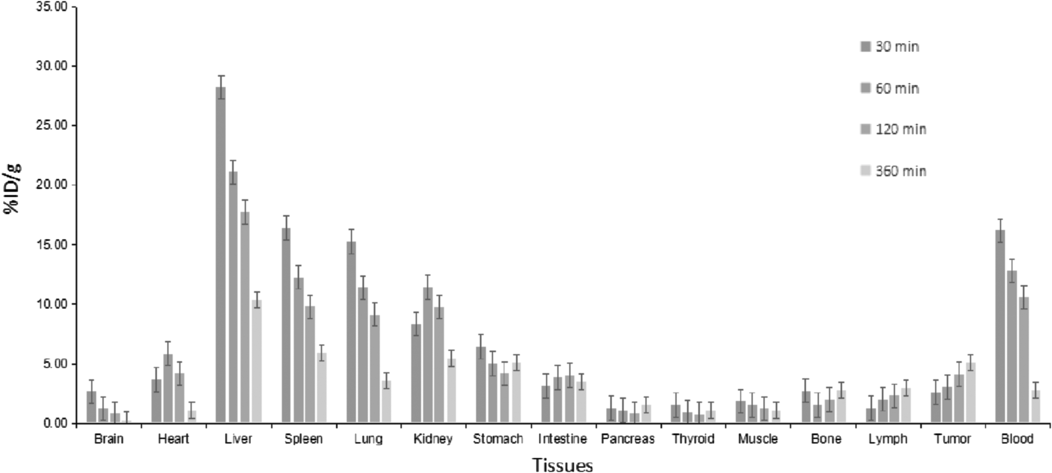
The distribution for comparison of 99mTc-nGO-PEG-FA in different tissues after injection at different time courses.
The radioactive uptake of each tissue is shown in Table 3. In the early stage of injection of 99mTc-nGO-PEG-FA, the tissues of liver, spleen, kidney, and blood had a high radioactive uptake. The radioactive uptake of tumor tissue was increased gradually with the increased time course and reached the peak at 6 h, and then decreased slowly. It was shown that 99mTc-nGO-PEG-FA was removed quickly in the blood, and the uptake was significantly reduced in the heart and blood at 6 h, whereas it was relatively high in liver, spleen, lung, and other organs.
The imaging of 99mTc-nGO-PEG-FA in nude mice
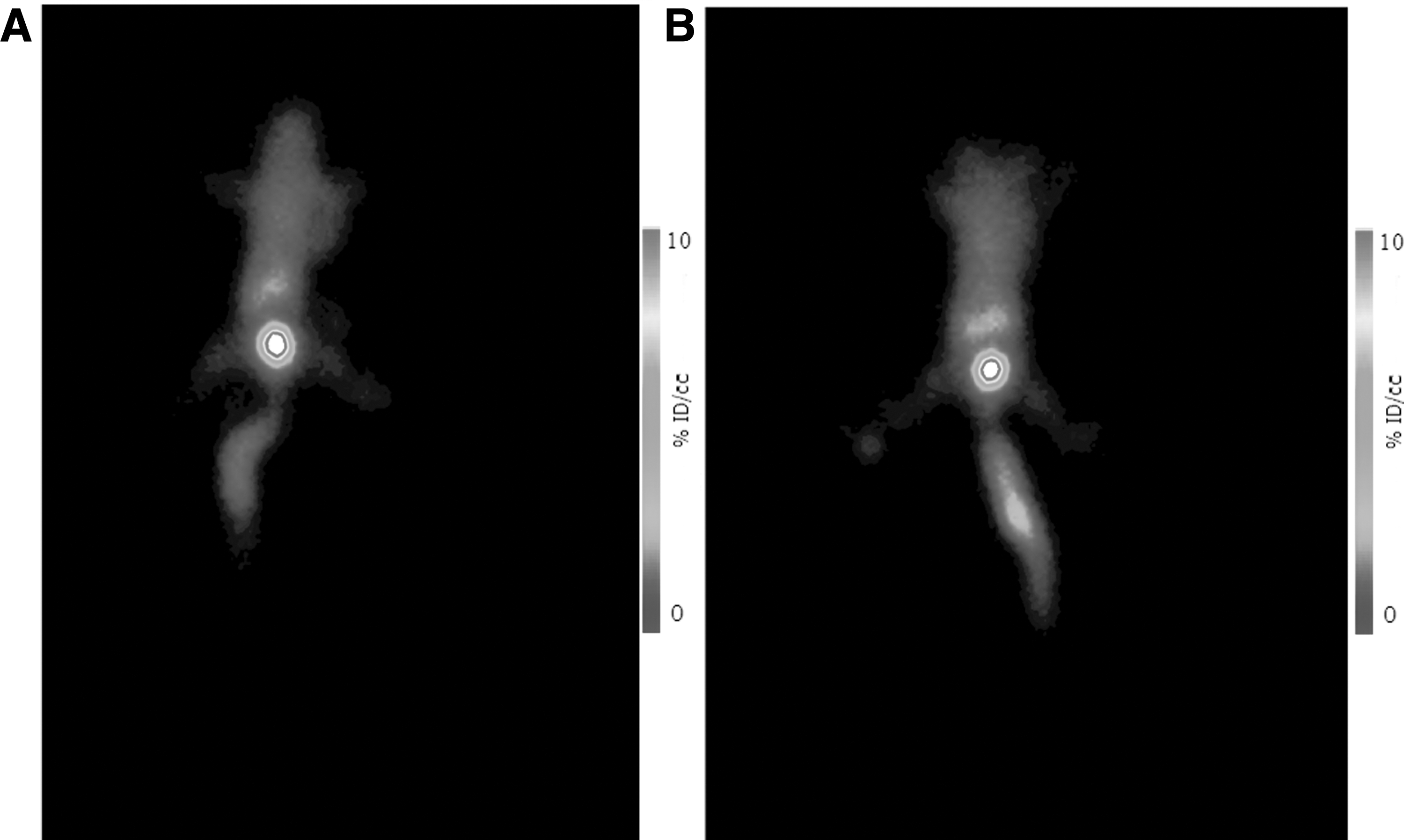
The radioactivity in tumor tissue was observed at 30 min after I.V. injection of mice with 99mTc-nGO-PEG-FA and 99mTc-nGO-PEG-FA+ blocking with folate; the radioactive concentration was increased with the prolonged time. The tumor was clearly imaged at 4 and 6 h, and there was high radioactive distribution in liver, spleen, lung, kidney, and bladder, whereas the rest of the organs had no significant radioactive distribution (Figs. 13 –16).
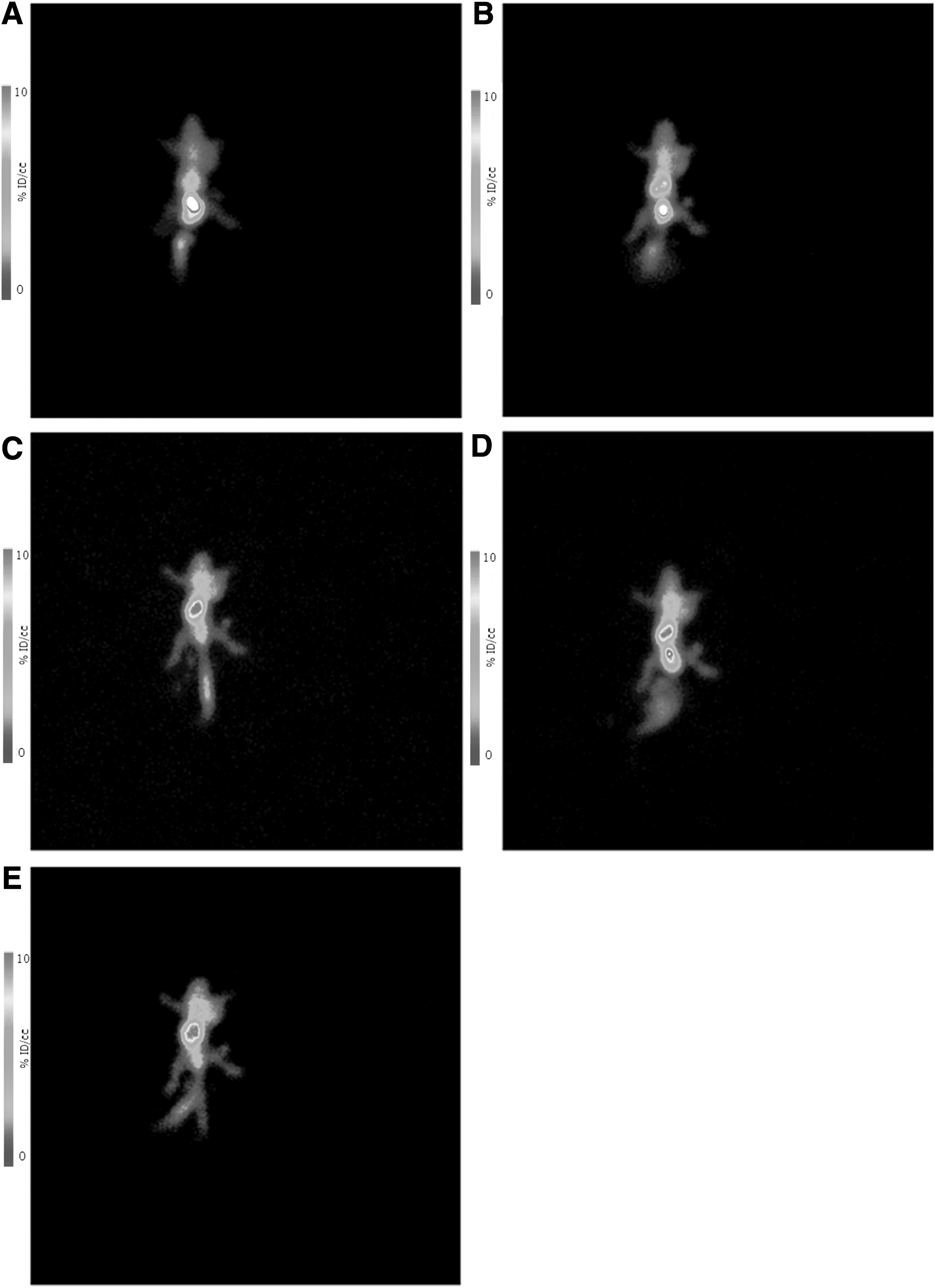
SPECT imaging of nude mice at different time course after Injection of 99mTc-nGO-PEG-FA.
SPECT imaging of nude mice after injection of 99mTc-nGO-PEG.
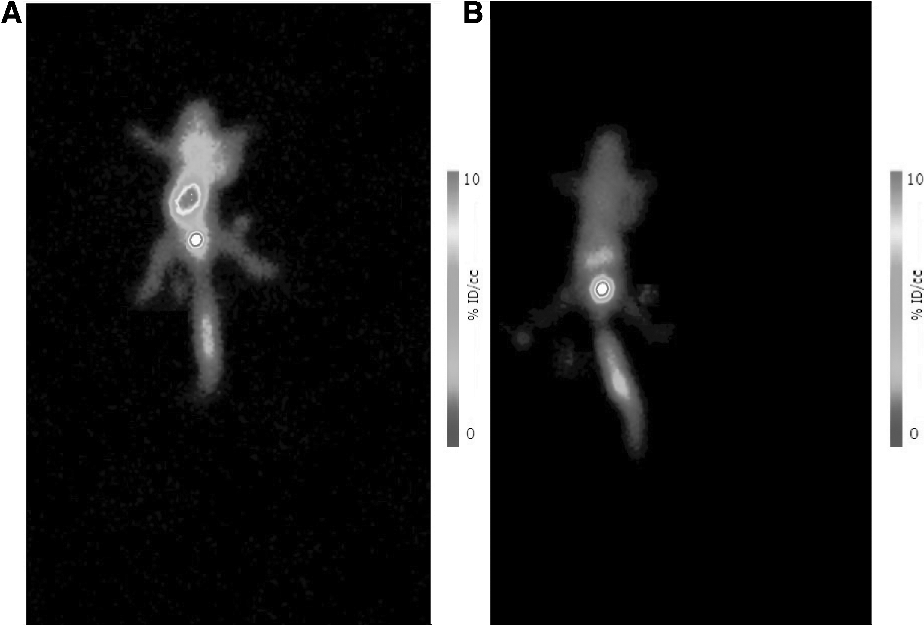
SPECT imaging of nude mice blocking with folate.
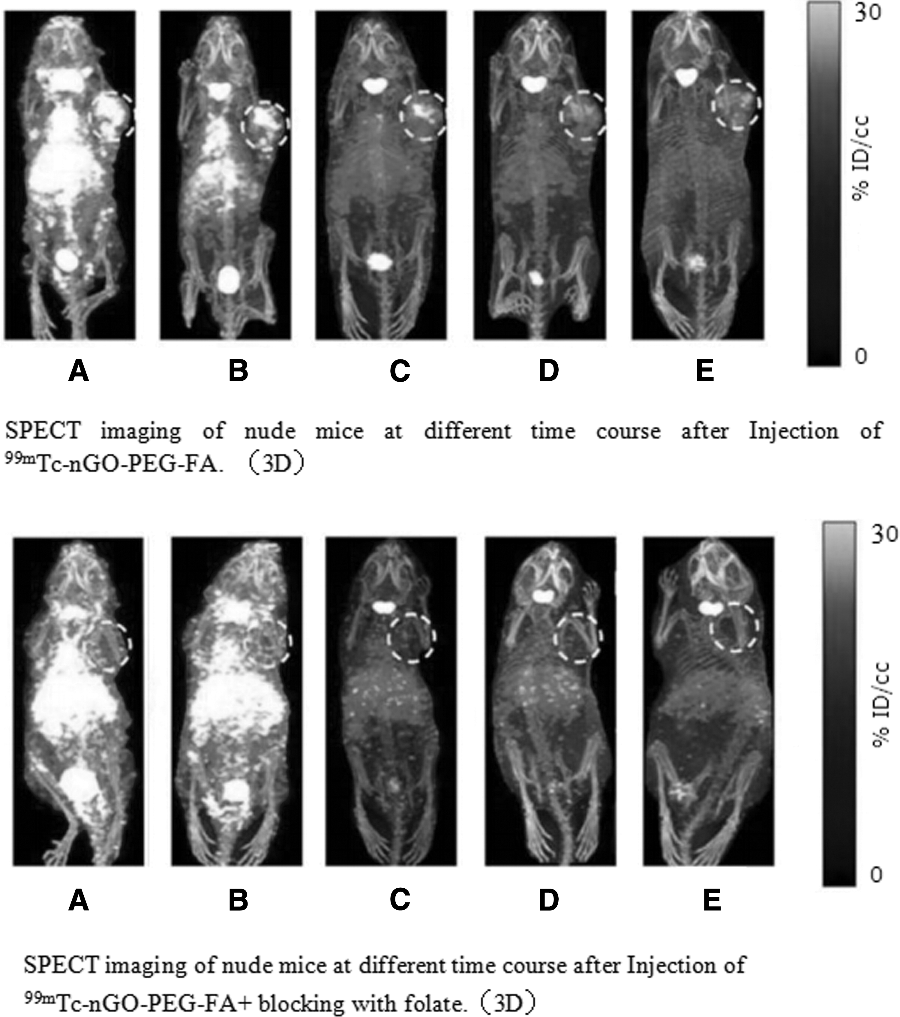
SPECT imaging of nude mice at different time course after injection of 99mTc-nGO-PEG-FA and 99mTc-nGO-PEG-FA+ blocking with folate.
Discussion
The surface of the graphene oxide contains a variety of oxygen-containing reactive groups, which provide the possibility of covalently modifying graphene oxide. PEG is a polymer with good hydrophilicity and biocompatibility, thus it has been widely used to modify various nanomaterials. The nanomaterials modified by PEG have good biocompatibility, and a significant reduction for their nonspecific binding to biomolecules and cells, which improves their pharmacokinetic and tumor targeting in vivo. 7,10,11
nGO-PEG-FA contains multiple N atoms and NH2 groups. It was possible that one or more nGO-PEG-FA chelated 99mTc by 4N or NH2. In this study, nGO-PEG-FA was labeled with 99mTc by a direct labeling method, and the labeling rate was more than 90%, and the labeled product had good stability. In addition, Patu8988 cells were used to perform the binding assay of 99mTc-nGO-PEG-FA, and it was shown that the specific cell binding rate was (3.15 ± 0.31)% when 8.0 × 10 3 cells per well were incubated with 370 kBq/mL 99mTc-nGO-PEG-FA, suggesting that has certain ability to bind pancreatic cancer cells.
Six hours after injection of 99mTc-nGO-PEG-FA, it was shown that the imaging in the tumor was clear and radioactivity within the tumor tissue was 5.11 ± 1.23%ID/g. Meanwhile, there were also more radioactive uptake in the liver, spleen, and lung, while the heart and blood had less radioactive uptake. The imaging of tumor was clear at 4.0–6.0 h after injection, suggesting that 99mTc-nGO-PEG-FA is targeted to pancreatic cancer. There was low level of radioactivity in the thyroid, indicating that the labeled product was stable in vivo and there is no obvious phenomenon of de-technetium.
In addition, although the radioactive accumulation in lung was abundant, the radioactivity per unit volume was low due to the low weight and large volume of lung tissue. The resolution of the SPECT instrument used in the study was about 1.0 cm, while the tumor size in the nude mice was larger and there was necrosis in the center of the tissue. This may be one of the reasons causing low image quality.
In general, 99mTc-nGO-PEG-FA can be easily used as labeling material with a high labeling rate, which can specifically bind to pancreatic cancer cells and has the characteristics of high affinity to propancreatic cancer in vivo. Because this material can be rapidly removed by the liver and kidney, it can be used as an imaging agent for pancreatic cancer.
Footnotes
Acknowledgments
This study was financially supported by Changzhou Health and Family Planning Commission Youth Project (Grant No. QN201717) and the Science and Technology Development Fund Project from Nanjing Medical University (Grant No. 2017NJMUZD037). The authors wish to acknowledge professors of Shanghai Normal University, for advice on experimental design and for their help in interpreting the significance of the results of this study.
Disclosure Statement
No competing financial interests exist.