
Abstract
Pathological angiogenesis can be a significant barrier to effective cancer therapy. Recent evidence suggests that Endostar may induce vascular normalization, thereby improving tumor perfusion and systemic chemotherapy. However, the molecular mechanism by which Endostar makes chemotherapy more effective remains to be fully elucidated. In this study, established 4T1 breast tumor-bearing animals treated with Endostar were evaluated at serial time points for treatment-associated changes in vascular architecture. As a result, Endostar induced a morphologically and functionally normalized vascular network. Combined Endostar and doxorubicin exhibited significant antitumor (34% of control size) and antimetastatic effects (29% of control metastatic nodules) in vivo. Finally, a two-dimensional gel electrophoresis and MALDIQ-TOF MS/MS-based proteomics approach was used to identify differentially expressed proteins involved in vascular normalization during Endostar administration. SRCIN1 was detected as one of the most significantly increased proteins. SRCIN1 is a novel Src-binding protein that regulates Src activation through C-terminal Src kinase, and attenuated Src activation during Endostar treatment was further confirmed by immunoblotting. Collectively, these data provided a molecular basis for vascular normalization, which were associated with the observed synergistic effect in vivo.
Introduction
To grow beyond a few millimeters in size, the solid tumors sprout neovasculature from existing ones for nutrients and oxygen as well as an ability to evacuate carbon dioxide and metabolic wastes. Angiogenesis is induced early during the multistage development of carcinogenesis and considered as one of the crucial hallmarks of cancers. 1 –3 Antiangiogenic drugs have a synergistic effect when used in combination with traditional chemotherapy or ionizing radiation. However, paradoxically destroying the vasculature may severely reduce the delivery of oxygen and drugs to the solid tumor, rendering therapeutics less effective. 4,5 Essentially, the blood vessels within tumors are typically dysfunctional, which is characterized by excessive and convoluted vessel branching, distorted and enlarged vessels, and abnormal endothelial cell proliferation. The abnormal tumor vasculature and the resulting aberrant microenvironment together form a physiological barrier to the drugs and immunocytes. 6 –8 Recent evidence suggests that judicious application of antiangiogenic agents, such as bevacizumab, results in morphologically less torturous vessels, tighter basement membrane that appears to be accompanied by functional changes, including reduced interstitial fluid pressure, improved oxygenation, and better penetration of drugs in the tumor, enhancing the efficacy of subsequent radiation and chemotherapy. The vascular normalization hypothesis has been well described in the literature. 9 –11
Endostatin is a carboxyl terminal proteolytic fragment of collagen XVIII, which is considered an endogenous inhibitor of angiogenesis. Endostar is a novel modified recombinant human endostatin, which was approved by the China Food and Drug Administration (CFDA) for the treatment of non-small cell lung cancer (NSCLC) in 2005. It has showed synergetic effect with cytotoxic drugs on a variety of tumor xenografts and cancer patients. 12,13 However, the molecular mechanism by which Endostar makes chemotherapy more effective remains to be fully elucidated.
Although multiple mechanisms of vascular normalization have been proposed, one of the challenges is to identify related proteins and which one is dominant. Proteomics, a study of the complete protein complements of the cell, is a promising approach in the identification of proteins that may be used as new targets for therapeutic intervention and as markers for early detection of cancers. 14
In this study, the authors used mouse 4T1 breast tumor model, which was highly tumorigenic and spontaneously metastasized to multiple distant sites. Endostar induced a morphologically and functionally normalized vascular network. Sequential combined doxorubicin exhibited significant antitumor and antimetastatic effects in 4T1 breast tumor model. Differentially expressed proteins, between paired endothelial cells and corresponding Endostar-treated cells, were identified by 2D gel electrophoresis. SRCIN1, a novel Src-binding protein, was chosen for validation and functional analysis. Data resulting from the study were expected to lead to an improved understanding of the mechanisms of vascular normalization induced by Endostar, which may ultimately promote the translation of experimental findings into clinical applications.
Materials and Methods
Reagents
Endostar (a novel recombinant human endostatin expressed and purified from Escherichia coli) was provided by Simcere Pharmaceutical Research Co., Ltd. (Nanjing, China).
Cell lines
Human umbilical vein endothelial cells (HUVEC) were isolated from human umbilical cord veins as previously described, and were cultured in EBM-2 medium with SingleQuots (Lonza, Basel, Switzerland) containing VEGF and other growth factors. 15 Murine breast cancer cell line 4T1 was purchased from the American Type Culture Collection and maintained in medium supplemented with 10% FBS.
Animal tumor model
BALB/c mice were purchased from the West China Experimental Animal Center. All experiments were in accordance with the protocols approved by the Animal Care and Use Committee of Sichuan University. BALB/c mice were inoculated s.c. with 5 × 10⁵ 4T1 breast cancer cells. After 5 d, mice bearing tumors were expanded for further study.
Immunofluorescence
Mice bearing 4T1 tumors were randomized into Endostar or control group. A dose of 5 mg/kg Endostar or normal saline was injected through the tail vein once a day. At different time points, mice were sacrificed and frozen sections of 4T1 tumor in each group were stained with the anti-CD31 antibody (BD Biosciences, San Jose, CA) using the method described previously. 16 Imaging was performed using a fluorescence microscope and captured in six random images from three different tumors.
Detection of tumor hypoxia
To visualize hypoxic regions in tumors, mice were injected intravenously with 100 μL of 10 mg/mL pimonidazole 15 min before sacrifice. Tumors were frozen for sectioning and handled according to the method previously described. 17 For each group, the hypoxic areas were counted in six randomly captured images from three different tumors.
Electron microscopy
At different time points, mice were sacrificed and tumor tissues in each group were fixed in 2.5% glutaraldehyde and examined for vascular ultrastructure using the scanning electron microscope. Six tissue areas were analyzed and used for quantitative assessment of vessel phenotype in each group.
Intratumoral drug concentration
At different time points in Endostar or normal saline group, doxorubicin was given intravenously at a dose of 5 mg/kg. Thirty minutes after doxorubicin administration, tumor tissue was harvested and prepared samples were subjected to high-performance liquid chromatography (HPLC) system. Doxorubicin concentration was measured with emission wavelength at 254 nm.
Tumor therapy studies
Treatments were initiated on day 5 postimplantation with 5 × 10⁵ 4T1 cells in BALB/c mice. Mice bearing 4T1 tumors were randomized into four groups, and the dosing schedules were (1) 0.9% saline; (2) Endostar 5 mg/kg/d intravenously on day 1–6 of each week; (3) doxorubicin 5 mg/kg intravenously on day 6 of each week; and (4) combined treatment. Treatments were administered every week for two cycles. Tumor length and width were measured every 3 d and tumor volume was calculated using the following formula: tumor volume (mm3) = (length × width 2 )/2. Twenty-two days after tumor cell inoculation, mice in each group were sacrificed. Tumors and lungs were harvested, and Hematoxylin and Eosin stain was used to indicate necrotic area in tumor tissue and metastatic nodules in lungs. Paraffin-embedded tumor sections were stained with an anti-Ki67 antibody (Thermo Scientific, Fremont, CA) to determine cellular proliferation in tumor tissues. The tumor cells with OnePlus nuclear staining was recorded Ki67 positive, and positive rate was calculated by dividing the Ki67-positive cells by the total cells. Cell apoptosis was detected by terminal deoxynucleotidyl transferase-mediated dUTP nick end labeling (TUNEL) assay according to the manufacturer's instructions (Promega, San Luis Obispo, CA). 18 The apoptotic index was calculated by dividing the number of TUNEL-positive cells (green fluorescence) by the total number of cells (blue fluorescence). For each group, the proliferative cells and apoptotic cells were counted in six randomly captured fields from three different tumors.
Two-dimensional electrophoresis
HUVEC were incubated in EBM-2 medium or EBM-2 medium with 100 μg/mL Endostar for 6 d. Two-dimensional electrophoresis was done as described previously with some modifications. 19 Briefly, cells were lysed in lysis buffer and applied to immobilized pH gradient strip using the passive rehydration method. After 16 h of rehydration, the strips were transferred to an isoelectric focusing (IEF) cell (Bio-Rad Laboratories, Richmond, CA). Once IEF was finished, the strips were equilibrated twice and transferred onto 12% SDS-PAGE gels for the second-dimension electrophoresis. The protein spots in gels were visualized by Coomassie Brilliant Blue R-250 (Merck, Darmstadt, Germany) staining. Only those spots that changed significantly (>2.0-fold) were selected for mass chromatographic analysis.
Mass chromatographic analysis
Spots of interest were excised and destained with 100 mM ammonium bicarbonate and 30% acetonitrile (ACN), then rehydrated in trypsin solution (Promega, Madison, WI) and digested for 20 h. Each digested protein was spotted onto the MALDI target, and matrix solution (saturated solution of CHCA in 50% ACN and 0.1% trifluoroacetic acid) was applied to the dried sample. The mass spectra were analyzed using 4800 Plus MALDI TOF/TOF instrument (Applied Biosystems, CA). For protein identification, combined peptide mass fingerprinting and the MS/MS queries were performed automatically by searching the protein database using the Mascot 2.2 software.
Western blot analysis
HUVEC were incubated in EBM-2 medium or EBM-2 medium with 100 μg/mL Endostar for 6 d. Cells at each time point were lysed with buffer containing 1% Triton X-100, 1% deoxycholate, and proteinase inhibitor cocktail (Sigma, St. Louis, MO). Cellular protein from each sample was applied to SDS-PAGE gels and probed with specific antibodies (Cell Signaling Technology, Danvers, MA), including GAPDH, SRCIN1, Src, phospho-Src (Tyr527), Erk1/2, and phospho-Erk1/2 (Thr202/Tyr204). Blots were developed with horseradish peroxidase-conjugated secondary antibodies and chemiluminescent substrate on Kodak X-ray films. 20
Statistical analysis
Data were presented as mean ± SD. Data were analyzed statistically using one-way ANOVA. p < 0.05 was considered statistically significant.
Results
Endostar promotes vessel normalization and decreases tumor hypoxia
We first used immunofluorescence to evaluate the effect of Endostar on microvessel density at different time points within established 4T1 tumor. As assessed by CD31 staining, tumors in Endostar group at each of these time points showed a progressive and significant decrease in tumor vessel density, and microvessel density in control group did not change significantly over time with tumor growth (Fig. 1A). The findings from this study demonstrated that the hypoxic tumor area in control tumors was significantly increased, being most notable on day 6. Interestingly, despite the apparent decrease in tumor vessel density, there was a less hypoxic area fraction in Endostar-treated tumors (Fig. 1B). To further investigate ultrastructure of tumor vasculature with Endostar treatment, the authors used scanning electron microscopy to assess the high-resolution images of tumor vasculature. Microphotographs showed that fewer abnormal vessels were found in Endostar-treated tumors, which were characterized by enlarged blood vessels (Fig. 2A, B). They also investigated how the changes in tumor vessel would affect tumor penetration of systemically administered doxorubicin. At day 6 after Endostar administration, HPLC analysis showed a more than twofold increase of doxorubicin concentration in Endostar-treated 4T1 tumors compared with control group (Fig. 2C).
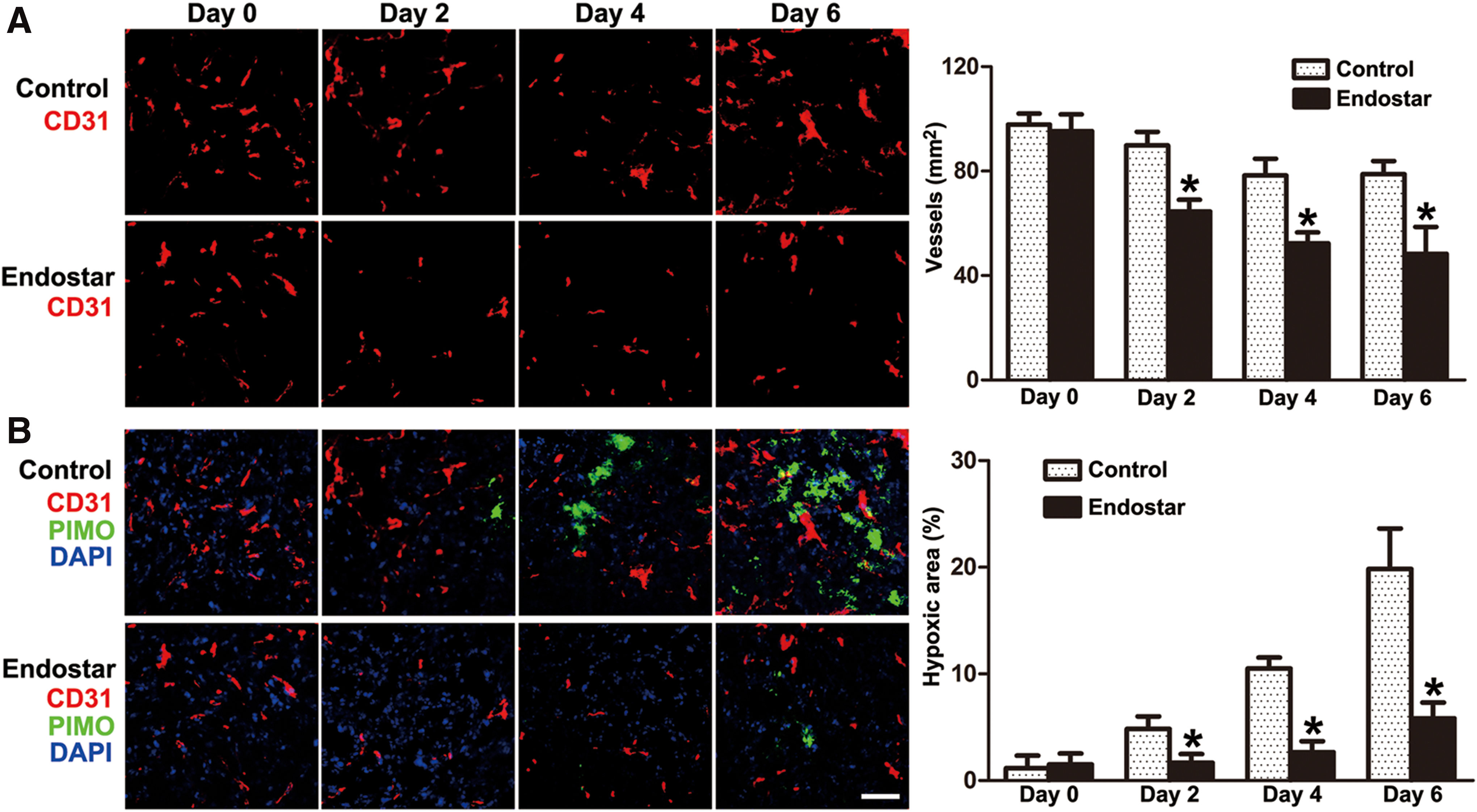
Endostar treatment depleted vasculature and improved hypoxia within established 4T1 breast tumor.

The effects of Endostar on the vascular ultrastructure and function within established 4T1 breast tumor.
The synergic antitumor effects of Endostar and doxorubicin
The standard management of breast cancers includes regimens based on doxorubicin, one of the anthracycline antibiotics. The structural normalization of tumor vasculature following Endostar treatment led us to ask whether subsequent administration of doxorubicin could enhance the antitumor response. Monotherapy with doxorubicin increased 4T1 tumor growth delay. However, pretreated with Endostar, doxorubicin administration resulted in a significant enhancement of the antitumor response compared with saline, Endostar, or doxorubicin monotherapy (Fig. 3).
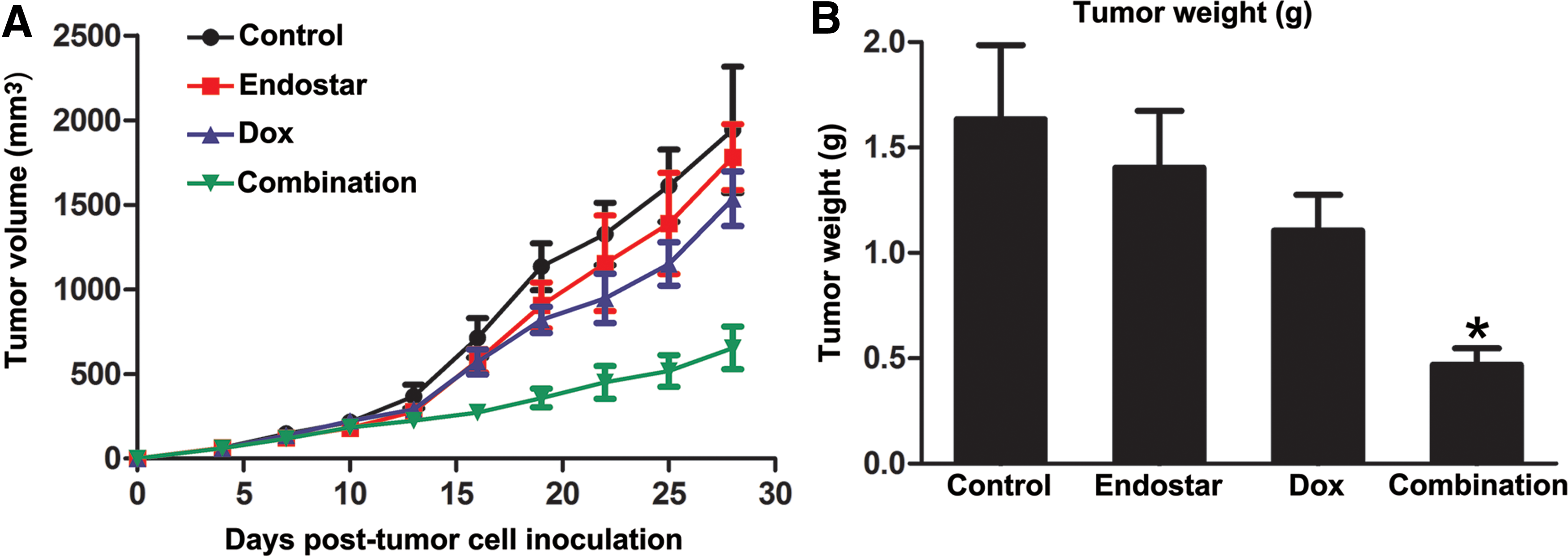
Effects of the combination of Endostar and doxorubicin on 4T1 breast tumor growth.
After mice were sacrificed, tumor samples were excised and Hematoxylin and Eosin stain was applied to investigate the pathological changes. Apparent necrosis was observed in the center of tumor tissues in mice treated with Endostar plus doxorubicin, but no apparent necrosis was observed in the other groups (Fig. 4A, D). To further understand the mechanisms underlying tumor growth suppression, the authors investigated tumor proliferation and apoptosis changes in each group. Tumor tissues of mice treated with Endostar plus doxorubicin exhibited much less staining for Ki67 (Fig. 4B, E). Accordingly, TUNEL assay demonstrated massive apoptosis in the combined treated tumors, in sharp contrast to the occasional apoptotic cells in control and monotherapy tumors (Fig. 4C, F). Thus, they demonstrated that tumor growth suppression might be caused by reduced proliferation and enhanced tumor cell apoptosis.

The combined treatment inhibited cell proliferation and induced tumor apoptosis in vivo.
The 4T1 breast cancer cell is widely used in vivo as a model of metastasis because of their highly metastatic potential. The 4T1 metastatic lesion could be found in various organs, most frequently in lungs. Therefore, they evaluated the synergic effect of combined therapy on the formation of distant lung metastases. Their result indicated that the number of the metastatic colonies in the whole lungs was much less in combined treatment group than that in other groups, as confirmed by pathological examination (Fig. 5).
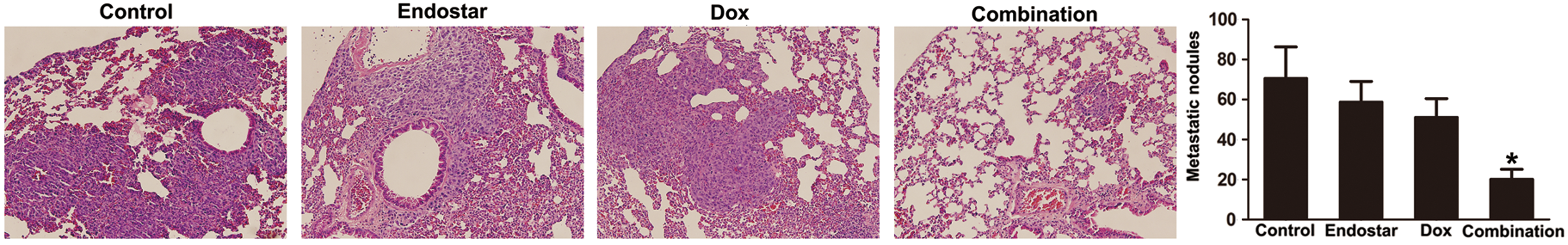
Mice were sacrificed 28 d after tumor cell inoculation. Metastatic nodules were observed under the dissecting microscope and subsequently evaluated by Hematoxylin and Eosin staining. The figure showed representative histological view of lung metastases in each group. Endostar in combination with doxorubicin had more profound inhibitory effects on lung metastasis formation than each therapy applied separately (n = 4; ANOVA; *p < 0.05). Color images available online at
Identification of differentially expressed proteins in Endostar-treated HUVEC
The protein expression profiles in Endostar-treated and untreated HUVEC were obtained by two-dimensional electrophoresis. A total of 28 spots were identified as differentially expressed, and of these 18 proteins were upregulated, whereas 10 proteins were downregulated in Endostar-treated HUVEC. Master gel images were shown and the representative spots were labeled with arrows (Fig. 6). Twenty-eight different proteins from two-dimensional electrophoresis gels were successfully identified by mass chromatographic analysis, and the details of the proteins identified were listed (Table 1). Among the identified proteins, SRCIN1, a novel Src-binding protein that regulates Src activation through C-terminal Src kinase, exhibited a high expression in Endostar-treated HUVEC.
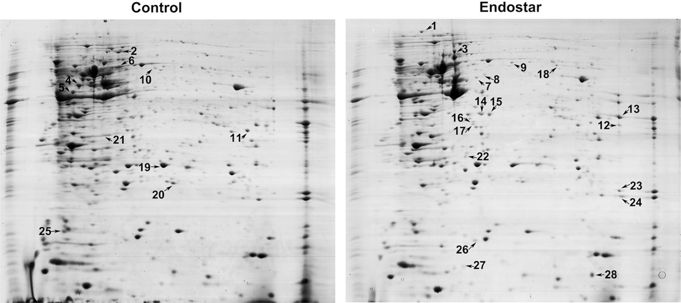
Proteomics analysis of Endostar-treated HUVEC and corresponding untreated HUVEC using two-dimensional electrophoresis gels. Whole cell lysates were separated by two-dimensional electrophoresis and visualized by Coomassie Blue staining. Arrows indicate identified protein spots significantly altered between Endostar-treated HUVEC and untreated HUVEC. HUVEC, human umbilical vein endothelial cells.
Validation of the increased SRCIN1 and attenuated Src signaling by Western blotting analysis
To confirm the altered expression of SRCIN1 in Endostar-treated HUVEC, a validation experiment was carried out by Western blotting analysis. The SRCIN1 expression was increased significantly, which was consistent with the observations in two-dimensional electrophoresis analysis. Furthermore, Endostar significantly suppressed the Src activity and its downstream signaling (Fig. 7). The SRCIN1 protein in HUVEC was seldom changed under the control medium treatment (Supplementary Fig. S1; Supplementary Data are available online at
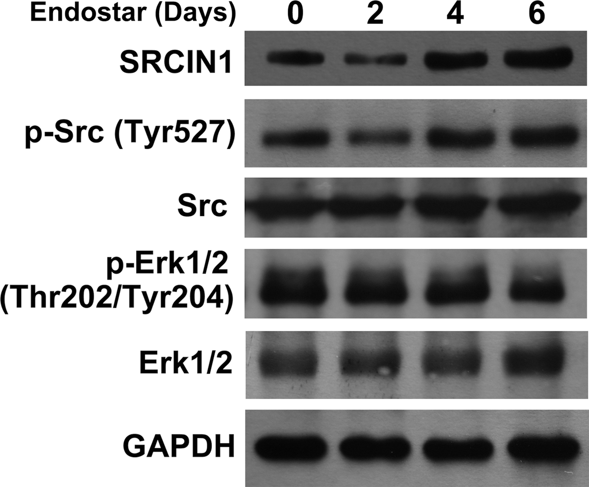
Endostar increased SRCIN1 expression, whereas inhibited Src kinase activity and its downstream signaling molecules. GAPDH expression was served as the control to show that similar amounts of protein were loaded in each lane.
Discussion
The abnormal structure of tumor vessels leads to a microenvironment within solid tumors, which is characterized as elevated interstitial fluid pressure, poor tumor perfusion, and severe hypoxia and acidosis. 21,22 Although antiangiogenic agents may have the potential to change the aberrant tumor vasculature and enhance the efficacy of cytotoxic chemotherapy, the process of vascular normalization seems to be transient with a relatively narrow time window. 8 A well-designed strategy should prune away immature vessels and cause minimal damage to normal tissue vasculature. Otherwise, these agents may destroy the tumor vasculature and negatively affect the efficacy of adjuvant therapy. Therefore, it is critical to determine how the use of antiangiogenic agents can be most effective, especially when used in combination with cytotoxic chemotherapeutics. In this study, the authors continuously monitored the time course of molecular and structural changes in the vasculature of 4T1 murine breast tumor in response to Endostar. They have shown that Endostar induced a transient remodeling of the intratumoral vasculature, reaching peak at day 6. Despite causing an overall decrease in intratumoral microvessel density, it resulted in more efficient oxygenation of tumors in mice with established orthotopic breast cancer model. They had also shown that one of the consequences of remodeling the intratumoral vasculature with Endostar was the significant improvement in therapeutic effects of systemic chemotherapy. Compared with the vehicle control, Endostar combined with doxorubicin reached more than 60% inhibition against 4T1 tumor models in BALB/c mice. These data are also consistent with other tumor models and recent data from patients with soft tissue sarcomas. 23 –25 While the underlying mechanism has yet to be fully elucidated, they have identified SRCIN1 to be potentially important for vascular normalization.
Endostatin is an endogenous inhibitor of endothelial cell and a tumor suppressor, and Endostar is the modified form of endostatin, with better stability and solubility. In China, Endostar has been approved in the treatment of advanced NSCLC in combination with chemotherapy. 26 Previous studies have indicated that a number of mechanisms contribute to the antiangiogenesis activities of Endostar, including inhibited Wnt/β-catenin, suppressed Ras/Raf kinases, and decreased ERK-1 and p38 activity. 27,28 In this study, the two-dimensional gel electrophoresis and MALDIQ-TOF MS/MS-based proteomics approach were used to identify differentially expressed proteins involved in vascular normalization during Endostar treatment. Thus, SRCIN1 was detected as one of the most significantly increased proteins. SRCIN1, also known as p140 cas-associated protein (p140CAP), contains two regions of highly charged amino acids, two proline-rich regions, and two coiled-coil domains. Recently, SRCIN1 has been shown to act in tumor progression, suppressing migration and invasion of breast cancer cells by regulating Csk and Src kinase activity. 29 Src activity is regulated by tyrosine phosphorylation at two sites with opposing effects. Phosphorylation of Tyr416 upregulates enzyme activity, and phosphorylation of Tyr527 in the carboxyterminal renders the enzyme less active. SRCIN1 controlled this process tightly by activating Csk kinase, which phosphorylated Tyr527 and prevented Src activity. 30 Further results confirmed that Endostar upregulated SRCIN1 protein, induced Src phosphorylation, and suppressed activation of various downstream signaling substrates, such as extracellular signal-regulated kinases (ERK).
Our study showed that Endostar could upregulate SRCIN1 protein in vascular endothelial cell influencing Src signaling pathway, which may be a novel target for vascular normalization. Meanwhile, their findings highlight an integrative approach to cancer therapy wherein Endostar therapy is precisely scheduled with chemotherapy to maximize the synergistic effects against tumors.
Footnotes
Acknowledgments
The authors thank Dr. Guobo Shen for help with article revision. This project was supported by Sichuan University Science Foundation for Youths (2017SCU11048).
Disclosure Statement
No competing financial interests exist.
References
Supplementary Material
Please find the following supplemental material available below.
For Open Access articles published under a Creative Commons License, all supplemental material carries the same license as the article it is associated with.
For non-Open Access articles published, all supplemental material carries a non-exclusive license, and permission requests for re-use of supplemental material or any part of supplemental material shall be sent directly to the copyright owner as specified in the copyright notice associated with the article.