
Abstract
Background:
Magnetic nanoparticles are increasingly applied in clinical area for drug delivery, also in magnetic induction hyperthermia (MIH), and so on. The present research would prepare appropriately modified superparamagnetic iron oxide nanoparticles (SPIONs) to conduct MIH and transfect the anticancer cytokine TNF-α into cells.
Materials and Methods:
The SPIONs were surface-modified by 3-aminopropyltriethoxysilane (APTS) and/or protamine sulfate (PRO). The combined MIH using SPIONs and gene therapy were applied to treat Hep G2 cells and tumor model transplanted in nude mice.
Results:
The PRO-SPIONs (the surfaces were sequentially modified by APTS and PRO) showed high transfection efficiency for TNF-α gene with no obvious cytotoxicity. It also exhibited great temperature rising performance under alternating current magnetic field. The combined MIH and gene therapy using PRO-SPIONs/TNF-α complex could reduce cell variability of Hep G2 cells and tumor size transplanted in nude mice.
Conclusions:
The PRO-SPIONs efficiently transfect the TNF-α gene into Hep G2 cells. Cells expressed more TNF-α when they were exposed to alternating current magnetic field only once. Combined MIH and gene therapy for treatment in vivo exhibited better effects than MIH or gene therapy alone. The combined MIH and gene therapy is promising in liver cancer treatment.
Introduction
Nanotechnology, molecular biology, and their combined utilization have greatly accelerated the progress of finding new cures for cancer. The small size and modified surface of nanoparticles facilitate their biodistribution and circulation in organisms. Nanodrug carriers, smaller than 200 nm, will accumulate primarily in the tumor rather than in the normal tissue due to their structure differences. Iron oxide nanoparticles have magnetism and excellent biocompatibility. They have been used in various biomedical applications, such as cancer hyperthermia, drug delivery, diagnostic sensing, and magnetic resonance imaging. The superparamagnetic iron oxide nanoparticles (SPIONs) are particularly favored for its low toxicity and reactive surface, which can be readily modified with biocompatible coatings and other imaging, targeting, and therapeutic molecules. 1 –3
Magnetic induction hyperthermia (MIH) is a promising noninvasive thermal activation therapy approach for tumors. Tumor cells are more vulnerable at 40°C–45°C (hyperthermic temperature) than normal tissue cells. SPIONs generate heat by the oscillation of their magnetic moments when exposed to external alternating current magnetic fields. MIH will elevate their temperature to 42°C–45°C. The tumor cells will be killed by the hyperthermic temperatures, while normal cells almost get no injuries. Therefore, MIH can reduce severe damage to the normal tissue cells in comparison with the conventional radiotherapy and chemotherapy while treating cancer. 3 –5
Gene therapy is a promising and more and more popular approach for cancer treatment. DNA vectors encoding specifically designed anticancer molecules can be systemically administered and delivered to the tumor tissues. The expression of these carefully tailored transgenes can meet specific cancer therapeutic needs, such as tumor selectivity, reduced toxicity to normal cells, and robust expression of anticancer molecules. Preclinical gene therapy tests have been carried out on liver cancer, pancreatic cancer, gliomas, and so on. 6,7 Recently, the combination of magnetofection and targeted gene therapy has got inspiring results. Genes reversibly bound onto superparamagnetic nanoparticles can be fastly and efficiently directed within the host under high-energy magnetic fields. 8 –10 The nonviral magnetofection vehicles are much more safer than the viral counterparts to some extent. 11
The authors' previous studies have shown that SPIONs can be used as a gene delivery system. 12 In the present study, the authors modified the surface of SPIONs with protamine sulfate (PRO). The gene of TNF-α, a potent anticancer cytokine, along with CMV promoter was bound to the surface modified SPIONs. The results of in vitro and in vivo studies indicated that the SPIONs/TNF-α gene complex could reduce cell variability of Hep G2 cells and human tumor size transplanted in nude mice. The combined MIH and gene therapy is promising in liver cancer treatment.
Materials and Methods
Materials
Ferrous chloride tetrahydrate (FeCl2·4H2O), ferric chloride hexahydrate (FeCl3·6H2O, >99%), ammonium hydroxide (25 wt% NH3 in pure water), 3-aminopropyltriethoxysilane (APTS), PRO, glutaraldehyde, and 3-(4,5-dimethylthiazol-2-yl)-2,5-diphenyltetrazolium bromide (MTT) were obtained from Sigma. All other reagents and solvents were analytical grade. Polymer transfection regent VigoFect was purchased from Vigorous Biotechnology Company (Beijing). Hep G2 human hepatoma cell line and SW480 human colon cancer cell line were provided by the Chinese Academy of Medical Sciences (Beijing, China).
Preparation of SPIONs (naked SPIONs, APTS-SPIONs, and PRO-SPIONs)
Naked SPIONs
Fe3O4 magnetic nanoparticles (MNPs) were prepared following the previous method 12,13 with modifications. Specifically, FeCl3·6H2O and FeCl2·4H2O (ration of Fe2+ to Fe3+ was 1:2) were dissolved in pure water under nitrogen protection. The solution was vigorously agitated at 85°C. Then, 25% ammonia solution with the volume of 1/20 of the solution was added into the solution. Nanoparticles were allowed to grow under continuous stirring before it was cooled to room temperature. At last, the nanoparticles were isolated from the solution by magnetic decantation and washed with pure water and ethanol.
APTS-SPIONs
The naked SPIONs (nSPIONs) were dispersed in ethanol. APTS was added slowly under argon protection and stirring at high temperature. The solution was left to maturing for 24 hours at room temperature before the particles were isolated from the solution by magnetism and washed with ethanol. The obtained APTS-SPIONs were dried under vacuum.
PRO-SPIONs
2.9% glutaraldehyde solution was prepared in phosphate buffer (100 mM, pH 8.0). APTS-SPIONs were dispersed in glutaraldehyde solution by stirring for 2 hours. The nanoparticles were activated by gentle shaking on a thermoshaker (Haimen). The activated MNPs were then collected under magnetic field and washed with pure water before they were sprayed onto a clean surface to let them completely dry. The activated nanoparticles were added into PRO in phosphate buffer (100 mM, pH 7.4). Subsequently, the suspension was gently shaked overnight at 4°C. Following the immobilization steps, the MNPs bound with proteins were rinsed with phosphate buffer and stored at 4°C.
Characterization of the SPIONs
Transmission electron microscopy
Water-diluted SPIONs suspension was dropped on a carbon-coated copper grid and the solvent was allowed to evaporate. Transmission electron microscopy (TEM) with a 200 keV HITACHI H-7650 microscope was used to examine the morphology and size of the SPIONs. The diluted suspension of sample in pure water in a special zeta-potential cuvette with 0.01 M KNO3 (pH 7.4) was used to measure the zeta potential.
Fourier transform infrared spectra
To examine the phase and the surface composition of the SPIONs, Fourier transform infrared (FTIR) spectra were carried out between 4000 and 400 cm−1 in a Nicolet Magna-IR 560 spectroscope. The powder of the SPIONs was dispersed in 2% KBr and pressed to pellets before measure.
Thermogravimetric analysis
Thermogravimetric analysis (TGA; Q5000 V3.5 Build 252) was used to determine the contents of Fe3O4 in the APTS-SPIONs and PRO-SPIONs. The SPIONs were heated from 10°C to 800°C at a heating rate of 10°C/min in air. The vibrating sample magnetometry (DMA 1600) was used to measure the magnetization of the nanoparticles.
Cell culture and treatment
Hep G2 cells or SW 480 cells of monolayers were cultured in Dulbecco's modified Eagle's medium supplemented with fetal bovine serum (10%) under the condition of 37°C, 5% carbon dioxide (CO2), and 100% humidity. When the cell density reached 60%–80%, the SPIONs were added into the medium and incubated for 24 hours before the SPIONs were removed. Residual MNPs were removed by washing with phosphate buffer.
Quantification of intracellular uptake of the SPIONs
Hep G2 cells were incubated with SPIONs of 0.2 mg/mL for 2, 4, 8, 16, and 24 hours, respectively. Cells were collected after washing with phosphate buffer and digesting with trypsin-EDTA and centrifuged to obtain cell pellet. The cell pellet was dissolved in HCl (37%) at 70°C–80°C for 30 minutes. Iron concentration was quantified by inductive coupled plasma using Thermo Scientific iCAP 6500.
Iron detection
Iron was detected by Prussian blue staining. After preincubation with SPIONs, Hep G2 cells were fixed with ice-cold methanol for 3 minutes and with 2% potassium ferrocyanide trihydrate for 15 minutes. Subsequently, the cells were counterstained with 0.5% neutral red for 5 minutes. The cells were washed with pure water and air dried. An Olympus BX51 microscope was used to observe the results.
Localization of the APTS-SPIONs and PRO-SPIONs
APTS-SPIONs and PRO-SPIONs of 0.1 mg/mL were respectively incubated with the Hep G2 cells for 4 hours. The cells were washed with phosphate buffer and collected before they were fixed by 2.5% glutaraldehyde in phosphate buffer (0.2 M) for 1 hour and 1% osmium tetroxide for 2 hours at 4°C. The cells were washed with phosphate buffer. The cells were embedded in Epon following dehydration in an alcohol series. TEM at 50 kV was used to observe the 70 nm-thick sections.
Cytotoxicity
The cell viability and proliferation of the cells were determined by MTT method to reflect the cytotoxicity of the SPIONs. In brief, cells seeded in 96-well culture plates were incubated with 5 mg/mL MTT in culture medium for 4 hours after preincubation with the SPIONs. The culture medium was removed and dimethyl sulfoxide was added to dissolve the formazan crystals for 10 minutes. The absorbance of each well at 570 nm was read on a microplate reader (Bio-Rad 680).
Plasmid
Plasmid pCDNA4-GFP, expressing GFP as a reporter, was used as a transfection control. Plasmid pCDNA4-GFP-TNFα was used to express tumor therapy GFP-TNFα fusion protein. Both plasmids contain CMV promoter controlling the expression of fusion protein.
Magnetofection and polyfection
Plasmid DNA was complexed to 8 μL VigoFect reagent in 200 μL of 150 mM sodium chloride each and incubated for 15 minutes. Four hundred microliters of the complexes were added into the culture medium. For magnetofection, 10 μg plasmid DNA was bound to 10 μL APTS-SPIONs or PRO-SPIONs and incubated at 4°C for 1 hour. The resulting complexes were called solution I. Solution I was diluted in 200 μL of 150 mM sodium chloride and complexed to 8 μL VigoFect reagent in 200 μL of 150 mM sodium chloride, then incubated for 15 minutes. A total volume of 400 μL complexes were added to culture medium and allowed to aggregate for 15 minutes by placing a permanent magnet under the cell culture dishes. After transfection, cells were incubated in 5% CO2 humidity atmosphere for further treatment.
Fluorescence microscopy study
Magnetofection and polyfection procedures were conducted as described before. Twenty-four hours after transfection, cell nuclei were stained with Hoechst 33258 for 30 minutes followed by fixation with 4% paraformaldehyde for 30 minutes. Fluorescence was observed by confocal laser scanning microscopy (OLYMPUS FluoView FV1000, Japan).
Preparation of liver tumor model
All animal experiments were approved by the Tsinghua University Animal Study Committee and conducted in compliance with the guide for the care and use of laboratory animals of Tsinghua University Animal Study Committee's requirements. Hep G2 cells have been used to generate tumor model in BALB/c nude mice and to evaluate the effects of test substances on tumor growth. 14,15 The liver cancer tumor model was established as previously described with some modifications. 14 Specifically, 1 × 10⁷ Hep G2 cells were subcutaneously injected into the right flank of the 4 or 5-week-old male BALB/c nude mice to induce tumor. The tumors were allowed to grow to 0.4 ± 0.05 cm3 before subsequent treatment. These mice were randomly divided into four groups: control group (Group Blank), MIH group (Group PRO-SPIONs), gene therapy group (Group TNF-α), and combined MIH and gene therapy group (Group PRO/TNF-α). The normal saline or SPIONs were injected into the tumor as previously described with some modifications. 14,16 An amount of half of the tumor volume of solution or mixture was injected into the tumor at multiple points to insure its uniform dispersion in the tumor. For Group Blank, normal saline was injected. For Group PRO-SPIONs, SPIONs of 100 mg/mL were injected. For Group TNF-α and Group PRO/TNF-α, PRO-SPIONs/pcDNA4/GFP/TNF-α complexes or PRO-SPIONs/pcDNA4/GFP complexes were injected, respectively. The plasmid used was of 45 μg per mice. Two and 4 days after the injection, anesthetized mice from Group PRO-SPIONs and Group PRO/TNF-α were exposed to alternating magnetic field (110 kHz, 11 mT) and the tumor temperature would increase. The tumor temperature, monitored by copper-constantan thermocouple, was kept at 43°C for 30 minutes. Thirty days after the injection, the tumors were dissected and examined. If the mice showed obvious anxiety-like behavior or if the weight of the tumor excessed 10% of the body weight, the mice would be euthanized and data would be discarded.
Statistical analysis
The data are presented as mean ± SEM. The statistical significance was analyzed by the Student's t-test (p < 0.05 was considered as of significance).
Results
Characterization of the SPIONs
The shape, size, and aggregation properties of SPIONs are shown in TEM photography (Fig. 1A1–C1). Round particles can be seen. The surface of the nSPIONs is clean. The APTS-SPIONs have white outer layers, while the PRO-SPIONs have black dots on their surfaces. The PRO-SPIONs have the largest size. The particle size ranges of nSPIONs, APTS-SPIONs, and PRO-SPIONs are 5–10, 10–15, and 15–20 nm, respectively.
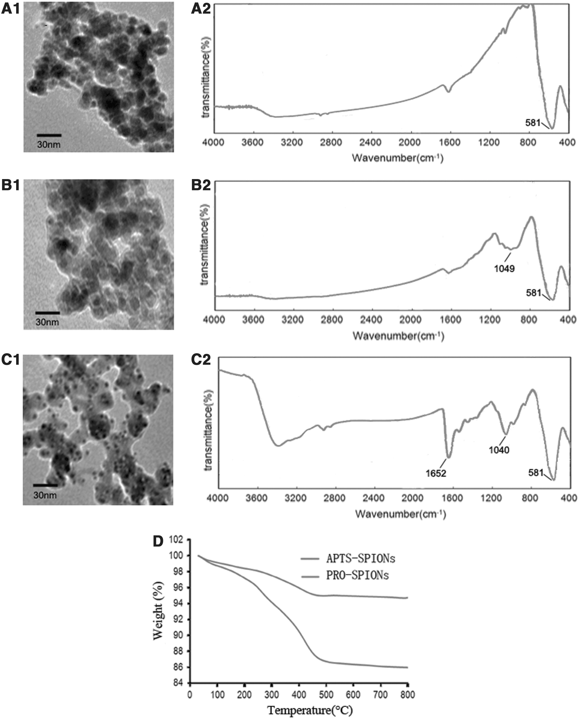
Characterization of the SPIONs. TEM images of nSPIONs
nSPIONs, APTS-SPIONs, and PRO-SPIONs have different FTIR spectra (Fig. 1A2–C2). There are strong peaks between 581 and 1652 cm−1 for all the three SPIONs. All the three nanoparticles' peak fluctuation range is from 400 to 581 cm−1. APTS-SPIONs and PRO-SPIONs have strong peaks at similar positions, 1040 and 1049 cm−1, respectively, while nSPIONs have no strong peaks between 800 and 1200 cm−1. PRO-SPOINs also have a strong peak at 1652 cm−1.
TGA was carried out to analyze the surface formation and the content of Fe3O4 in the APTS-SPIONs and PRO-SPIONs. As is shown in Figure 1D, the weight lost increased along with the temperature from 10°C to 800°C. The weight loss for the APTS-SPIONs and PRO-SPIONs were about 5% and 14%, respectively. The nanoparticles lost their coating and only Fe3O4 was left at temperatures above 450°C.
Properties of the SPIONs in vitro
Under MIH, the SPIONs reduced the cell viability and proliferation of the SW480 and Hep G2 cells (Fig. 2A). PRO-SPIONs showed more inhibition effects on the cancer cells compared with ATPS-SPIONs. The iron content changed with the coincubation time of SPIONs with Hep G2 cells (Fig. 2B). The iron content at 2 hours was set as 100%. The iron content reached the peak at 4–8 hours. Prussian blue staining was carried out to detect iron in Hep G2 cells after their coincubation with ATPS-SPIONs and PRO-SPIONs (Fig. 2C1, C2). The iron content increased when the nanoparticles were modified by PRO. Both the APTS-SPIONs and PRO-SPIONs localized primarily in the plasma (Fig. 2D1, D2). However, PRO-SPIONs mainly accumulated around the nucleus. The cell viability and proliferation of the cells were determined by MTT method to reflect the cytotoxicity of the PRO-SPIONs (Fig. 2E). The cell viability decreased to about 10% even at the highest PRO-SPIONs concentration, no significant difference with the control group.
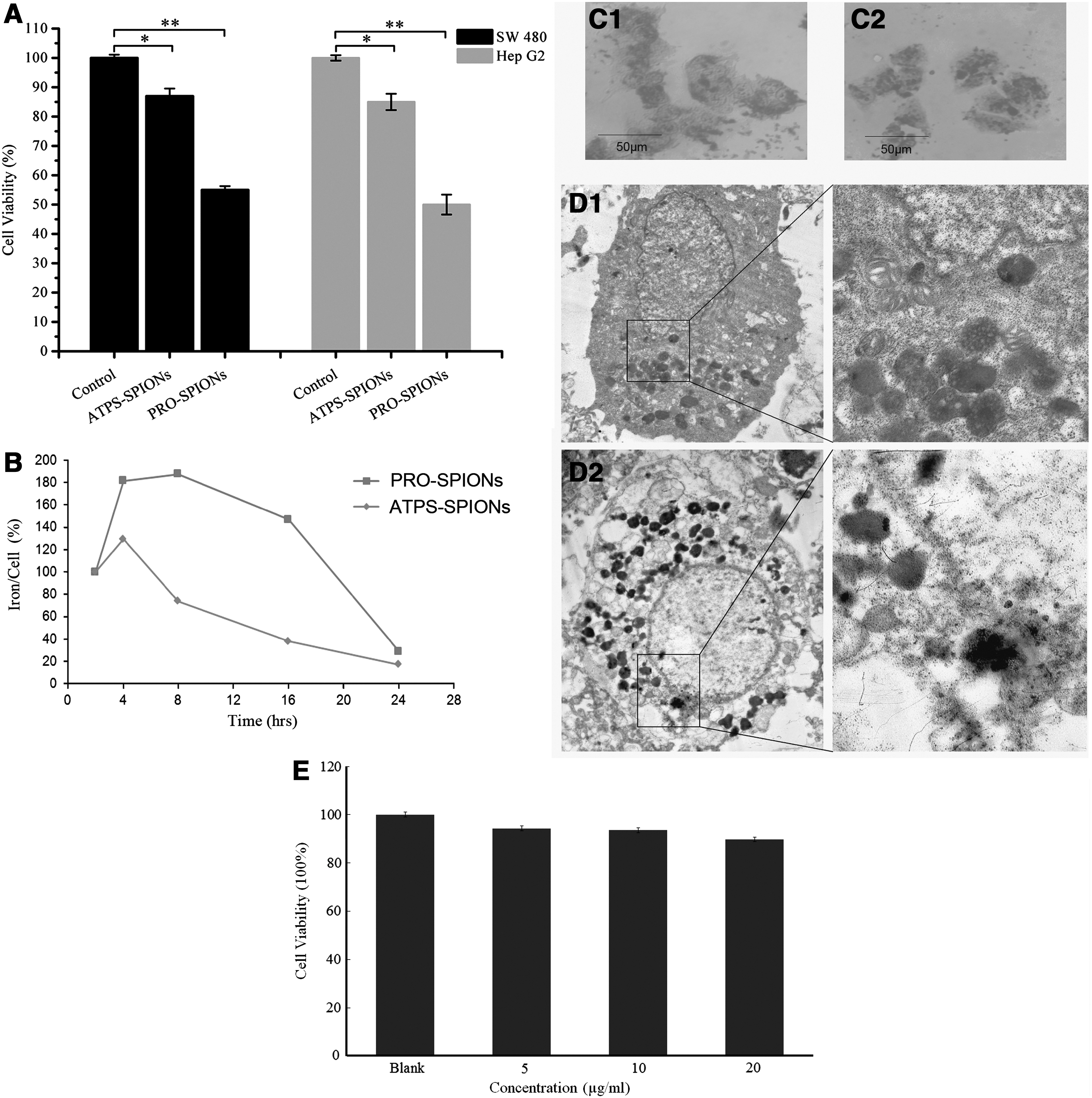
Properties of the SPIONs in vitro.
The influence of magnetofection and alternating current magnetic field on TNF-α expression
The authors used different methods to transfect plasmid pcDNA4/GFP/TNF-α into Hep G2 cells. The cells were then exposed to alternating current magnetic field (110 kHz, 11 mT) once (24 hours after transfection) or twice (24 and 36 hours after transfection). Green fluorescence intensity of GFP is correlated with TNF-α expression. The green fluorescence is the strongest when commercial VigoFect was used together with PRO-SPIONs and the cells were exposed to alternating current magnetic field only once (Fig. 3E). However, the fluorescence weakened when the cells were exposed to alternating magnetic field one more time.

The expression of plasmid pCDNA4-GFP-TNFα.
Combined application of MIH and gene therapy in tumor model
The PRO-SPIONs injected into the tumor exhibited good temperature-increase performance (Fig. 4A). Under alternating magnetic field, the temperature in the tumor increased to 43°C within 2 minutes. The tumor temperature was kept at 43°C ± 0.5°C for 30 minutes by adjusting the current intensity. No significant increase was observed in the temperature of the mice rectum. The tumor volume was measured every 3 days. The tumor volume increased along with the time in Group Blank, in which the mice were injected with normal saline (Fig. 4B). MIH, gene therapy, and the combined therapy all can effectively inhibit the tumor growth (Fig. 4B, C). In Group PRO/TNF-α, the tumor almost stopped to grow. At the end of the experiment, the tumors were dissected. The PRO-SPIONs mainly accumulated in the tumor other than normal tissues (Fig. 4D).
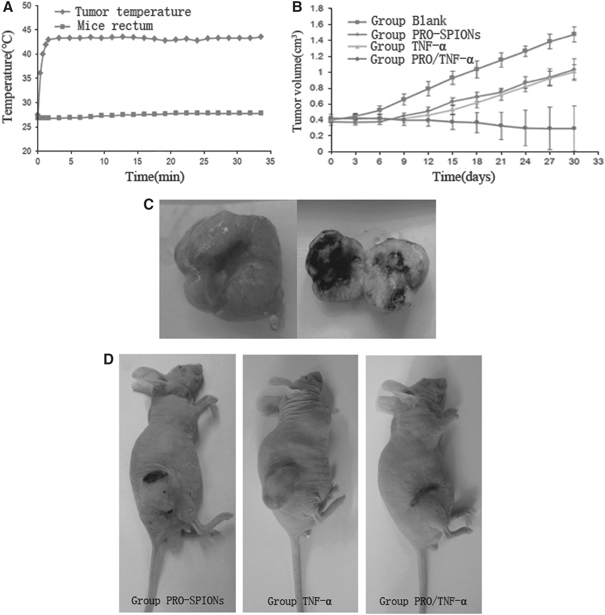
Combined application of magnetic induction hyperthermia and gene therapy in tumor model.
Discussion
Cancer is one of the most lethal diseases threatening human beings' lives. MNPs are drawing more and more attentions because of their unique properties and promising applications in medicine. As therapeutic tools for tumors, MNPs have been evaluated extensively as targeted drug carriers. MNPs can direct therapeutic agents specifically to the tumor site, which overcome the limitations of conventional chemotherapies. The drug dosage and side-effects can then be dramatically reduced. 17 –19 Fe3O4 based SPIONs, one of the most promising MNPs, has all the attractive properties of MNPs. Moreover, the temperature of Fe3O4 can rise rapidly under alternating current magnetic field. As a result, the temperature of the tumor rises, while the temperature of normal tissue remains steady. The tumor site directing and temperature rise properties make Fe3O4 nanoparticles more effective in targeted treatment of tumors.
Usage of SPIONs depends largely on the preparation processes. The authors used coprecipitation method to prepare the SPIONs to obtain uniform size of 5–10 nm. These SPIONs are paramagnetic and accumulational (Fig. 1A–C). Appropriate modification of the surfaces can obviously increase the SPIONs' delivery efficiency and performances. In the present research, the authors respectively coated APTS and PRO on the SPIONs and study their properties and performances. FTIR was carried out to determine whether APTS or PRO has been coated onto the surface of nSPIONs. It was reported that there is a characteristic peak at around 570–590 cm−1 for the naked Fe3O4 due to the Fe-O groups. The nSPIONs, APTS-SPIONs, and PRO-SPIONs, which the authors prepared, have the characteristic peak at 581 cm−1 caused by Fe-O-Fe stretching vibration. The APTS-SPIONs have the Si-O peak at 1049 cm−1. 1,13,20,21 The PRO-SPIONs have an absorption peak at 1652 cm−1, which is the characteristic peak of amide bond in PRO. PRO exhibits higher coupling rate to the SPIONs than APTS in the TGA (Fig. 1D). As a result, PRO can modify the surface more efficiently than just APTS and the content of Fe3O4 will be much lower in application, which reduces the toxicity. The intracellular iron content decreased with time since 8 hours after incubation of PRO-SPIONs with Hep G2 cells (Fig. 2B). The authors presumed that proliferation of the cells contribute to this phenomenon. The SPIONs were separated into daughter cells after division and the SPIONs in a single cell reduced. Their results were in accordance with previous reports. It was reported that SPIONs go into cell cytoplasma by cytophagy to form secondary endosome enveloped by membranes. The endosomes are evenly distributed into the daughter cells. 22,23 They finally selected PRO to coat on the surface of Fe3O4 nanoparticles. The PRO-SPIONs distributed more concentrated around the nucleus and promoted transfection of the conjugated TNF-α gene and its expression. Several advantages of PRO-SPIONs may explain the results. The PRO-SPIONs' size only increases modestly, and this does not influence its transfection efficiency. PRO carries abundant positive charges, which compress the electronegative DNA. 24 As a result, the DNA/SPIONs complexes become small and can be more easily transfected into the cells. Protamine also contains nuclear localization signal sequence which targets nucleus. The results showed that PRO-SPIONs have elevated zeta-potential, and this property facilitates its combination with DNA and protect DNA from enzymolysis. 25 On the basis of the above merits, PRO-SPIONs effectively transfect DNA into Hep G2 cells under magnetic field and promoted gene expression. PRO-SPIONs may be used as a novel effective gene transfection carrier. Inspiringly, protamine is found to be safe and has been approved by U.S. Food and Drug Administration (FDA) as insulin sustained release preparation to neutralize excessive heparin. 26 In the animal experiments, PRO-SPIONs accumulated only in the tumor without penetrating into normal tissues. This will increase the safety during PRO-SPIONs' application.
Combined MIH and gene therapy for treatment of liver cancer model in their research exhibited better effects than MIH or gene therapy alone. TNF-α is a potent anticancer cytokine. TNF-α gene therapy has been investigated abundantly, and there have been some exciting applications. TNFerade is a replication incompetent adenoviral vector that delivers TNF-α gene under the transcriptional control of a radiation inducible promoter. TNFerade is currently in stage III trials. 27 After being exposed to alternating magnetic field once, the transfection efficiency was enhanced. However, the transfection efficiency was weakened when the cells were exposed to alternating magnetic field twice. This may be because the first time exposure transfected abundant TNF-α gene into the cell. Maybe being exposed to the alternating magnetic field twice exerted much more pressure on the cells and caused the decrease of the cell viability. Thus, the cells could not express the gene, as previously said. The results show that MIH or gene therapy alone did not totally block the tumor growth, while the combined therapy caused the tumor to shrink. The temperature of 43°C can activate the CMV promoter, 28 which drives TNF-α expression in this research.
A novel type of SPIONs has been prepared by coprecipitation and surface-modified sequentially by APTS and PRO. The gene of the anticancer cytokine TNF-α is conjugated onto the PRO-SPIONs. The PRO-SPIONs efficiently transfect the TNF-α gene into cells. As the PRO-SPIONs accumulated solely in the tumor site, when the tumor temperature rises under alternating current magnetic field, normal tissues stay undisturbed. The combined MIH and gene therapy is promising in liver cancer treatment.
Footnotes
Acknowledgment
This study was supported by the China National Natural Science Foundation (No. 81071885; to X.W.) and (No.10775085; to J.T.).
Disclosure Statement
No competing financial interests exist.