
Abstract
Background:
Hybrid positron emission tomography (PET)-magnetic resonance imaging (MRI) systems have been taken in use as new clinical diagnostic tools including detection and therapy planning of cancer. To reduce the amount of contrast agents injected in patients while fully benefitting both modalities, dual-modality probes are required.
Material and methods:
This study was first aimed at developing a hybrid PET-MRI probe by labeling superparamagnetic iron oxide nanoparticles (SPIONs) with 64Cu using a fast and chelator-free conjugation method, and second, to demonstrate the ability of the agent to target sentinel lymph nodes (SLNs) in vivo using simultaneous PET-MRI imaging.
Results:
High labeling efficiency of 97% produced within 10–15 min was demonstrated at room temperature. 64Cu-SPIONs were chemically stable in mouse serum for 24 h and after intradermal injection in the hind paw of C57BL/6J mice, demonstrated specific accumulation in the SLN. Simultaneous PET-MRI clearly demonstrated visualization of 64Cu-SPIONs, in dynamic and static imaging sequences up to 24 h after administration.
Conclusion:
The use of a single hybrid probe and simultaneous hybrid imaging provides an efficient, complementary integration of quantitation and is expected to improve preoperative planning and intraoperative guidance of cancer treatments.
Introduction
Positron emission tomography (PET) is a proven imaging technique used extensively for cancer diagnostics and therapy monitoring. After administration of radiopharmaceuticals of high specific activity, the tracer accumulates in the lesion or organ of interest, providing functional or high sensitivity (picomolar range) quantitative molecular images. However, PET images suffer from a relatively low spatial resolution (2–5 mm) and lack anatomical details of the studied subject.
Magnetic resonance imaging (MRI) is a noninvasive method to generate high resolution two-dimensional or three-dimensional (3D) morphological or functional images with excellent soft tissue contrast. On the other hand, for many diagnostic MRI procedures contrast agents need to be used to enhance sensitivity and to visualize lesions.
The synergistic combination of PET and MRI have important advantages in several clinical applications, for example oncology, 1 –4 neurological diseases, 5,6 and vascular or cardiac inflammation imaging. 7,8 PET-MRI not only provides high resolution multiparametric quantitative images, but it also improves lesion characterization, in particular, if an appropriate bimodal contrast agent is used. The simultaneous PET-MRI data acquisition is highly appreciated in translational research, especially for pharmacokinetic and biodistribution studies of new radiolabeled-biomarkers and bimodal contrast agents. 9
Currently, few preclinical PET-MRI systems are available for true simultaneous data acquisition, in part because of physical and technical difficulties to fit a miniaturized PET insert into the MRI system. The reduction of the size of the PET detectors leads to a small field of view (FOV), radio frequency (RF) interferences with the electronics may occur, hardware RF heating can lead to drop in PET detector stability and the attenuation correction in PET is challenging. 10
The aims of this study were first, to develop a bimodal PET-MRI contrast agent, based on 64Cu-labeled superparamagnetic iron oxide nanoparticles (64Cu-SPIONs) and second to use this new contrast agent in a proof of concept study for visualizing the sentinel lymph nodes (SLNs) in a mouse model (C57BL/6J) up to 24 h, using a simultaneous PET-MRI system available at Brookhaven National Laboratory (New York, NY). 11,12 The SLNs are of particular interest because they are the first sites where malignant cancer cells spread in several cancer types, for example breast cancer, prostate cancer, and malignant melanoma. Therefore, the localization and visualization of the SLNs is of major importance in cancer staging and therapy. 13
We previously demonstrated the feasibility of labeling SPIONs with 99mTc (half-life t1/2 = 6.02 h) for sequential SPECT/MR imaging 14 and with 68Ga (t1/2 = 67.71 min) for PET-MRI and Cherenkov luminescence imaging of the SLN. 15 64Cu (t1/2 = 12.7 h) has the advantage of a longer half-life than previously used radionuclides and permits the execution of dynamic imaging and biodistribution studies for a longer time period. The simultaneous PET-MRI data acquisition strategy provides a perfect match of temporal and spatial information to demonstrate the stability and reliability of the agent.
64Cu-SPIONs have been reported to be produced by using various acyclic and macrocyclic chelates for conjugation, such as 1,4,7,10-tetraazacyclo-dodecane-1,4,7,10-tetraacetic acid (DOTA), 1,4,7-triazacyclononane-1,4,7-triacetic acid (NOTA), and 2-[1,4,8,11-tetraazacyclotetradecane tetraacetic acid (TETA). 16 –19 Other studies described the synthesis of 64Cu(II)-Bis(dithiocarbamatebisphosphonate) and its conjugation with SPIONs for PET-MRI imaging. 20
Herein, we report a new chelator-free, time-efficient radiolabeling method of 64Cu-SPIONs with high specific activity and demonstrate the feasibility of the agent in vivo using simultaneous PET-MRI imaging to visualize SLNs in a preclinical setting.
Materials and Methods
Nanoparticles
SPIONs were generously provided by Genovis AB, Sweden and produced as previously described by Kjellman et al. 21 Briefly, the iron oxide monodispersed core is mainly composed of iron (III) oxide-hydroxide and coated with funtionalized polyethylenglycol (amino-PEG) and stored in 1 mL 150 mM NaCl. The iron concentration was measured to be 3.4 mg/mL using inductively coupled plasma–optical emission spectrometry. 21 The average hydrodynamic diameter of SPIONs was measured to be 27 nm (standard deviation 3 nm), as determined by dynamic light scattering using a Malvern Zeta Sizer Nano Series (Malvern Instruments Ltd., Worcestershire, UK).
Tracer selection
Table 1 presents the most commonly used PET radionuclides for labeling radiopharmaceuticals for clinical or research applications. The majority of PET radionuclides such as 18 F or 68Ga have a short half-life and need to be produced locally, by either cyclotron or using a generator. 64Cu can be produced with high specific activity using medical cyclotrons via the fusion-evaporation reaction 64Ni(p, n) 64Cu, or in a nuclear reactor. 22,23 Due to its relatively long half-life, it is possible to transport 64Cu from the site of production to remote locations. In addition, 64Cu has relatively low positron energy and thus a short positron range in tissue, permitting higher spatial resolution PET imaging.
Because of its favorable positron emission characteristics, half-life, and chemical properties we decided to use 64Cu to label SPIONs for our PET-MRI imaging and biodistribution studies. There are many reports describing the conjugation chemistry, as previously mentioned. 19 However, radiolabeling with the conventional methods was found to be time consuming and we therefore developed an alternative faster and direct labeling process.
PET-MRI system
The PET-MRI system used for the studies presented here has previously been evaluated and described in detail. 12,13 It is based on the Rat Conscious Animal PET (RatCAP) scanner, 24 –26 modified to be MR-compatible with a custom-built quadrature RF transceiver coil and integrated into a Bruker Biospec 9.4 T MRI scanner, using ParaVision 5.0.
Briefly, the PET insert consists of 12 lutetium oxyorthosilicate (LSO) block detectors forming a ring. The blocks are divided into 4 × 8 arrays, each individually coupled to Hamamatsu S8550 avalanche photodiodes. Inside the detector ring fits the custom built RF coil and the 3D printed animal bed centered in the MRI scanner. The PET front-end electronics uses an application-specific integrated circuit to minimize space and interconnection requirements. The events are recorded in list mode via a Field-Programmable Gate Array (FPGA)-based data acquisition module and transmitted through fiber-optic cables to an acquisition computer situated outside the MRI room.
Phantom studies
An evaluation of the PET-MRI system was performed using a Deluxe Mini Jaszczak image quality phantom filled with 18 F-flouride (∼15 MBq). The phantom was placed on the animal bed and centered in the FOV of both the PET and MRI scanners. First PET and then simultaneous PET-MRI data acquisition was performed while time resolution, singles and coincidence detection count rates were compared. To assess the image registration accuracy, PET and 3D MRI fast low angle shot (FLASH) sequences were acquired and coregistered using PMOD (PMOD Technologies, Zurich, Switzerland) and custom Matlab® (The MathWorks, Natick, MA) based software.
64Cu-SPION radiolabeling and quality control
64Cu was purchased from Mallinckrodt Institute of Radiology (Washington University School of Medicine, St. Louis, MO) in the form 64CuCl2 (28 μL in 0.5 HCl) and diluted in ammonium acetate buffer (40 μL, pH 5.5, 0.2 M).
SPIONs (80 μL, 3.4 mg Fe/mL) diluted with ammonium acetate were injected into a sterile, vacuum collecting vial (5 mL in volume). Subsequently, the 64Cu-acetate solution (∼20 μL, 47 MBq) was added into the same vial for radiolabeling, and stirred for 30 s. The pH was monitored with pH test strips (three steps; Sigma, USA and Merck, Germany). The reaction mixture was incubated at room temperature for 10 min.
Then, the radiolabeled 64Cu-SPIONs were separated from free 64Cu using a magnetic separation column (LS-column; Miltenyi Biotec, Bergisch Gladbach, Germany). By attaching a permanent magnet to the column and filtering the reaction mixture, 64Cu-SPIONs were trapped within the column, while the unbound 64Cu and buffer solution flowed through. Additional 2 mL ammonium acetate was added to the column to “wash” the nanoparticles, before removing the magnet. The 64Cu-SPIONs were eluted in ammonium acetate buffer solution (the iron concentration was estimated to be ∼1.4 mgFe/mL postlabeling).
The labeling efficiency was calculated as follows: eluted 64Cu-SPIONs activity/total activity × 100% (Eq. 1); the activity was measured by using a dose calibrator (CRC-25PET; Capintec Inc., USA).
In vitro stability measurements were performed using instant thin layer chromatography (ITLC, silica gel-impregnated glass fiber sheets, 2.5 × 10 cm; Life Science) and 50 mM ethylenediaminetetraacetic acid (EDTA) in ammonium acetate/methanol (50/50; v/v) as mobile phase. Samples (0.7–1 μL) of 64Cu-SPIONs in buffer solution were taken 6, 12, and 24 h postlabeling, dropped on silica gel sheets and developed for 15 min. The silica gel strips were dried, placed under a phosphor storage plate (Multisensitive, medium; Perkin Elmer, Wellesley, MA), and exposed for 5 min. Then, the plate was scanned by a phosphor imager (Perkin Elmer) and the labeling yield was quantified using OptiQuant image analysis software (Perkin Elmer).
The radiochemical stability was assessed by incubating the 64Cu-SPIONs in mouse serum. Samples (0.7–1 μL) were taken at 6, 12, and 24 h and the radiolabeling yield quantified using the ITLC method already described. EDTA challenge was performed incubating 64Cu-SPIONs with solution of EDTA 1:300-fold excess for 10 min. The solution was then filtered using the magnetic separation columns, and the radiolabeled yield was calculated with the Equation (1).
Animal studies
Animals in this study were used in compliance with local and national regulations and protocols were approved by the Institutional Animal Care and Use Committee of Brookhaven National Laboratory.
Six mice (n = 6), C57BL/6, 3 months old, female (Charles River Laboratories) were injected intradermally in the right hind paw with 64Cu-SPIONs (1.5–6 MBq, ∼0.01 mL in volume, ∼0.014 mg Fe). Two animals were imaged prone, using dynamic acquisition mode. Acquisition began 5–10 min after the injection and continued for 60–180 min; and shorter duration PET-MRI scans were subsequently performed after 3 h (n = 4), 6 h (n = 2), and 24 h (n = 6) postinjection. All animal procedures were performed under anesthesia by inhalation of a 1.8%–2% isoflurane-air mixture in addition to an intraperitoneal dose of 0.02 mg glycopyrrolate in 0.2 cc 0.9% NaCl.
Simultaneous PET-MRI data acquisition
The PET data were collected with an energy window of 350–700 keV and coincidence timing window of 40 ns.
The MRI images were acquired using two different sequences. FLASH 3D image sequence using the following parameters: slice thickness 0.123 mm, spacing between slices 0.125 mm, repetition time 16.5 ms, echo time 4.3 ms, flip angle 10°, and matrix size 256 × 256. For rapid acquisition with refocused echoes (RARE) sequence, we used the image parameters: slice thickness 0.6 mm, spacing between slices 0.4 mm, repetition time 2500 ms, echo time 40 ms, echo train length 8, pixel bandwidth 394, flip angle 180°, number of averages 6, and matrix size 128 × 128.
Image reconstruction and coregistration
A custom data acquisition software 27 was used to monitor the PET events; singles, coincidence rates in real time, and to visualize the sinograms. The images were reconstructed using a 3D maximum likelihood-expectation maximization iterative algorithm with normalization (detector efficiency) and random correction. Finally, the PET images were produced with 1.08 × 1.08 × 1.19 mm3 voxels, and were coregistered to the MRI images using both PMOD and custom MATLAB based software using the registration parameters determined from the phantom images.
Biodistribution
All animals were euthanized as per protocol after the last imaging session (24 h postinjection) and organs of interest including lymph nodes (LNs), liver, spleen, kidneys, and intestine and injection sites were excised, weighed, and assessed for radioactivity. Background corrected count rates of the samples were acquired on an automatic LKB Wallac gamma counter (CompuGamma CS) using an energy window of 400–600 keV. The counts rates were corrected for 64Cu decay and positron branching fraction and converted to activity (Bq) using a standard efficiency value for 511 keV. The data were then decay corrected to the time of the injection, and is reported as percentage of injected activity per mass unit of organ (% IA/g).
Results
Phantom studies
Both PET and MRI systems visualized the cylindrical phantom containing 18 F (∼15 MBq) in 0.9% NaCl solution with good accuracy. Figure 1A presents the transverse MRI, PET, and the fused PET-MRI images of the phantom as a result of the simultaneous data acquisition. No distortions were observed in the merged PET-MRI images. Although the PET has a more limited axial FOV of 18 mm in comparison with MRI, which covers 27.4 mm, the system is able to accommodate small animals such as mice.

PET-MRI phantom studies.
In Figure 1B and C, the spatial resolution in MRI and PET system was evaluated. All five rods in the phantom (1–5 mm) could be easily distinguished in the MRI images indicating a spatial resolution of ∼1 mm, while the PET images only displayed four rods with a spatial resolution of ∼2 mm. Accordingly, the PET-MRI system is capable of detecting the smaller LNs (popliteal, iliac, and lumbar) in mice, which are within a size range of 1.7–4.8 mm.
64Cu-SPIONs radiolabeling and quality control
The labeling efficiency as determined by the magnetic separation and ITLC was 97%. Using the ITLC system, 64Cu-SPIONs are retained at the application point, while free 64Cu moved with the solvent front at an Rf value of 0.9. The in vitro stability of the agent was >95% in radiolabeled yield, in both buffer solution and mouse serum for 24 h. A small degradation of the complex was observed after the EDTA challenge, resulting in a 95% radiolabeled yield. The magnetic filtration method was shown to be a fast and effective method to separate radiolabeled 64Cu-SPIONs from the reaction mixture and using Equation (1) a fast and accurate result of labeling yield can be calculated.
Dynamic imaging
Dynamic PET-MRI images revealed rapid (within 5–10 min) lymphatic drainage and transport of the 64Cu-SPIONs from the injection site (right hind leg paw) into the SLN (popliteal node) (Fig. 2). The example images presents the transverse, sagittal, and coronal slices through the mouse including the sentinel (popliteal) LN marked with a red hair cross in the MRI images (first row). The SLN appear as a “black,” low signal intensity structure in the T2-weighted MRI images reflecting the accumulation of the 64Cu-SPION particles within the node. 64Cu-SPIONs in tissue causes shortening of the transverse relaxation of the spins and induces local inhomogeneity in the magnetic field; consequently, the signal intensity in SLNs is significantly decreased. In this particular case, the SLN can be easily visualized due to the “white,” triangle-shaped fatty tissue surrounding the node. In the corresponding PET images (second row) the SLN is clearly visualized in all three directions. However, the PET images lack anatomical information and it is not possible to accurately localize the SLNs in vivo without the anatomical template. Therefore, fused PET-MRI images (third row) are preferred to both detect and anatomically localize the SLNs.
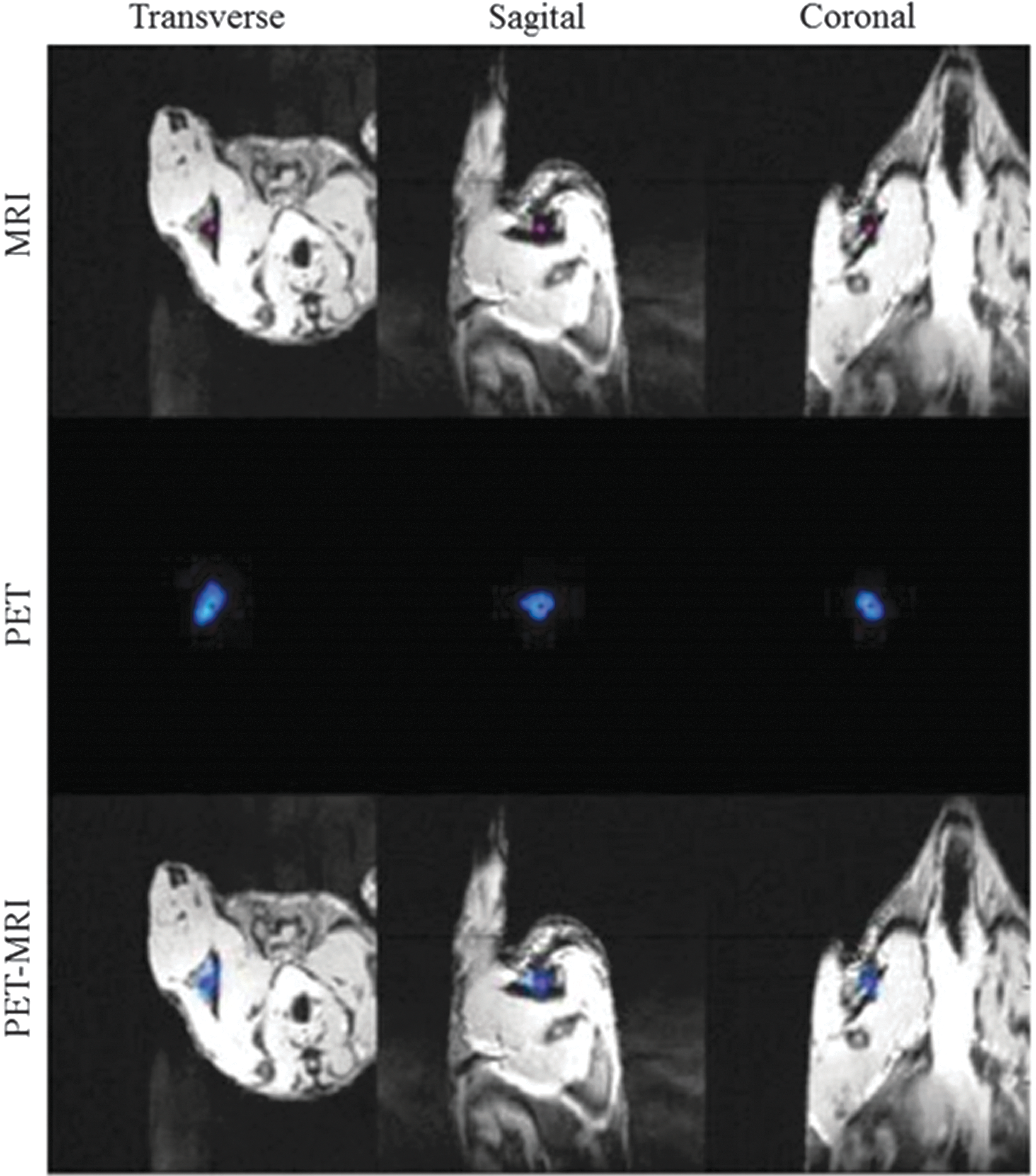
Transverse, sagittal, and coronal T2 weighted MRI, Dynamic PET and PET-MRI fused images visualizing the SLN after subcutaneous administration of 64Cu-SPIONs in the back hind paw of the mouse, ∼10–15 min after subcutaneous administration of the agent. 64Cu-SPIONs accumulated in the SLN (marked with a red cross in MRI images), cause shortening of the transverse relaxation of the spins and introduce inhomogeneities in the magnetic field; consequently, SLNs appear as dark due to the signal loss in tissue in MRI images. PET images are clearly depicting the SLNs in all three orthogonal planes, but lack anatomical information. Therefore, the fused PET-MRI images are preferred for exact localization and high accuracy detection of SLNs. MRI, magnetic resonance imaging; PET, positron emission tomography; SLNs, sentinel lymph nodes; SPIONs, superparamagnetic iron oxide nanoparticles. Color images available online at
Three hours postinjection, as the 64Cu-SPIONs were transported from the SLN and further along the efferent lymphatic vessels; activity was detected in inguinal and lumbar lymph nodes. Activity was also observed in the intestine tissue.
Simultaneous PET-MRI imaging
The combination of high-sensitivity PET for detection of 64Cu-SPIONs and MRI with high spatial resolution including excellent soft tissue contrast provides accurate, anatomical localization of the LNs (Fig. 3). Figure 3 shows an example of MRI, PET, and PET-MRI images of a mouse presented in three orthogonal planes (transverse, sagittal, and coronal), 24 h after intradermal administration of 64Cu-SPIONs into the right hind paw. Accumulation of the agent in the SLN (popliteal) and inguinal LN is evident due to the “negative” contrast enhancement, also marked with white arrows in the transverse and coronal MRI images. The signal loss was not so evident in the intestines presented in the sagittal plane until examining the PET images. PET clearly depicts the LNs and clearly demonstrates the presence of 64Cu-SPIONs in, for example, large intestine, the ultimate elimination path of the agent, 6 h postinjection. Fused PET-MRI images enable both detection and localization of the LNs with high accuracy at 3, 6, and 24 h postinjection. As the SPIONs concentration increases in the most tiny LNs (1.7–2.5 mm in diameter) it may lead to “blooming” or susceptibility artifacts at late scan-times as observed in both FLASH and RARE images (Fig. 3, transverse plane, first row). Therefore, SPIONs alone might not be ideal for the localization of SLNs and as demonstrated here, 64Cu-radioisotope and additional PET imaging in addition to MRI is highly beneficial.

Visualization and anatomical localization of the SLNs in vivo using simultaneous PET-MRI data acquisition, 24 h after subcutaneous administration of the 64Cu-SPIONs in the back hind paw of the mouse. Signal loss can be observed in T2 weighted MRI images (first row) within and around the LNs in transverse and coronal plane. PET images lack anatomical information; however, they clearly depict the LNs and show high activity levels in the large intestine, sagittal plane (second column). Fused PET-MRI images (third column) demonstrate high accuracy in both detection and localization of SLNs and accumulated activity in other organs, for example, intestine that could not be detected without PET. MRI, magnetic resonance imaging; LNs, lymph nodes; PET, positron emission tomography; SLNs, sentinel lymph nodes; SPIONs, superparamagnetic iron oxide nanoparticles. Color images available online at
Biodistribution
The 64Cu-SPIONs show a high overall clearance from the injection site (∼80%), and high accumulation and retention in the SLNs (4.8 ± 1.73% IA), 24 h after intradermal administration in the right hind paw in mice. Activity was also detected in spleen (1.3 ± 0.3% IA), kidneys (6.3 ± 0.96% IA), and liver (11 ± 1.88% IA) and at the injection site (17.52 ± 1.92% IA). The mean uptake for organs of interest is presented in Figure 4. A quite high amount of activity was also found in gastrointestinal tract, in the small intestine (27 ± 2.86% IA) and large intestine (23 ± 1.92% IA), the ultimate elimination path of the contrast agent.
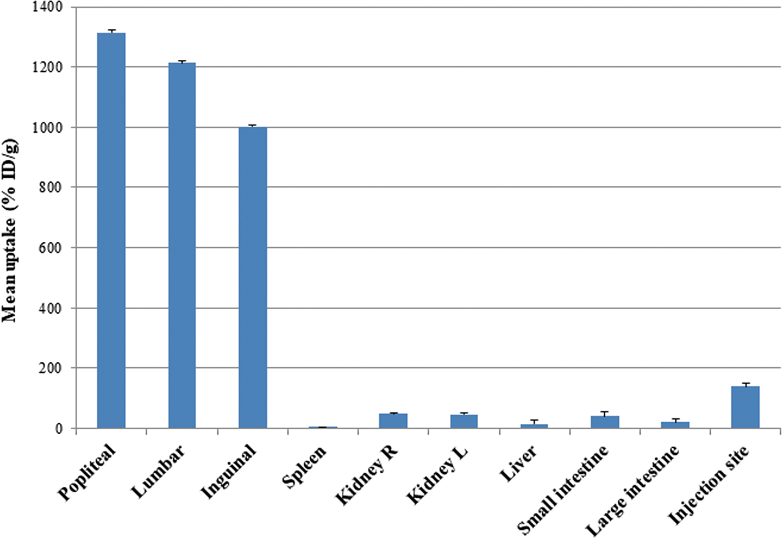
Biodistribution data of 64Cu-SPIONs in 6 C56 black mice after subcutaneous injection in the right hind paw and sacrificed at 24 h postinjection. The mean uptake is expressed as percentage of injected activity per gram. SPIONs, superparamagnetic iron oxide nanoparticles. Color images available online at
Discussion
In the past few years, hybrid PET-MRI systems have been developed for clinical and preclinical use. As a consequence, it has become evident that a double-modality contrast agent able to visualize the target of interest by both modalities may be required, not only to streamline the process for patients, but also to assure that the PET-MRI images reflect the same biological process. PET tracers are usually small molecules, the standard procedure is to attach or incorporate the radioisotopes into generally larger MRI contrast agents (molecules) such as gadolinium diethylenetriamine pentaacetic acid, liposomes, dendrimers, or SPIONs for SLN imaging. 28,29
SPIONs have the great advantage to be produced within a narrow size range of 20–50 nm optimal for SLN imaging. In addition, possess a large surface-to-volume ratio making them optimal platforms for assembly of multimodality contrast agents as diagnostic and therapeutic site-specific delivery vehicles.
Current, SLN imaging agents used in the clinic such as 99mTc-Nanocoll or 99mTc labeled sulfur colloids for scintigraphic imaging are not target specific, revealing little information about SLN's status and are lacking in anatomical details in regards to accurate localization of SLNs in patients. SPIONs on the other hand, have shown a great potential to differentiate between normal and metastatic LNs in patients with breast cancer, prostate cancer, pelvis and urological cancer with superior sensitivity and diagnostic specificity compared with conventional MRI. 30 –32
In this study, a new dual modality contrast agent was developed using a time efficient, direct conjugation of 64Cu-SPIONs for PET-MRI detection and localization of SLNs.
It was demonstrated that a high (∼97%) radiolabeling efficiency can be achieved by adding 64CuCl2 to functionalized PEG coated SPIONs in ammonium acetate buffer solution (pH 5.5). Cheng et al. did report that chelator-free radiolabeling of iron-based nanoparticles with an oxophilic exterior using 89Zr was possible, in a PET/MR study of lymphatic drainage. 33 The 64Cu-SPIONs were found stable in vitro and in vivo up to 24 h. The labeling procedure takes ∼10 min, much faster than other authors reported when using bifunctional chelators such as NOTA, DOTA, TETA, or DTCBP. 33 In this study, the labeling is carried out at room temperature, which minimizes the risk of destroying the coating or structure of the nanoparticles.
Dynamic and simultaneous PET-MRI imaging demonstrates the feasibility of 64Cu-SPIONs to visualize SLNs in mice with good accuracy. Both modalities can visualize the accumulation of 64Cu-SPIONs in SLNs within minutes after intradermal administration of the agent in the hind paw of the mice (Fig. 3). However, due to the negative contrast enhancement on MRI and lack of morphological information in PET, fused PET-MRI images are more informative as shown in Figures 3 and 4. The contrast enhancement in the images is concentration dependent; therefore the amount of injected 64Cu-SPION and timing of image acquisitions must be carefully chosen. The amount of 64Cu-SPION injected in this study (1.5–6 MBq, 0.014–0.02 mg of Fe in ∼0.01 mL in volume) was optimal for early detection (3–6 h); however, the injected amount can easily be reduced to minimize the susceptibility artifacts in MRI images for later time points. Generally, ∼10 mg Fe/kg is recommended (mouse weight ∼20 g) when the agent is administrated intravenously. 17 Here, we demonstrate that SLNs are clearly visualized by injecting ∼10–15 times less SPIONs by intradermal administration of 64Cu-SPION, thereby reducing the risks for toxicity.
The biodistribution of 64Cu-SPIONs in vivo is influenced mainly by the size of the nanoparticles, injection route, and technique. Our formulation demonstrated a rapid retention and slow clearance of 64Cu-SPIONs in SLNs. Approximatively 5% of the IA was still present in SLNs, at 24 h postinjection enabling imaging and evaluation of SLNs even in >1 d protocols. Individual variations in the biodistribution between subjects were observed, probably resulting from differences in the 64Cu-SPION dose administered intradermally. The reproducibility of the intradermal injections is also important because minor deviation can lead to differences in uptake, as we have seen in 1 of 6 animals. The mean uptake in SLNs was found to be 4.8% IA, with the lowest value of 0.5% IA and highest value of 26% IA measured at 24 h postinjection. This can be compared to the mean value of 2% IA for 68Ga-SPIONs at 3 h postinjection 14 and 1.72% IA for 99mTc-SPIONs at 5 h after subcutaneous injection in the hind paw of white Wistar rats. 15 A significant amount of 64Cu-SPIONs was observed in the small and large intestine of the mice 24 h postinjection, which may indicate that the radiolabeled SPIONs are ultimately eliminated through the intestines.
Conclusion
Here, we report the development of a hybrid PET-MRI probe based on 64Cu-SPIONs. In contrast to other studies reported, the tracer was conjugated without chelating agents, within 10 min at room temperature. The agent was shown to (1) be stable at physiological conditions, (2) produce a strong contrast between targeted and normal tissue in T2-weighted MRI images, and (3) enable detection and visualization of sentinel nodes in vivo using simultaneous PET-MRI imaging up to 24 h postinjection.
Footnotes
Acknowledgments
This study was performed with generous support from the Swedish Cancer Foundation, the Swedish Science Council, Mrs Berta Kamprad's Foundation and the Gunnar Nilsson's Foundation. The SPIONs were provided by Genovis AB, Lund, Sweden.
Disclosure Statement
No competing financial interests exist.