
Abstract
Objectives:
As acid–base imbalance is involved in many pathological processes, the capability to image tissue pH alterations in the clinic could offer new ways to detect disease and respond to treatment. In this study, the authors show that tissue pH can be imaged in vivo with 11C-labeled bicarbonate (H11CO3 −) buffer and positron emission tomography (PET).
Methods:
H11CO3 − was produced by on-column NaOH adsorption. Biodistribution of H11CO3 − in normal mice was determined. In addition, uptake studies and inhibition experiments of H11CO3 − in the S180 fibrosarcoma-bearing mice and the inflammatory mice were investigated with PET imaging. The tumor and inflammatory interstitial pH was measured by a needle pH microelectrode.
Results:
PET imaging demonstrated the high uptake of H11CO3 − in mice tumor tissues and inflammatory tissues, which showed that the average tumor or inflammatory interstitial pH was significantly lower than the surrounding tissue. Administration of sodium bicarbonate in the drinking water increased the measured tumor pH, while the uptake of H11CO3 − in mice model tissues had no change. Similarly, administration with ammonium chloride (NH4Cl) decreased the pH, whereas the unchanged uptake of H11CO3 − in mice model tissues was also found. However, after administration of acetazolamide, the low uptake of H11CO3 − in mice model tissues was observed.
Conclusions:
H11CO3 − solution is an endogenous bicarbonate buffer tracer that can be injected into patients without toxicity. H11CO3 − PET can be used clinically to image pathological processes that are associated with acid–base imbalance, such as cancer and inflammation.
Introduction
M
Current methods to assess acid–base balance in patients are limited to systemic monitoring (e.g., blood gasses, urine pH, and so on) and cannot evaluate regiospecific imbalances that may occur. Delineation of pHi is impossible with electrode-based techniques due to the scale of the electrode compared with the tissue. 5 In recent years, noninvasive measurement of tissue pH has been developed that can assess the pHi and/or pHe of tissues. The most recent advances in the in vivo measurement of tumor pH focus on molecular probes developed for magnetic resonance spectroscopy (MRS) and magnetic resonance imaging (MRI), nuclear medicine imaging, and optical imaging. 6
Several magnetic resonance-based methods for the estimation of pH in vivo have been developed in recent years. These approaches include the use of T1-weighted MRI with extracellular molecular probes that exhibit pH-dependent 1 H and gadolinium (Gd3+) relaxation enhancement, 3,6,7 1 H-MRS, 3,6,7,19F-MRS, 3,8,9 Y-MRS, 6,7 and 31P-MRS 6,7 pH- dependent chemical shift measurements of exogenous extracellular species, and 13 C-MRS chemical shift imaging of hyperpolarized 13 C-labeled bicarbonate 4 and [1- 13 C]pyruvate. 8,9 Recently, 64Cu-labeled (1,4,7,10-tetraazacyclododecane-1,4,7,10- tetraacetic acid) (DOTA) pH low insertion peptide (pHLIP) (64Cu-DOTA-pHLIP) 5,6 and 18F (or 68Ga)-labeled pHLIP 10 positron emission tomography (PET) probes, fluorescent pHLIP probes, 10,11 and 99mTc-labeled pHLIP (99mTc-AH114567) 12 single-photon emission computed tomography (CT) probes have been developed for pH imaging in vivo. At present, however, these techniques cannot be readily translated to the clinical use. In addition, although 11 C-labeled probes such as 11 CO2 and 11 C-labeled dimethyl oxazolidinedione based on the distribution of neutral and ionized species have been used for patient PET imaging, 5 there are some limitations for hindering their further clinical use. 2
Given that H11CO3 − solution is an endogenous HCO3 − buffer, the authors propose that this PET tracer can image the lesion pH in vivo, which can be readily translated into clinical PET imaging. In this study, the authors describe H11CO3 − buffers as nontoxic PET probes that exploit the endogenous mammalian physiological buffering system for the in vivo imaging of acid–base imbalance, such as tumor and inflammatory lesions.
Materials and Methods
General
All chemicals obtained commercially were used without further purification unless otherwise indicated. Sep-Pak plus C18 cartridges were obtained from Waters Corporation (Milford, MA), which were preconditioned with ethanol (10 mL) and water (10 mL) before use. Radioactivity was determined using a calibrated ion chamber (Capintec CRC-15R; Capintec, Inc., Ramsey, NJ) or a γ counter (GC-1200; USTC Chuangxin Co. Ltd., Zonkia Branch, China). PET-CT images were performed using a GEMINI GXL scanner (Philips, Netherlands).
Cell culture and animal models
The S-180 fibrosarcoma cell line and the Kunming mice were obtained from the Laboratory Animal Center of Sun Yat-Sen University. Mice were housed 5 animals per cage, 5 week old and weighed 18–24 g, under standard laboratory conditions at 25°C and 50% humidity. S-180 fibrosarcoma cells (1.0–2.0 × 10⁷) were injected subcutaneously into the right shoulder blade and allowed to grow for 1–3 weeks till the tumor reached 5–10 mm (diameter). The mice were subcutaneously injected with 0.2 mL of turpentine oil (Sigma-Aldrich) into the shoulder area, and inflammation was allowed to develop for 3–4 d before the PET study. The Institutional Animal Care and Utilization Committee of the First Affiliated Hospital, Sun Yat-Sen University (approval no. 2012.001), approved the studies. All efforts were made to minimize animal suffering, to reduce the number of mice, and to utilize alternatives to in vivo techniques, if available.
Automated synthesis of 11 C-labeled bicarbonate
11 C-Carbon dioxide ( 11 CO2) was prepared using an IBA Cyclone 10/5 cyclotron (IBA Technologies, Louvain-la-Neuve, Belgium) by the 14 N(p,α) 11 C nuclear reaction, delivered to the radiochemical laboratory, and trapped in a loop ring cooled at −160°C with liquid nitrogen. The synthetic route of 11 C-labeled bicarbonate was shown in Scheme 1. The automated synthesis was performed using PET-CS-II-IT-I 11 CH3I synthesizer and PET-CS-I-IT-I 11 C-tracer synthesizer (Beijing PET Co., Beijing, China) (Fig. 1). In brief, after the cooling bath was removed and the trap was heated to ambient temperatures, the 11 CO2 under a helium flow (20 mL/min) was bubbled into a Sep-Pak plus C18 cartridge loaded with 0.50 M NaOH solution. The cartridge was eluted with 0.025 M HCl or 0.3 M NaH2PO4 solution (pH 3–4) in bottle 1 through a 0.22 mm sterile filter, to give the desired form of 11 C-labeled bicarbonate solution. By varying pH of the 11 C-labeled bicarbonate solution, 11 C-labeled injection was either as 11 C-CO2 at a pH below 5 or as H 11 CO3– 11 CO2 solution within a pH range of 7–8 or as H 11 CO3 − (or 11 CO3 2−) at a pH above 8. 13 Radiochemical purity of 11 C-labeled bicarbonate was detected with silica plate-thin layer chromatography (TLC) eluted with methanol ( 11 C-carbonate or 11 C-bicarbonate, Rf = 0).
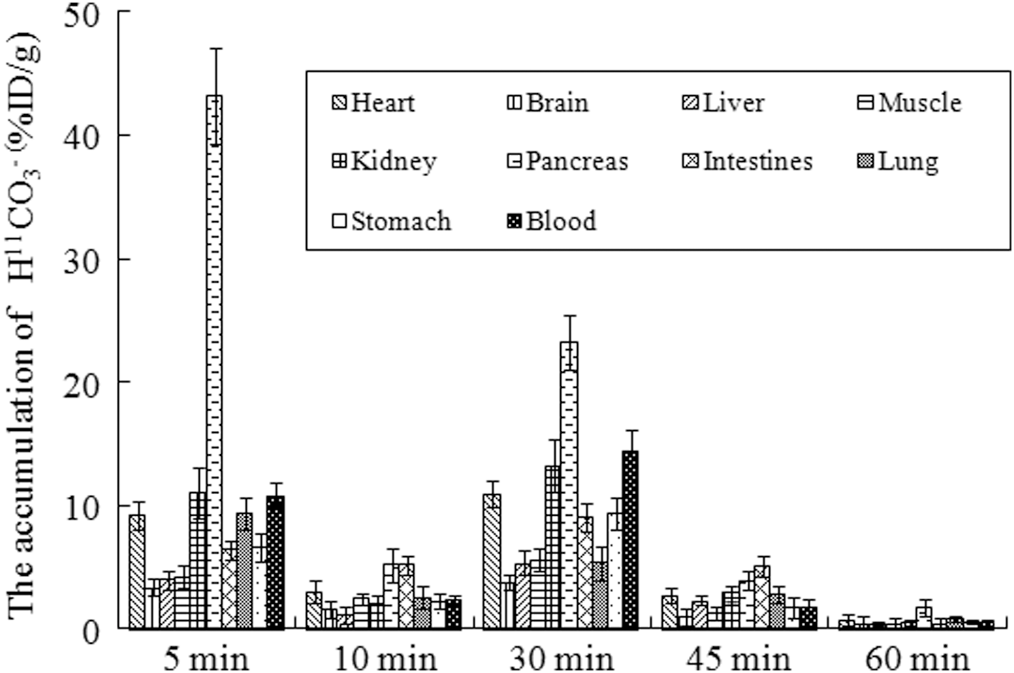
Biodistribution of H11CO3 − solution (pH 7–8) in normal mice.
Ex in vivo biodistribution
The 20 normal Kunming mice were divided into five groups (n = 4 per group), each mouse was injected with ∼3.7 MBq (∼100 μCi) of 11 C-labeled bicarbonate in 100–200 μL of 0.9% NaCl solution through the tail vein. At 5, 10, 30, 45, and 60 min postinjection of the radiotracer, the mice (n = 4 per each time point) were sacrificed by cervical fracture and dissected. Blood was obtained through the eyeball vein; tissue samples of interest (blood, brain, heart, lung, liver, kidney, pancreas, stomach, small intestine, and muscle in the right thigh) were rapidly dissected and weighed, and radioactivity was counted with a gamma (γ) counter. All measurements were background-subtracted and decay-corrected to the time of injection and then averaged. Data were expressed as a percentage of the injected dose per gram of tissue (% ID/g).
Intraperitoneal administration of NaHCO3/NH4Cl
Two groups of S-180 fibrosarcoma-bearing mice or inflammatory mice (n = 4, each group) were used. One group was administered a single dose of either 0.7 mL 1 M NaHCO3 or 0.7 mL 1 M NH4Cl (intraperitoneal [i.p.] administration) and denied access to drinking water for the next 4 h, whereas the other group did not receive any treatment and had uninterrupted access to ad libitum water. 14 At 5 min and 4 h after treatment with NaHCO3 or NH4Cl, PET imaging of tumor-bearing mice was performed with H 11 CO3 − buffer.
Intravenous administration of acetazolamide
Two groups of S-180 fibrosarcoma-bearing mice or inflammatory mice (n = 4, each group) were used. One group was administered a single dose of 50 mg/kg (intravenous administration) at a concentration of 50 mg/mL acetazolamide (ACT) (carbonic anhydrase [CA] inhibitor) and denied access to drinking water for the next experiment. At 45 min after treatment with ACT, PET imaging of tumor-bearing mice was performed with H11CO3 − buffer.
PET imaging
The mice were injected with 3.7 MBq (100 μCi) of H 11 CO3 − solution in 0.2 mL of 0.9% NaCl solution through the tail vein. They were anesthetized with 5% chloral hydrate solution (6 mL/kg) before imaging and remained in anesthesia state during the study. The CT scan was used for attenuation correction and localization of the lesion site. Mice were visually monitored for breathing and any other signs of distress throughout the entire imaging period. PET images were acquired at four-time points (10, 30, 45, and 60 min) postinjection. PET imaging was divided into six groups (three subgroups, and n = 4, each subgroup), including the tumor-bearing mice imaging (H 11 CO3 − solution pH <5, pH 7–8, and pH >8), the antitumor therapy with cyclophosphamide (H 11 CO3 − solution pH >8), the inflammatory imaging (H 11 CO3 − solution pH <5, pH 7–8, and pH >8), the NaHCO3-treated inflammatory mice (H 11 CO3 − solution pH >8.0 and pH <5.0), the tumor-bearing mice treated with ACT (H 11 CO3 − solution pH <5, pH 7–8, and pH >8), and inflammatory mice treated with ACT (H 11 CO3 − solution pH <5, pH 7–8, and pH >8). The CT scan was used for attenuation correction and localization of the lesion site. A region of interest was placed on each tumor, inflammatory lesion, and a region of femoral muscle. After PET imaging, the animals were sacrificed, the tissues of tumors, inflammation region of the right thigh muscle, and the normal right thigh muscle were excised and weighted, and the radioactivity of these tissues was measured with the gamma counter.
Determination of pH in tumors, inflammatory lesions, and normal muscle
pH in tumor tissues, inflammatory lesion, and contralateral muscle of the model mice were measured using MI-419 needle (pin diameter is 0.8 mm) pH microelectrode and MI-402 reference microelectrode (Microelectrodes, Inc.). After peeling off the tumor, the inflammatory lesion, and the contralateral normal muscle of mice, the needle of pH microelectrode was inserted into the central portion of these tissues and the reference microelectrode placed in the subcutaneous area to measure tissue pH.
Statistical analysis
All data are expressed as mean ± standard deviation. Statistical analysis was performed with SPSS software, version 20.0 (SPSS, Inc.), for Windows (Microsoft). Comparisons between conditions were performed using the unpaired Student t test. A p-value of <0.05 was considered to indicate statistical significance.
Results
Radiosynthesis of H 11 CO3 − solution
Automated synthesis gave high radiochemical yield above 95% for H 11 CO3 −– 11 CO3 2− solution (pH >8.3) and over 80% for H 11 CO3 −– 11 CO2 solution (pH 7–8), but gave relative low yield of <50% for 11 CO2-based solution (pH <6), with the total synthesis time of 8 min. Radiochemical purity of 11 C-labeled bicarbonate was detected with TLC eluted with methanol, with 11 C-carbonate or 11 C-bicarbonate Rf = 0. Three forms of 11 C-bicarbonate injected solution with pH <5, pH 7–8, and pH >8.3 were used for the next experiments.
Biodistribution of H 11 CO3 −
Three forms of 11 C-labeled bicarbonate had the similar biodistribution in mice (data not shown). A representative biodistribution of H 11 CO3 − solution (pH >8.0) is shown in Figure 1. Most tissues showed high uptake of H 11 CO3 − at 5 and 30 min after injection, and all the tissues demonstrated low uptake of H 11 CO3 − at 10, 45, and 60 min after injection. The pancreas had the highest uptake of H 11 CO3 − at 5, 30, and 60 min postinjection. The blood, kidney, and heart possessed the moderate uptake at 5 and 30 min postinjection. The lung showed the moderate uptake of H 11 CO3 − at 5 min postinjection and low uptake at other time point postinjection. Brain, liver, and muscle had low uptake of H 11 CO3 − within all observed time. In addition, H 11 CO3 − demonstrated slow clearance from most tissues.
Small-animal PET imaging
PET images of tumor-bearing mice, cyclophosphamide-treated tumor-bearing mice treated, and inflammatory mice are shown in Figures 2 and 3. PET imaging showed that tumor and inflammatory lesion had a high uptake of H 11 CO3 − in the S180 fibrosarcoma-bearing mice (Fig. 2) and the turpentine-induced inflammatory lesion-bearing mice (Fig. 3). The ratios of tumor to muscle were 2.4, 1.7, 1.3, and 1.0, and the ratios of inflammatory lesion to muscle were 1.5, 1.7, and 2.1. The three forms of H 11 CO3 − solution exhibited the similar results. However, the uptake of H 11 CO3 − (pH >8) in the tumor (pH 6.95 ± 0.05) was lower than that in untreated tumor (pH 7.00 ± 0.05) (Fig. 2).
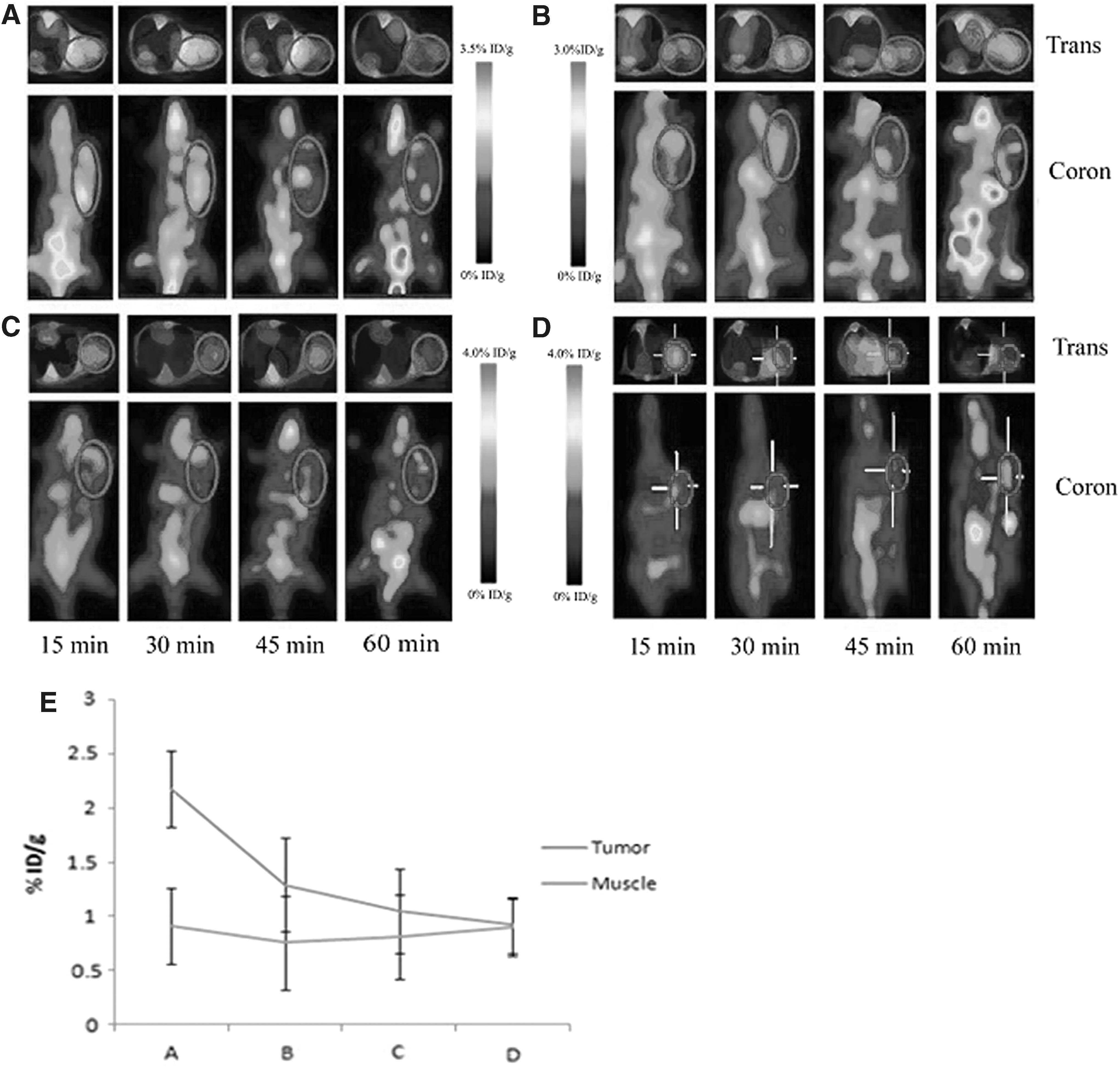
PET images of the tumor-bearing mice with H
11
CO3
− solution [
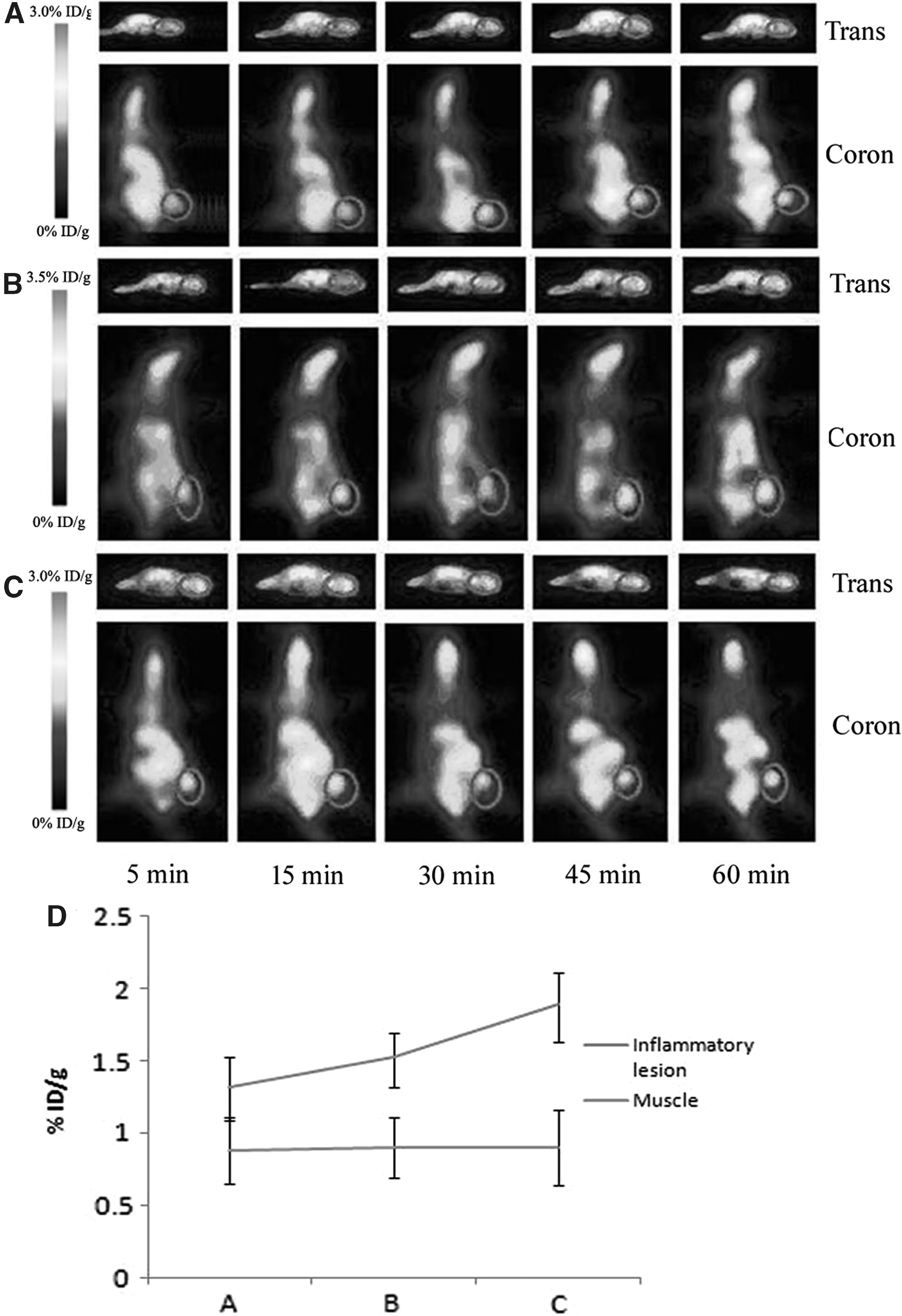
PET images of the inflammatory lesion-bearing mice with H
11
CO3
− solution [
Uptake inhibition experiment
After i.p. injection of NaHCO3 for 5 min and 4 h, PET imaging showed that the inflammatory lesion had high uptake of H 11 CO3 − (pH <5.0 and pH >8.3) after 15 min postinjection of the tracers (Fig. 4). The ratios of inflammatory lesion to muscle were 1.34, 1.40, 1.37, and 1.54. In addition, the group of the NaHCO3 (or NH4Cl)-treated tumor demonstrated the similar results as the NaHCO3-treated inflammatory lesion (data not shown).
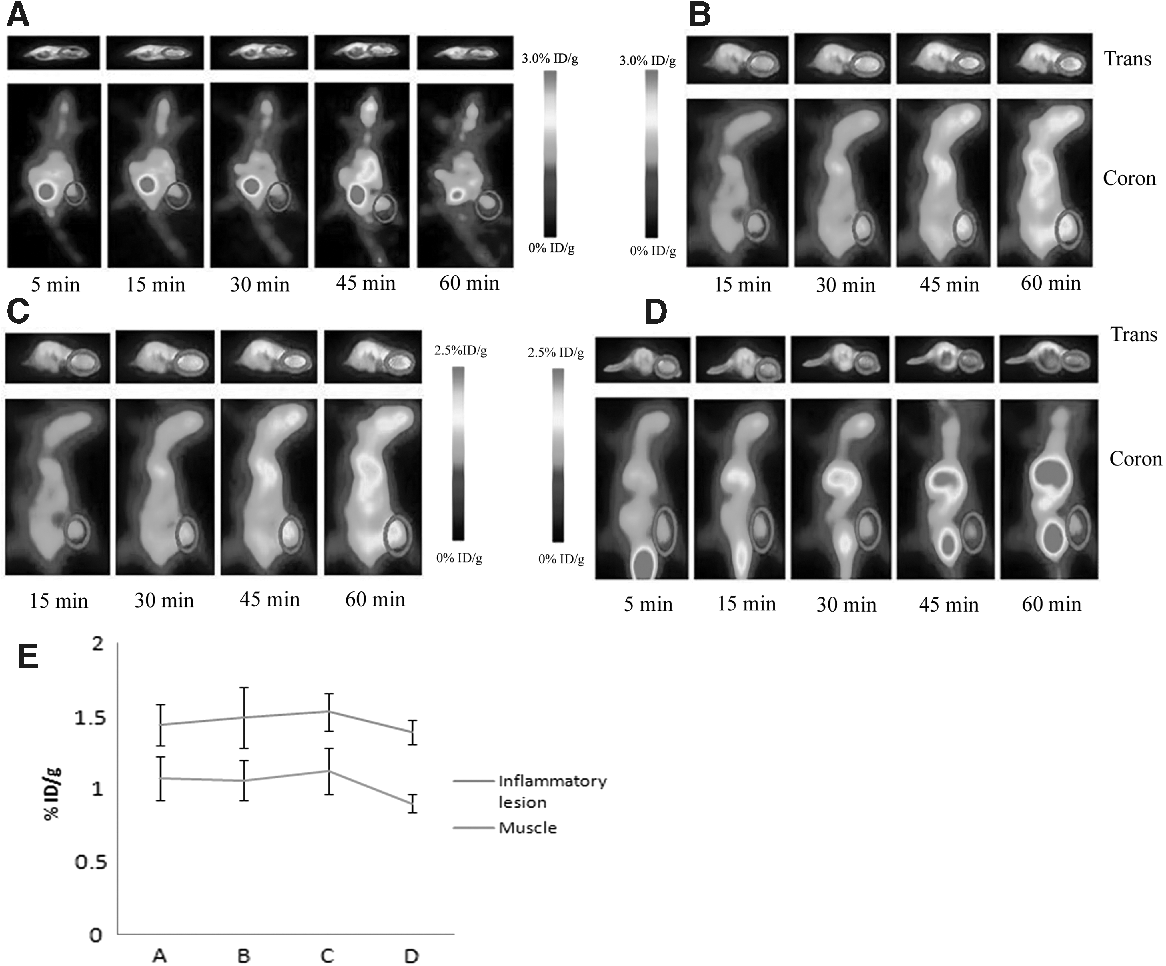
PET images of inflammatory lesion-bearing mice with H
11
CO3
− solution [
After administration of ACT, low uptake of H 11 CO3 − was found in tumor-bearing mice at 30 min postinjection of H 11 CO3 − solution (pH <5.0 and pH >8.3) (Fig. 5). However, PET images showed that the uptake of H 11 CO3 − solution (pH 6–7) in inflammatory lesion had no significant difference between before and after administration of ACT (Fig. 5) (p > 0.05).
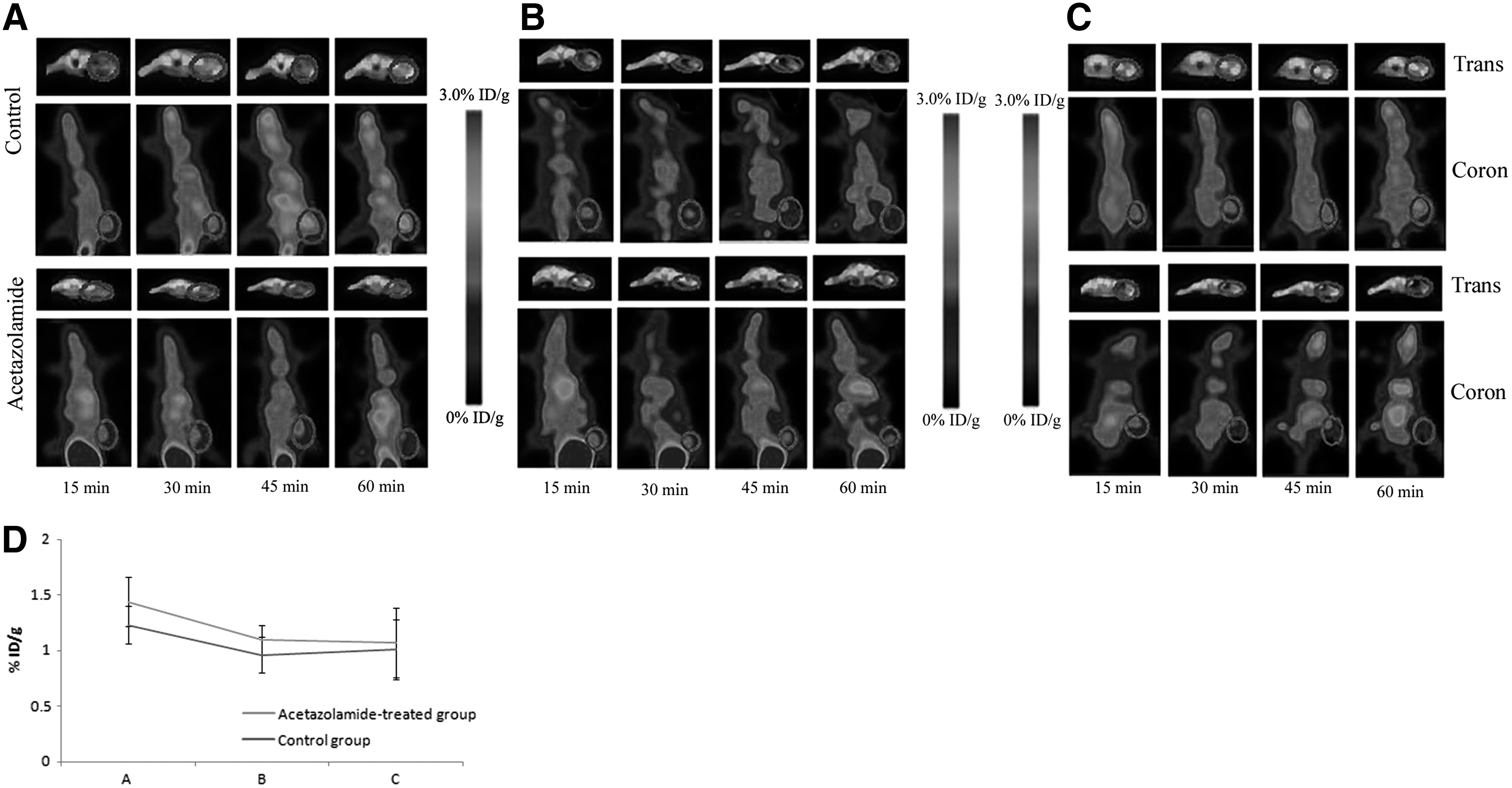
PET images of inflammatory lesion-bearing mice in the control group and the acetazolamide-treated mice postinjection of H
11
CO3
− solution [
In addition, the uptake of H 11 CO3 − (Fig. 6A, pH <5; Fig. 6B, pH >8.3) in tumor of the control group was low, but the uptake of H 11 CO3 − (pH >8.3) in ACT-treated tumor was significantly lower than that in the control group (Fig. 6) (p < 0.05).

PET images of the tumor-bearing mice in the control group and the acetazolamide-treated group postinjection of H
11
CO3
− solution [
Ninety minutes postinjection of NaHCO3 and NH4Cl in tumor-bearing mice and inflammatory lesion-bearing mice, PET images showed that the uptake of H 11 CO3 − in the NaHCO3-treated group was low, whereas it was still high in the NH4Cl-treated group.
PET images showed that tumor had low uptake of H 11 CO3 − after administration of ACT at 30 min postinjection of H 11 CO3 − solution (pH <5.0 and pH >8.3) (Fig. 6). However, PET images indicated that the uptake of H 11 CO3 − (pH 6–7) in inflammatory lesion had no significant difference between untreated group and ACT-treated group (Fig. 5). That is, the uptake of H 11 CO3 − solution (Fig. 6A, pH <5; Fig. 6B, pH >8.3) in tumor was high, but the uptake of H 11 CO3 − (pH >8.3) in ACT-treated tumor was significantly lower than that in the tumor group (Fig. 6) (p < 0.05).
Tumor and inflammatory tissue pH measurement
The average pH values in tumor, inflammatory lesion, and contralateral normal muscle of the model mice are shown in Table 1. Both tumor tissue and inflammation lesion were a bit acidic (pH 6.95) compared with contralateral normal muscle, which was a bit alkaline (pH 7.33). After anticancer therapy with cyclophosphamide, the pH value in tumor was nearly neutral (pH 7.00), while the contralateral normal muscle was also a bit alkaline (pH 7.27).
n = 5, mean ± SD.
SD, standard deviation.
Administration of NaHCO3 in the drinking water increased the measured tumor pH to 7.21 ± 0.03 (Table 2) and the inflammatory tissue pH to 7.23 ± 0.04 (Table 3), while gavage with NH4Cl over 90 min decreased the tumor pH to 6.51 ± 0.04 (Table 2) and the inflammatory lesion pH to 6.48 ± 0.03 (Table 3). Similarly, administration of ACT decreased the tumor pH to 6.62 ± 0.02 (Table 2) and the inflammatory lesion pH to 6.65 ± 0.05 (Table 3).
Mean ± SD.
ACT, acetazolamide.
Mean ± SD.
Discussion
11 C-bicarbonate buffer was prepared from the on-column reaction of 11 CO2 with 0.50 M NaOH solution loaded in a Sep Pak plus C18 cartridge, followed by neutralization with dilute hydrochloric acid or NaH2PO4 solution (pH 3–4). There existed three forms of 11 C-bicarbonate buffer, as 11 C-CO2 at a pH below 5 or as H 11 CO3 −– 11 CO2 buffer within a pH range of 7–8 or as H 11 CO3 −– 11 CO3 2− at a pH above 8.3. 12 11 CO2-based bicarbonate solution (pH <5.0) gave low radiochemical yield (<50%) because 11 CO2 is easy to escape from the solution. H 11 CO3 −– 11 CO2 buffer (pH 7–8) contained small amounts of 11 CO2, resulting in high radiochemical yield (>80%). H 11 CO3 −– 11 CO3 2− solution gave the highest radiochemical yield of over 95%, due to almost no 11 CO2 produced from the 11 C-bicarbonate solution (pH >8.3). Compared with 11 CO2-based solution (pH <5.0), the radiochemical yield of H 11 CO3 −– 11 CO2 solution (pH 7–8) and H 11 CO3 −– 11 CO3 2− solution (pH >8.3) is very high. Thus, 11 C-bicarbonate solutions (pH 7.0–8.0 and pH >8.3), as a nontoxic PET tracer, are easy to perform automated radiosynthesis for translation into clinical use.
Biodistribution profiles of three forms of 11 C-bicarbonate solution (pH <5, pH 7–8, and pH >8.3) in mice were similar. The pancreas had the highest uptake, the brain had low uptake, and rapid clearance from all organs or tissues was observed after 30 min postinjection of the 11 C-bicarbonate solution. Similar biodistribution profiles of three 11 C-bicarbonate tracers could be contributed to interconversion between bicarbonate ion and CO2 in the blood to form the similar H 11 CO3 −– 11 CO2 buffer postinjection of the tracers. All organs or tissues appeared to be high uptake of 11 C-bicarbonate in mice at 5 and 30 min postinjection, respectively. The possible reason was that the red cell membrane and the capillary endothelium were much more permeable to neutral molecules (CO2) than HCO3 −. 15 11 CO2 gave rise to the first high uptake at 5 min after injection, while H 11 CO3 − gave rise to the second high uptake at 30 min after injection.
The uptake mechanism of three 11 C-bicarbonate tracers in an acidic environment possibly involves the following aspects. As the pHe of solid tumor is acidic, 1 –5 the main form of 11 C-bicarbonate in an acidic environment is 11 CO2. CO2 is a weak acid with a pKa of 6.12. As 11 CO2, but not 11 C-bicarbonate ion, readily diffuses across intact blood–brain barrier (BBB) or cell membrane, regional cerebral 11 C activity following 11 CO2 inhalation reflects the regional cerebral pH (rpH) of the combined intra- and extracellular cerebral compartments; the rpH being weighted toward pHi. Tumors with an intact or a disrupted BBB are also expected to have a raised 11 CO2 solubility compared with normal tissue. Thus, PET with 11 CO2, like 11 C-bicarbonate solution (pH <5), can be used for measurement of tumor rpH. A theoretical disadvantage of the 11 CO2 approach is that fixation of 11 C activity in the form of nonvolatile substrates can occur. 14 In addition, 11 C-bicarbonate anion can easily diffuse across a disrupted blood–tumor barrier into cancer cells, besides that they enter tumor mainly existing as 11 CO2 in an acidic environment in the presence of the enzyme CA. 13,14 Therefore, 11 C-bicarbonate (pH 7–8 or pH >8.3) is a potential endogenous imaging agent for PET imaging of tumor.
PET images showed that the tumor had high uptake of three 11 C-bicarbonate tracers at 15 and 30 min after administration, while tumor showed a low uptake after 45 min postinjection. The inflammatory lesion had high uptake of H 11 CO3 − in PET images within all the observed time. The uptake of H 11 CO3 − (pH >8.3) in the tumor after antitumor therapy was lower than that in the untreated tumor, but a little high uptake of H 11 CO3 − (pH >8.3) in the treated tumor was also found at 60 min postinjection, which possibly was due to the balance of H 11 CO3 −– 11 CO2 in vivo after 60 min postinjection. The pH measurements showed that the tumor and the inflammatory lesion possessed are a bit acidic (6.95), while the contralateral normal thigh muscle is a bit alkaline (7.33). Compared with the untreated tumor (pH 7.00), pH in the cyclophosphamide-treated tumor was a bit alkaline (pH 7.30), which was closed to the pHe of normal tissues. 4 However, the limitations of pH measurement in this study are as follows. There must be sampling errors during the measurement of needle use and reproducibility of pH meters, and a lot more research is needed.
After i.p. injection of NaHCO3 in inflammatory mice for 5 min and 4 h, PET imaging showed that the uptake of 11 C-bicarbonate (pH <5 and pH 8.3) in inflammatory lesion was still high. In addition, the similar results were observed in the NaHCO3 (or NH4Cl)-treated tumor and in the NH4Cl-treated inflammatory lesion. The pH measurement demonstrated that administration of NaHCO3 over 90 min increased tumor pH and inflammatory tissue pH to >7.2, while gavage with NH4Cl over 90 min decreased tumor pH and inflammatory tissue pH to <6.6. These results indicated that NaHCO3 could not make tumor pH and inflammatory tissue pH increase by >7.0, and NH4Cl could not make tumor pH and inflammatory tissue pH decrease to <7.0 for a short time of 5 min. In addition, after treatment with NaHCO3 or NH4Cl for a long time of 4 h, NaHCO3 or NH4Cl could fully clear from the mice body and result in renewing tumor pH and inflammatory tissue pH to balance (pH <7.0). Therefore, high uptake of 11 C-bicarbonate (acid injectate and alkaline injectate) in tumor and inflammatory lesion was possibly closely associated with acidic environment.
PET images also showed that the uptake of 11 C-bicarbonate solution (pH >8.3) in tumor and inflammatory lesion after administration of ACT was significantly higher than that in untreated tumor and untreated inflammatory lesion, especially at 45 min postinjection of the tracers. Compared with 11 C-bicarbonate (pH >8.3), the uptake of 11 C-bicarbonate (pH <5) in tumor and inflammatory lesion had no obvious change before and after treatment with ACT. Mammalian animals have robust pH buffer systems that maintain pH within a narrow optimal pH range (7.35–7.45), and the HCO3 −–CO2 system is the principal system. Bicarbonate (HCO3 −) is rapidly exchanged reverse reaction with CO2 that is catalyzed by the enzyme CA. Absence of CA or CA inhibition (such as ACT) slows down the interconversion between bicarbonate and CO2. Based on the reason that administration of ACT could obviously decrease tumor pH and the inflammatory tissue pH, low uptake of 11 C-bicarbonate (pH >8.3) in the ACT-treated tumor and ACT-treated inflammatory lesion was found. Therefore, the authors could deduce that uptake of 11 C-bicarbonate (alkaline injectate) in the tumor and inflammatory lesion was closely related to acidic environment in the presence of CA.
Conclusions
In conclusion, PET with H 11 CO3 − can be used to image the lesion pH in vivo. 11 C-bicarbonate solution as a noninvasive and nontoxic endogenous PET tracer can be easy to perform automated radiosynthesis for translation into clinical use. Thus, H 11 CO3 − could be used to image the lesion pH alteration and the response to therapy in clinical setting.
Footnotes
Acknowledgments
This work was supported, in part, by the National Natural Science Foundation of China (No. 81571704, No. 81371584, No. 81671719, No. 81701723), the Science and Technology Foundation of Guangdong Province (No. 2016B090920087, 2014A020210008, No. 2013B021800264, 2013B010404018), and the Science and Technology Planning Project Foundation of Guangzhou (No. 201504301345364, No. 201510010145, No. 201604020169).
Disclosure Statement
No competing financial interests exist.