
Abstract
Background:
Recently, the direct intratumoral (i.t.) injection of anticancer agents has been investigated. A newly synthesized Antineoplaston A10 analog 3-(4-methoxybenzoylamino)-2,6-piperidinedione (MPD) showed an antitumor activity in human breast cancer cell line. Unfortunately, MPD suffered from poor water solubility.
Materials and Methods:
Pseudoternary phase diagram of oil (isopropyl myristate), surfactant (Tween 80), cosurfactant (ethanol), and water was plotted. MPD microemulsion (MPDME) was developed and characterized for particle size (PS), polydispersity index (PDI), zeta potential (ZP), and morphology (transmission electron microscopy). MPDME and MPD solution (MPDS) were radiolabeled with technetium 99m (99mTc) using stannous chloride dihydrate (SnCl2.2H2O). Molecular docking of MPD and 99mTc-MPD was performed to study the interaction with DNA.
Results:
The impacts of intravenous (i.v.) and i.t. injections of 99mTc-MPDME and 99mTc-MPDS on biodistribution were studied. The developed MPDME showed spherical droplets with mean PS (74.00 ± 5.69 nm), PDI (0.25 ± 0.03), and ZP (33.90 ± 0.90 mV). Labeling yield of 99mTc-MPDME and 99mTc-MPDS was 97.00% ± 0.60% and 92.02% ± 0.45%, respectively. MPD and 99mTc-MPD showed almost same binding affinity with DNA binding site. Biodistribution results showed that i.t. injection of 99mTc-MPDME significantly enhanced tumor retention compared to i.v. route.
Conclusions:
Herein, the authors concluded that microemulsion could be used as i.t. injectable delivery vehicle to improve targeting and tumor retention of MPD.
Introduction
Although intravenous (i.v.) route of administration is considered the conventional route of cancer treatment, the concentration of the anticancer drug reaching the tumor site is low and the systemic toxicity is high. Hence, to minimize the morbidity and mortality rate of neoadjuvant chemotherapy, various authors explored an alternative route of anticancer drug administration. Intraperitoneal chemotherapy has shown a promising antitumor effect on peritoneal carcinomatosis, ovarian carcinoma, advanced gastric carcinoma, and peritoneal carcinomatosis. 1 –3 In addition, intra-arterial chemotherapy was tried in breast cancer and hepatocellular carcinoma, while the intra-arterial transcatheter occlusion of abdominal tumor was tried in 1976. 4,5 However, the intra-arterial injection method has some disadvantages. 6,7 First, this technique requires extremely selective catheterization which is highly dependent on operator's skill and equipment. Second, the radionuclide reaches the tumor site by a nonspecific route. This means that normal tissue is irradiated in the same way as the tumor. Third, because of the nonspecific distribution, large quantities of radionuclides must be used. 8 In addition, local toxic effects to vessels, nerves, connective tissue, muscle, and skin have been observed in studies of regional perfusion and intra-arterial infusion. 9 Similar complications have not been reported in the relatively few clinical studies of intratumoral (i.t.) chemotherapy. 9
I.t. route is the most straightforward method of bringing the therapy into the tumor while avoiding a high dose to the normal tissue. 8 Moreover, it was stated that i.t. injection of cytotoxic drugs might be one of the most promising approaches for targeting solid tumors to maximize the cytotoxic activity at the tumoral region and minimize systemic side-effects. 10 There are many barriers in solid tumors, such as high interstitial fluid pressure and a stiff extracellular matrix. 11,12 These barriers greatly hinder the penetration and delivery of drug throughout the tumor. Direct i.t. administration can not only overcome the major barriers to systemic delivery but also can take advantage of those obstacles to promote prolonged tumor retention and prevent rapid drug clearance.
Throughout the years, microemulsions (MEs) have been considered as potential drug delivery systems. 13,14 MEs are transparent and thermodynamically stable systems that are spontaneously formed when water, oil, and surfactant with a cosurfactant are properly mixed. 15 MEs have low viscosity and are able to solubilize both hydrophilic and lipophilic drugs. 16 Therefore, MEs have been developed as novel parenteral drug delivery vehicles for both water-soluble and oil-soluble drugs. 17,18 The structure of ME provides a considerable barrier to the dissolved drug in the inner phase toward its rapid diffusion to the external phase and beyond especially when the drug solubility in the inner phase is high but is poorly soluble in surfactant/cosurfactant (S/Cos) interphase and the external phase. 19 Prolonged release effect of the injected ME has an impact on the drug safety and efficacy. 19
ME can be administered subcutaneously, intramuscularly, or intravenously. Various patents concerning calcium antagonists, steroids, fluorocarbons, and different lipophilic drugs have been reported. 20 The behavior of ME at the injection site was investigated by observing the release of technetium 99m (99mTc) pertechnetate following injection in rabbits. Imaging the injection site with a γ-camera showed that the disappearance of 99mTc-pertechnetate in aqueous solution was about 10-fold faster than that in ME formulation. 21
Antineoplaston (ANP) is a group of chemical compounds found naturally in urine and blood. They are made up of peptides and amino acids. ANP A-10 (3-phenylacetylamino-2,6-piperidinedione) was the first identified ANP compound. 22 It was found that ANP A10 inhibits methylation (introduction of a methyl group) in the DNA and RNA of cancer cells, thus inducing malignant cells to differentiate and enter a normal cell cycle. 23 In a previous study, a new ANP A10 analog 3-(4-methoxybenzoylamino)-2,6-piperidinedione (MPD) was synthesized, and the interaction between MPD and DNA of tumor cells was reported in comparison with ANP A10. 24
Hence, MPD is hypothesized to have the same active site and follow a similar mechanism of action of ANP 10. In addition, the glutarimide moiety (2,6-piperidinedione) in MPD structure with the intact imide group (OC-NH-CO) can act as carrier molecule (vector), which transports biologically active substituents (functional groups) through cell membranes. This is due to a striking similarity of the physicochemical properties of glutarimide and uracil derivatives as revealed by quantum chemical calculations and spectroscopic studies. 25 –27 Thus, glutarimide moiety can interact with specific receptors involved in transport of uracil and thymine nucleosides and it may easily cross biological membranes, including nuclear membranes. Molecular ring of glutarimide is quite resistant to hydrolytic cleavage since it can be stabilized by formation of hydrogen bonding with biomolecules. 28
The role of γ scintigraphic methods is well recognized for detecting and following the biodistribution of newly developed drugs or formulations. 29 The radiolabeled compounds have been used in the drug release studies because radioactivity can be measured using liquid scintillation detector simply. 30 99mTc has been used to label many kits in the last decade and has several properties and features that make it a suitable radionuclide for nuclear imaging, with a half-life of 6 h and energy of 140 keV which are sufficient for the diagnostic procedures. Moreover, 99mTc is easily available by elution of molybdenum-99 (99Mo)/99mTc generator in normal saline. 31,32
In this article, the authors hypothesized the direct relationship between the i.t. administration of 99mTc-MPD microemulsion (99mTc-MPDME) and the high retention inside the tumor. Related to this concept, the cytotoxicity will be increased and the localization in normal healthy tissues will be attenuated. To verify this hypothesis, the radiolabeling of MPDME and MPD solution (MPDS) with 99mTc was done using stannous chloride dihydrate (SnCl2.2H2O) as a reducing agent. The binding affinity of MPD, as well as 99mTc-MPD with the DNA, was studied, using in silico docking, to explore the cytotoxic activity and to obtain additional validation of the experimental results. Moreover, the impact of the i.v. and i.t. injection of 99mTc-MPDME on the biodistribution in comparison with 99mTc-MPDS was studied.
Materials and Methods
Materials
MPD was kindly provided by the Department of Pharmaceutical Organic Chemistry, Faculty of Pharmacy, Mansoura University, Egypt. 99mTc was eluted from 99Mo/99mTc generator (Elutec, Brussels, Belgium). Ehrlich ascites carcinoma (EAC) was provided by National Cancer Institute (Cairo, Egypt). Isopropyl myristate (IPM), Tween 20, Tween 40, Tween 80, ethanol (95%), ammonium hydroxide, methanol, and stannous chloride dihydrate (SnCl2·2H2O, M.wt. = 225.6) were purchased from Sigma Aldrich Chemical Co. (St. Louis, MO). All other chemicals and solvents were of analytical grade and were used as received.
Methods
Development of formulations
Construction of pseudoternary phase diagrams
To formulate ME systems, pseudoternary phase diagram was developed using water titration technique. 33 Oil phase (IPM) and surfactant (Tween 80)/cosurfactant (ethanol) (S/Cos) mixture were mixed at weight ratios of 1:9, 2:8, 3:7, 4:6, 5:5, 6:4, 7:3, 8:2, and 9:1. Water was added drop by drop to each mixture with stirring. The corresponding pseudoternary phase diagram was constructed with Tri-plot software (Ver. 4.1.2, D. Graham and N. Midgley, Loughborough University, Leicestershire, England). Aqueous MPDS was not prepared due to the limited solubility of the drug in water. Alternatively, MPD was suspended in ethanol and stirred until it was completely dissolved.
Characterization of ME formulation
Determination of particle size, polydispersity index, and zeta potential
The average particle size (PS), polydispersity index (PDI), and zeta potential (ZP) of the selected formulation were determined by Nanotrac Wave II (Malvern Instruments, Malvern, United Kingdom) using Photon dynamic light scattering technique (DLS). All measurements were done in triplicate at ambient temperature, and the mean values obtained were reported. PDI was determined based on measuring the time dependent fluctuation of scattering of laser light by the nanoparticles undergoing Brownian motion. 34 The measurement of ZP was based on the direction and velocity of nanoparticles exposed to an electric field. 35
Transmission electron microscopy
The morphological feature of the selected formulation was examined through transmission electron microscopy (TEM; Joel JEM 1230, Tokyo, Japan). One drop of the formulation was loaded onto a copper grid, and the excess was removed with a filter paper. The formulation was negatively stained with one drop of phosphotungstic acid aqueous solution (2% w/v) for 3 min. Examination of the grid was achieved using a transmission electron microscope.
In silico docking studies
Docking studies were performed using AutoDoc 4.1 along with LGA algorithm parameter. The crystal structures of DNA (ID: 3GJH) were obtained from the Protein Data Bank (PDB;
Radiolabeling studies
Radiolabeling of MPDME, MPDS, and the ME without MPD (placebo) was performed by direct labeling method. 36,37 Briefly, the required concentration of MPDME, MPDS, or placebo was transferred to a penicillin vial, and the required stannous chloride solution concentration was added. One milliliter freshly eluted 99mTcO4 − (400 MBq) was added to the above solution. The factors affecting labeling yield namely effect of substrate concentration, SnCl2.2H2O concentration, reaction time, pH, and temperature were studied. In addition, the in vitro stability was studied at the optimum condition in normal saline and serum. The formulations were sterilized by filtering through 0.2 μm pore sterile filter (Millipore Corporation, Bedford, MA). Each experiment was repeated thrice, and differences in the data were evaluated. Statistical analysis of the results was done by one-way analysis of variance using SPSS software 19.0 (SPSS, Inc., Chicago, IL). Post-hoc analysis was done using Tukey's honest significant difference test. The difference at p ≤ 0.05 was considered significant.
HPLC analysis
HPLC analysis was done by injection of 10 mL of the sample, after 0.22 μm Millipore filtration, into the column (alphaBond RP-C18, 300 × 3.9 mm; Merck, Darmstadt, Germany) and UV spectrophotometer detector (SPD-6A; Shimadzu, Kyoto, Japan) adjusted to the 262 nm wavelength. The mobile phase consisted of a mixture of methanol and water (60:40 v/v), and the flow rate was adjusted to 1 mL/min. Using the fraction collector, fractions of 1 mL were collected separately up to 30 min and then they were counted by a well-type γ counter (Scalar Ratemeter SR7; Nuclear Enterprises Ltd.). The method was validated for accuracy, selectivity, linearity, and precision.
Paper chromatography analysis
The labeling efficiency of 99mTc-MPDME and 99mTc-MPDS was determined using paper chromatography. The paper chromatography was performed using acetone as the mobile phase. Approximately 1–2 μL of the reaction mixture was applied at a point 1 cm apart from one end of a paper chromatography strip. The strip was eluted in acetone where free pertechnetate (99mTcO4
−) moved with the solvent to the top of the paper, and the radiocolloids (99mTc-RH) along with the labeled complex remained at the point of spotting. The amount of 99mTc-RH was determined using water: ammonium hydroxide: ethanol (5:1:2) mixture as mobile phase. The 99mTc-RH remained at the application point but both the free 99mTcO4
− and the labeled complex moved away with the solvent front. The percentage of labeling yield was calculated according to the following equation:
Electrophoresis analysis
Paper electrophoresis was performed using EC-3000 p-series programmable power (E.C. apparatus corporation, Frederick, MD). After filtration of the reaction mixture using 0.22 μm Millipore filter to remove 99mTc-RH, on Whatman paper sheet (3 cm width and 50 cm length), 3 μL of the reaction mixture was spotted far from the cathode edge of the paper sheet. Electrophoresis was carried out for 1.5 h at a voltage of 300 V using normal saline as an electrolyte solution. After 1.5 h, the paper was removed, dried, and cut into fragments; each fragment length was 1 cm and then each fragment was counted using a well-type γ counter (Scalar Ratemeter SR7; Nuclear Enterprises Ltd.). The percentage of labeling yield was calculated according to the following equation:
Tumor implantation
One hundred and forty-four female Swiss albino mice type weighing 25–30 g were purchased from the Institute of National Research Center, Cairo, Egypt. The animals were kept at constant nutritional and environmental condition throughout the experimental period and at ambient temperature with 12 h on/off light schedule. All over the experiments, standard water and food were allowed to mice. EAC cells were maintained in the female Swiss albino mice through weekly intraperitoneal transplantation. Under the aseptic condition, EAC cells were obtained by needle aspiration. The ascetic fluid was diluted using sterile normal saline. To produce solid tumor, 0.2 mL was injected intramuscularly in the right thigh while the left thigh was kept as control. The animals were kept for 10–15 d until the tumor development was apparent.
Biodistribution studies
All experiments were conducted in accordance with the guidelines provided in the Egyptian Atomic Energy Authority and were approved by the animal ethics committee, Labeled Compounds Department, and the protocol of the studies was approved by the Research Ethics Committee in the Faculty of Pharmacy, Cairo University (REC-FOPCU), Egypt. Four groups of female Swiss albino tumor bearing mice were used to study the biodistribution. Each group contained 36 mice. The tumors of groups 1 and 2 were infused intratumorally using infusion pump (KD Scientific, Holliston, MA) with 99mTc-MPDME and 99mTc-MPDS, respectively. The attached needle to the infusion pump was inserted into the center of the tumor as guided through direct palpation and visual placement.
The infusion rate was 0.5 mL/min, and the radioactivities of the infused preparations were 654 and 636 μCi/mL for groups 1 and 2, respectively. The total infused volume for each mouse's tumor was based on the tumor volume. Immediately after i.t. injection, the injected site was washed and covered with absorbing paper to retrieve 99mTc-MPDME and 99mTc-MPDS leaking out of the tumor. Groups 3 and 4 of the female Swiss albino tumor bearing mice were intravenously injected into the mice tail vein with 99mTc-MPDME and 99mTc-MPDS, respectively. The radioactivities of the injected preparations were 1276 and 1230 μCi/mL for groups 3 and 4, respectively. At 0.25, 0.50, 1, 6, 18, and 24 h after injection, each mouse was weighed and then anesthetized. The animals were sacrificed by cervical dislocation, and their tumors, tissues, and organs were harvested and rinsed with saline and then weighed. Radioactivity of each sample, as well as the background, was counted in γ counter. The percentage of injected dose per gram (ID%/g ± SD) in a population of six mice for each time point was reported. Samples of fresh blood, bone, and muscle were collected in preweighted containers and counted. It was assumed that blood, bone, and muscle were 7%, 10%, and 40% of total body weight, respectively. 38
Drug inhibition study
To confirm that 99mTc-MPDME was accumulated specifically with high affinity binding to the active site in the tumor, a total of 3 μg/mouse of nonlabeled MPDME was prepared under the same conditions as99mTc-MPDME and injected into the tail vein of female Swiss albino tumor bearing mice immediately before the i.v. injection of 99mTc-MPDME then mice were biologically evaluated for tumor uptake at 1 h postinjection.
Results and Discussion
Pseudoternary phase diagram
According to the developed pseudoternary phase diagram (Fig. 1), the selected ME formulation contained 15% IPM, 65% S/Cos (1:1), 20% water, and 0.5% w/v of drug. The mixture was observed for transparency then filtered through 0.2 μm pore sterile filter (0.22 μm; Millipore Corporation).
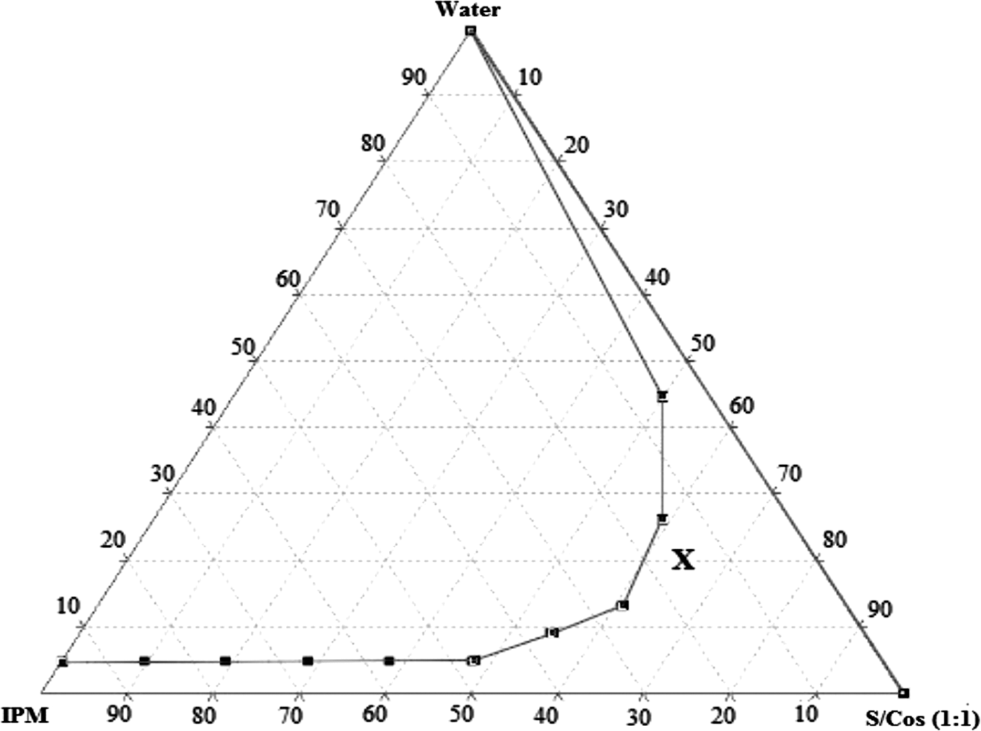
Pseudoternary phase diagram of IPM, water, and surfactant (Tween80)/cosurfactant (ethanol) (S/Cos) at 1:1. The investigated formula is marked (x). IPM, isopropyl myristate.
Characterization of ME formulation
PS, PDI, and ZP
The average PS, PDI, and ZP of MPDME were 74.00 ± 5.69 nm, 0.25 ± 0.03, and 33.90 ± 0.90 mV, respectively. Nano range of MPDME formulation facilitates drug delivery to tumor tissues through the enhanced penetration and retention (EPR) effect. 39 The obtained PDI and ZP values reflect the high homogeneity and stability of MPDME, respectively. 40, 41
Transmission electron microscopy
The TEM study was carried out to get more insight about the morphology of MPDME. It is clear from the TEM photograph (Fig. 2) that the particles were finely distributed, spherical in shape, and had a uniform droplet size distribution. In addition, the PS shown by the TEM micrograph was in a good agreement with that obtained by DLS.
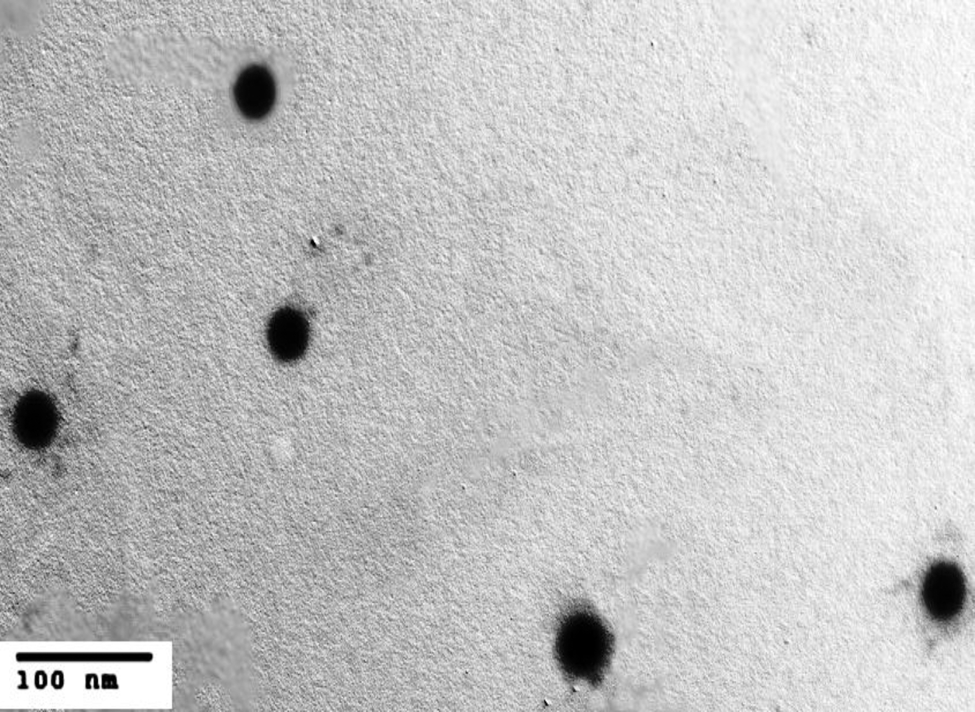
Transmission electron microscopy of the MPDME. MPD, 3-(4-methoxybenzoylamino)-2,6-piperidinedione; MPDME, MPD microemulsion.
In silico docking studies
To design effective chemotherapeutic agents with high anticancer activity, it is essential to explore the interactions of drug with DNA. 42 Preliminary biological evaluation showed the capacity of MPD to interact with DNA in a specific manner. 24 Structure of MPD and the proposed structure of 99mTc-MPD are shown in Figure 3A and B. MPD exhibited a binding affinity (−7.18 kcal/mol) with DNA and formed three hydrogen bonding with DG10, DG16, and DA17 at a bond distance of 2.19, 2.47, and 2.71 nm, respectively. Three π-bonds with DG11, DG16, and DA17 at a bond distance of 3.08, 2.78, and 2.71 nm, respectively, were formed (Fig. 4A). 99mTc-MPD showed almost the same binding affinity of MPD (−7.11 kcal/mol); the bond formation with interact residues was slightly different, including four hydrogen bonds with DA5, DG16, DA17, and DA18 at a bond distance of 2.45, 1.94, 2.14, and 2.85 nm, respectively. Two π-bonds with DA17 and DT20 at a bond distance of 2.28 and 3.05 nm, respectively, were formed (Fig. 4B). The molecular docking results showed a good binding affinity of MPD and 99mTc-MPD toward the active site. In addition, 99mTc did not influence the binding affinity or the interaction of MPD with DNA binding site.

Structure of MPD

The docking conformation of MPD
Labeling yield determination
MPDME and MPDS were radiolabeled using99mTc through direct labeling method in the presence of SnCl2.2H2O as a reducing agent to reduce the oxidation state of technetium (+7) in pertechnetate state to technetium (+4). 43 Standard solution of MPD showed a peak at retention time (4 min) (Fig. 5A). MPDME showed a peak of MPD at the same retention time (Fig. 5B). Therefore, the component of ME did not interfere with the analysis of the sample. The radiochromatogram (Fig. 5C) showed two peaks, one at a retention time (2 min) which corresponded to the free pertechnetate, while the second peak at a retention time (5 min) which corresponded to 99mTc-MPD.

HPLC chromatogram of MPDS
Factors affecting percentage of labeling yield
Effect of substrate concentration
The influence of substrate concentration on the labeling yield of 99mTc-MPDME and 99mTc-MPDS was studied. The reaction was performed at different MPD concentrations (10–300 μg/mL) (Fig. 6A, B). At low substrate concentration (10 μg/mL), the percentage of labeling yield was 91.97% ± 0.68% and 78.38% ± 0.16% for 99mTc-MPDME and 99mTc-MPDS, respectively. By increasing the substrate concentration, the labeling yield increased where the maximum labeling yield of 99mTc-MPDME (97.27% ± 0.22%) and 99mTc-MPDS (92.80% ± 0.36%) was obtained at substrate concentration of 25 and 20 μg/mL, respectively. A further increase in the substrate concentrations higher than the optimal concentrations caused a significant decrease in the labeling yield. This reduction in the labeling yield could be attributed to that the labeling reaction might be reversible, and thus, the increase in the substrate concentration led to forcing the reaction to go in the reversed direction and breakdown the labeled compound into the initial reactants and so the labeling yield decreased. 44
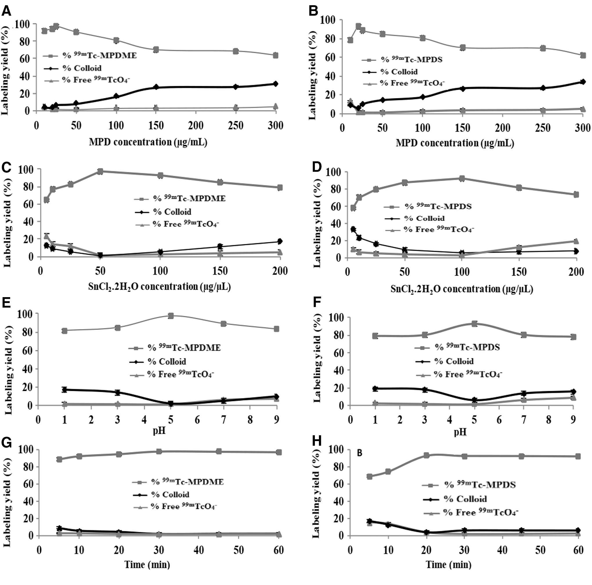
Labeling yield of 99mTc-MPDME and 99mTc-MPDS as a function of substrate concentration
Effect of SnCl2.2H2O concentration
Results showed that the percentage of labeling yield was dependent on SnCl2.2H2O concentration (Fig. 6C, D). The maximum labeling yield of 99mTc-MPDME (97.26% ± 1.80%) and 99mTc-MPDS (92.01% ± 0.48%) was achieved at SnCl2.2H2O concentration of 50 and 100 μg/mL, respectively. The concentrations of SnCl2.2H2O below the optimum levels showed low percentage of labeling yield for 99mTc-MPDME and 99mTc-MPDS. This might be due to the incomplete reduction of pertechnetate to form 99mTc-complex. Also at higher SnCl2.2H2O concentrations than the optimum concentration, there was a decrease in the labeling yield. This reduction might be due to the conversion of excess SnCl2.2H2O to 99mTc-stannous colloids. 45
Effect of pH of the reaction medium
The effect of pH was studied at pH range (1–9). At pH 1, the labeling yield of 99mTc-MPDME, as well as 99mTc-MPDS, was low (Fig. 6E, F). The maximum labeling yield of 99mTc-MPDME (97.39% ± 0.38%) and 99mTc-MPDS (92.77% ± 0.25%) was obtained at pH 5. When the pH of the reaction medium was shifted toward the alkaline side, the labeling yield decreased again. At pH 9, the labeling yield of 99mTc-MPDME and 99mTc-MPDS was 82.87% ± 0.66% and 78.13% ± 1.10%, respectively. This decrease might be attributed to the formation of 99mTc-RH, 99mTc-stannous colloids, and free 99mTcO4 −. 46
Effect of reaction time
As shown in Figure 6G and H, the maximum labeling yield of 99mTc-MPDME (97.39% ± 0.35%) and 99mTc-MPDS (92.43% ± 0.25%) was obtained at reaction time of 30 and 20 min, respectively.
Effect of reaction temperature
The reaction was performed at ambient temperatures, 40 and 60°C. As shown in Figure 7A and B, the maximum labeling yield of 99mTc-MPDME (97.23% ± 0.25%) and 99mTc-MPDS (92.54% ± 0.20%) was obtained at ambient temperature. At 60°C, the labeling yield of 99mTc-MPDME and 99mTc-MPDS was 90.43% ± 0.19% and 63.47% ± 0.10%, respectively. There was a significant decrease in labeling yield of 99mTc-MPDS by elevating the reaction temperature to 40 and 60°C. This decrease might be attributed to the thermal decomposition of 99mTc-MPDS complex at higher temperatures. In contrast, stability of 99mTc-MPDME remained intact at higher temperatures. This might be attributed to the thermodynamic stability of ME. 47
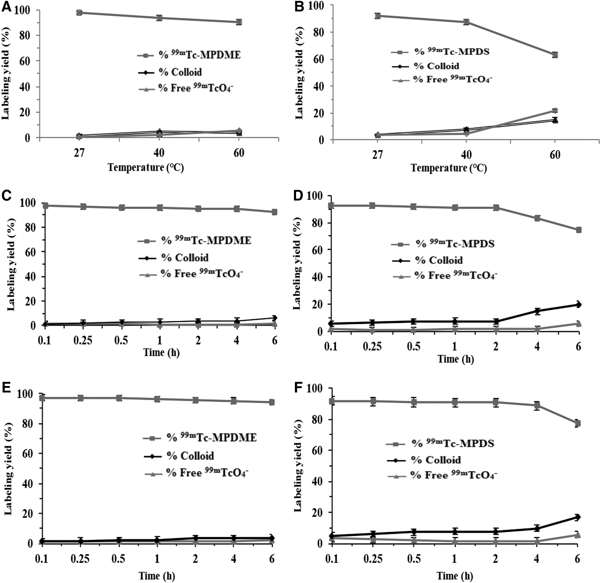
Labeling yield of 99mTc-MPDME and 99mTc-MPDS as a function of reaction temperature
Maximum labeling yield
The maximum labeling yield of 99mTc-MPDME and 99mTc-MPDS was about 97.00% ± 0.60% and 92.02% ± 0.45%, respectively. The labeling yield for the placebo ME was found to be less than 4%. Thus, the formulation excipients did not interfere with the radiolabeling of MPD. In general, the labeling yield of 99mTc-MPDME was higher compared with 99mTc-MPDS. This might be due to the ability of ME to solubilize both nonpolar and polar substances and to compartmentalize and concentrate the reactants. 48 In addition, ME has been proven to be a useful media for chemical reactions such as metal ligand complex formation. 49,50
In vitro stability studies
In vitro stability of 99mTc-MPDME and 99mTc-MPDS was studied to avoid the formation of the undesired by-products that may result from the ionizing γ-radiation (radiolysis) of the labeled compound. Stability studies of 99mTc-MPDME and 99mTc-MPDS in normal saline (Fig. 7C, D) and serum (Fig. 7E, F) showed more stability of 99mTc-MPDME compared to 99mTc-MPDS. This could be explained by the stability of ME system as mentioned earlier. 51
Biodistribution
EAC cells are considered excellent model to study the biological behavior of malignant tumor and drug assumed to produce an effect at the tumor site. 52 The i.v. injection of 99mTc-MPDME and 99mTc-MPDS showed a high liver and spleen uptake (Table 1). The target to nontarget (T/NT) (tumor muscle uptake/normal muscle uptake) ratio represents the main factor in the evaluation of sensitivity and selectivity of 99mTc-MPDME to the solid tumor. 99mTc-MPDME showed much higher T/NT ratio compared with 99mTc-MPDS (Table 1). For 99mTc-MPDME, T/NT ratio was 4.16 at 0.25 h post i.v. injection and remained high up to 24 h postinjection (3.00), while for 99mTc-MPDS the T/NT ratio was only 1.55 and 1.50 at 0.25 and 24 h, respectively, post i.v. injection. This result might be explained by the nanosize and long circulation time of 99mTc-MPDME. It is well known that nanoscale particles could target tumor tissues through the EPR effect if long circulation was achieved by avoiding macrophage's phagocytosis. 53,54 Although the tumor uptake of 99mTc-MPDME was higher compared with 99mTc-MPDS, the high accumulation of 99mTc-MPDME in the reticuloendothelial system and other healthy organs remains a major problem.
Values represent mean ± SD (n = 6).
Tc, technetium 99m; MPD, 3-(4-methoxybenzoylamino)-2,6-piperidinedione; MPDME, MPD microemulsion; MPDS, MPD solution.
Generally, the accumulation of intratumorally injected 99mTc-MPDME in the liver and spleen was significantly lower compared with 99mTc-MPDS (Table 2). It seems that after i.t. injection of 99mTc-MPDME, a low percentage was taken up by liver and spleen and then was cleared slowly through the intestine, as evidenced by the intestine uptake (2.30% ± 0.02%) at 24 h postinjection. In contrast, a high percentage of 99mTc-MPDS was rapidly accumulated in the liver and spleen and then quickly eliminated. Moreover, high tumor retention of 99mTc-MPDME following i.t. injection could be attributed to the sustained release of 99mTc-MPD inside the tumor. The ME provided a considerable barrier toward the rapid diffusion of 99mTc-MPD outside the tumor. 55 In contrast, 99mTc-MPDS showed very low tumor retention post i.t. injection, which indicates high leakage outside the tumor.
Values represent mean ± SD (n = 6).
Tc, technetium 99m; MPD, 3-(4-methoxybenzoylamino)-2,6-piperidinedione; MPDME, MPD microemulsion; MPDS, MPD solution.
The i.t. injection of 99mTc-MPDME showed a lower blood activity than i.v. injection route at each time point (Fig. 8A). Up to 24 h post i.t. injection of 99mTc-MPDME, the blood activity was significantly lower than that after i.v. injection. At 24 h, the blood activity was 7.68% ± 0.11% and 2.89% ± 0.03% post i.v. and i.t. injection of 99mTc-MPDME, respectively. These results indicate that a large portion of intratumorally injected 99mTc-MPDME was retained in the tumor site with a high residence time. In contrast, in case of 99mTc-MPDS, the blood activity following i.v. and i.t. injection was very close at each time point (Fig. 8B). The blood activity at 24 h post i.v. and i.t. injection was 4.47% ± 0.07% and 3.85% ± 0.02%, respectively. This might be attributed to the rapid absorption of 99mTc-MPDS through blood capillaries into the circulatory blood. 56,57

Percentage of the injected dose (%ID) in blood of 99mTc-MPDME
Drug inhibition study
Predosing the mice with nonlabeled MPDME led to reduction of tumor uptake of 99mTc-MPDME to drop from 2.04% ± 0.01% to 1.19% ± 0.67%. This shows the selectivity of 99mTc-MPDME for tumor site and its ability to bind selectively and accumulate specifically in the tumor.
Conclusions
MPDME was successfully prepared using water titration method at 15% IPM, 65% S/Cos (1:1), 20% water, and 0.5% w/v of drug. The developed MPDME displayed nano-spherical particles having suitable characters for i.v. or i.t. administration. Molecular docking results showed appropriate binding affinity of MPD and 99mTc-MPD with DNA binding site. MPDME and MPDS were radiolabeled efficiently with 99mTc, and the factors affecting the percentage of labeling yield were studied. The labeling yield and stability of 99mTc-MPDME exceeded that of 99mTc-MPDS. Biodistribution data demonstrated that i.t. injection of 99mTc-MPDME has significantly improved retention in the tumor and reduced localization in many healthy organs compared to i.v route. Moreover, the localization of the tracer in the tumor tissues with a high percentage encourages the use of 99mTc-MPDME as a promising tool for tumor imaging and radiodiagnostic applications. Further investigations are required to explore the possibility of using 99mTc-MPDME as a novel radiopharmaceutical for targeting the metastasis sites or unreachable tumors.
Footnotes
Disclosure Statement
The authors declare that they have no conflicts of interest to disclose. Moreover, this research did not receive any specific grant from funding agencies in the public, commercial, or not-for-profit sectors.