
Abstract
Radioimmunotherapy offers an effective way to direct ionizing radiation to cancer cells through attachment of radionuclides to antibodies while limiting negative effects of off-target irradiation. This, however, requires effective facile methods for attachment of therapeutic radionuclides onto antibodies. Herein, the authors report their efforts in evaluating N-succinimidyl S-acetylthioacetate (SATA), a commercially available reagent, for use as a bifunctional chelating agent (BCA) to attach 188Rhenium (188Re) onto h8C3, a humanized IgG antibody that can effectively target extracellular melanin present in malignant melanoma. Micro single photon emission computer tomography/computer tomography was used to determine an effective timeline for antibody uptake in B16-F10 tumor bearing C57BL6 mice guiding the selection of 188Re with its 16.9 h physical half-life. Radio instant thin layer chromatography coupled with radio high-performance liquid chromatography was used to assess radioisotope incorporation, as well as stability during the labeling process for SATA conjugated h8C3. It was determined that despite the relatively mild conditions used, incorporation of the SATA conjugate resulted in antibody instability during labeling requiring a different BCA to facilitate rhenium incorporation onto the antibodies.
Introduction
There has been a renewed interest in targeted radionuclide therapy of cancer. 1 The approval of 223Radium chloride (Xofigo) 2 for treatment of metastatic prostate cancer and of 177Lutetium-labeled somatostatin receptor binding peptides for neuroendocrine tumors (Lutathera), 3 as well as recent successes of 177Lu-PSMA-A617 4 compound in patients with metastatic prostate cancer, demonstrates the potential of targeted radionuclide with β and α emitters in treatment of cancers resistant to all other therapies.
188Rhenium (188Re) is a high energy β emitter (Emax = 2.12 MeV) that exhibits a 3.5 mm average tissue penetration depth and has a short physical half-life of 16.9 h. In addition, 188Re is a nonbone seeking and nonresidualizing radioisotope that does not linger in nontarget organs or blood, making 188Re particularly attractive for therapy. 5 When 188Re separates from the carrier protein molecule in vivo as a result of catabolism and oxidizes back to a chemically inert perrhenate anion, it is quickly excreted through the kidneys, leaving little time to cause significant toxicities. 6 To date, 188Re has been used in a variety of clinical trials of radiolabeled antibodies 5,7 and peptides. 8
The attachment of a radiometal to an antibody molecule requires a bifunctional chelating agent (BCA), which is attached to an antibody followed by extensive quality control procedures. The resulting conjugate can be stored under nondenaturing conditions for prolonged periods of time and used on demand when a patient dose needs to be prepared. In contrast to the multiplicity of commercially available BCAs for trivalent radiometals, there is a scarcity of such reagents for 188Re. In many cases, after 188Re is eluted from a 188W/188Re generator in the form of sodium perrhenate, it is reduced to the lower oxidation states with tin (II) chloride, and the antibodies are labeled with 188Re “directly” through binding of reduced 188Re to the generated sulfhydryl groups on the antibodies. 9 It is a laborious procedure which involves prolonged handling of radioactive compounds by the involved personnel. In an effort to develop a user friendly procedure for radiolabeling the antibodies with 188Re which also results in stable radiolabeled product, the authors have evaluated commercial agent N-succinimidyl S-acetylthioacetate (SATA) (Fig. 1) which adds sulfhydryl groups to proteins and other amine-containing molecules in a protected form as a potential ligand for radiolabeling humanized antibodies with 188Re. In this study, the authors describe the results of their in vitro and in vivo experiments with SATA and melanin-binding humanized antibody h8C3.
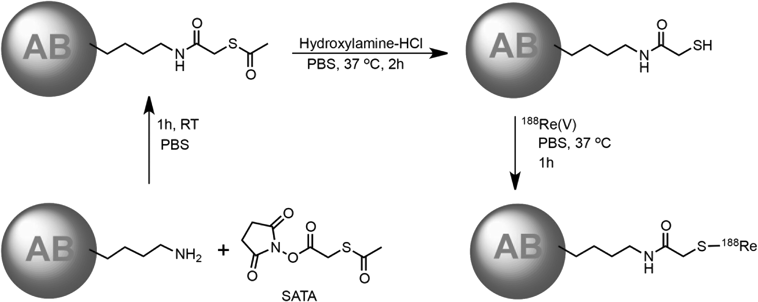
Structure of SATA and schematic of radiolabeling. SATA, N-succinimidyl S-acetylthioacetate.
Materials and Methods
Reagents and antibodies
All chemicals and reagents were purchased from Sigma-Aldrich unless otherwise stated. 188W/188Re generator (85 mCi) obtained from Oak Ridge National Laboratory (ORNL) was used for eluting 188Re activity. Alumina A Sep-Pak cartridges from Waters (Canada) were used for purification of eluted 188Re. 111Indium (111In) was obtained from Nordion (Canada). Pierce brand SATA and Ultrex grade hydrochloric acid (HCl) were obtained from Thermo Fisher (Canada). Melanin-binding humanized antibody h8C3 with IgG1 isotype was produced by Aragen. Human IgG isotype control antibody with IgG1 isotype was purchased from Creative Diagnostics. [(R)-2-Amino-3-(4-isothiocyanatophenyl)propyl]-trans-(S,S)-cyclohexane-1,2-diamine-pentaacetic acid (CHX-A”) BCA was purchased from Macrocyclics (USA).
B16-F10 melanoma tumor model
All animal studies were approved by the Animal Research Ethics Board of the University of Saskatchewan. For the imaging study 6 weeks old C57BL6 female mice obtained from Charles River Laboratories were injected subcutaneously with 5 × 10 5 B16-F10 murine melanoma cells in Matrigel (Corning) into the right flank.
Elution and purification of 188Re
188Rhenium in the form of sodium perrhenate was eluted from a 188W/188Re generator (ORNL) using 10 mL 0.9% saline as the eluent. The eluent was passed through an Alumina A Sep-Pak (Waters) to remove any potential 188W breakthrough. The Sep-Pak was preconditioned by passing 5 mL 0.9% saline and 5 mL air. To reduce the sodium perrhenate to the lower oxidation state the 0.5 mL of the eluent was added to a microcentrifuge tube containing 20 mg sodium gluconate followed by addition of 20 μL freshly prepared SnCl2 solution (20 mg/mL in 0.1 M Ultrex grade HCl). The pH was checked to ensure that it remained between 4.5 and 5.5, and the mixture was then heated to 37°C for 1 h with shaking. Reduction was monitored by spotting 1 μL of reaction mixture on two different silica gel instant thin layer chromatography (SG-iTLC; Agilent, Canada) 8 cm strips. One strip was developed with acetone to monitor rhenium reduction (unreduced perrhenate at the top with reduced rhenium gluconate at the bottom) and one with saline to ensure minimal colloid formation (reduced and unreduced rhenium at the top with colloids remaining at the bottom). 5,7 SG-iTLCs were read on a PerkinElmer 2470 Automatic Gamma Counter.
Conjugation of BCA SATA to h8C3 antibody
Conjugation of SATA to h8C3 was performed by following the manufacturer's guidelines. In short, the shipping buffer for the h8C3 antibody was exchanged into phosphate buffered saline (PBS) for conjugation using Amicon Ultra-15 Centrifugal Filters (50K MW cutoff; Fisher). The filter was prewashed with 5 mL PBS, 0.9 mg h8C3 was added to the filter and washed six times with 1.5 mL PBS. The portion containing the antibody was transferred to a microcentrifuge tube followed by 7 μL 2 mg/mL solution of SATA in dimethyl sulfoxide (DMSO) (made fresh, in a freshly opened ampule of DMSO) to reach 10 M excess of SATA over h8C3 antibody. The reaction mixture was incubated at room temperature for 1 h with shaking. After 1 h the reaction mixture was transferred back into an Amicon filter and washed six times with PBS to remove any of the uncreated SATA and DMSO. A Bradford assay was performed to determine protein recovery and concentration.
Deprotection of h8C3-SATA
Deacetylation solution was prepared by dissolving 1.75 g hydroxylamine (HA)-HCl in 10 mL PBS, adding 2.5 mL 0.5 M ethylenediaminetetraaceticacid (EDTA) and adjusting pH to 7.2 with NaOH followed by addition of ultra pure water, for a final volume of 50 mL. A thousand fold excess of HA solution was added to the SATA-conjugated antibody and incubated at 37°C for 2 h with shaking. After 2 h the reaction mixture was added to an Amicon Ultra 0.5 mL centrifugal filter (30K MW cutoff; Fisher) and washed four times with PBS to remove all traces of the HA solution.
Radiolabeling of deprotected h8C3-SATA
The activity of the freshly reduced 188Re was measured in a dose calibrator, and the desired amount of the 188Re was added to the freshly deprotected h8C3-SATA antibody in a microcentrifuge tube (specific activity of 5:1 mCi/mg) and incubated at 37°C for 1 h. Radiometal incorporation was determined by SG-iTLC using a saline eluent (top containing unlabeled 188Re, bottom containing protein conjugated 188Re) 10 and radio HPLC (high performance liquid chromatography). HPLC studies were performed using a TosoHaas size exclusion column with PBS at 1 mL/min as an eluent using a Waters 2695 HPLC system equipped with UV and radiation (Bioscan) flow-through detectors. SG-iTLCs were read on a PerkinElmer 2470 Automatic Gamma Counter.
Conjugation of BCA CHX-A” to h8C3 antibody
Ten times of conjugation buffer (0.05 M carbonate/bicarbonate, 0.15 M NaCl, 5 mM EDTA, pH 8.6–8.7), 5 mL, are combined with 0.5 M EDTA, pH 8.0 (0.5 mL) and were diluted to 50 mL in a 50 mL Falcon tube with deionized water to give the 1 × buffer. An Amicon Ultra 15 mL centrifugal filter (30K MW cutoff; Fisher) is loaded with 2 mg of the h8C3 antibody. The antibody is exchanged into the above conjugation buffer by performing 6 × 1.5 mL washes using an Amicon concentrator in a refrigerated centrifuge at 4°C. The final volume should be around 250 μL containing 2 mg of the antibody. As the buffer exchange is getting close to completion, a solution of bifunctional CHX-A” ligand with 2 mg/mL concentration is prepared by dissolving CHX-A” in conjugation buffer. The antibody is recovered from the Amicon, and 23.6 μL of 2 mg/mL CHX-A” solution in conjugation buffer is added to provide fivefold molar excess of CHX-A” over the antibody. The reaction mixture is incubated at 37°C for 1.5 h. The reaction mixtures is then purified into 0.15 M ammonium acetate buffer, pH 6.5–7.0, with 6 × 1.5 mL washes on Amicon concentrators in a refrigerated centrifuge at 4°C. The sample is stored at 4°C. A Bradford assay was performed to determine protein recovery and concentration.
Radiolabeling of antibody-CHX-A” conjugate with 111In
The radiolabeling of an antibody-CHX-A” conjugate 111In is performed to achieve the specific activity of ∼5 μCi/μg of the antibody. Six hundred microcurie of 111In chloride was added to 10 μL 0.15 M ammonium acetate buffer and added to a microcentrifuge tube containing 120 μg of the h8C3-CHX-A” conjugate in 0.15 M ammonium acetate buffer. The reaction mixture was incubated for 60 min at 37°C, and then the reaction was quenched by the addition of 3 μL of 0.05 M EDTA solution. The percentage of radiolabeling was measured by SG-iTLC using 0.15 M ammonium acetate buffer as the eluent (top containing unlabeled 111In, bottom containing protein conjugated 111In). SG-iTLCs were read on a PerkinElmer 2470 Automatic Gamma Counter.
Micro single photon emission computer tomography/computer tomography imaging
Micro single photon emission computer tomography/computer tomography (microSPECT/CT) images were collected on a MILabs VECTor 4 (Netherlands) microSPECT/CT scanner and processed using the comprehensive image analysis software package PMOD (version 3.9; PMOD Technologies, Inc., Switzerland). Imaging studies were conducted using 200 μCi 111In at a 5:1 mCi/mg specific activity with a CHX-A” conjugated h8C3. Two tumor-bearing mice were injected IV through tail vein and imaged in the prone position at 1, 4, 24, 48, 72, 96, and 216 h post injection. SPECT data were collected for 20 min using an Extra Ultra High Sensitivity Mouse (XUHS-M) collimator for 20–350 keV range using spiral trajectories. 11 All SPECT images were reconstructed using both 245 and 171 keV 111In gamma emissions on a 0.4 mm voxel grid with MILabs reconstruction software.
Results
MicroSPECT/CT imaging of h8C3 antibody is melanoma tumor bearing mice
The pharmacokinetics of the h8C3 antibody in murine melanoma was tested using microSPECT/CT by injecting 111In labeled antibody (h8C3-CHX-A”-111In); 111In radionuclide labeling was >90%. Specific uptake of the antibody in the tumor can be seen after 24 h, as well as the antibody remaining in the tumor for up to 216 h, at which point the mouse was humanely sacrificed due to tumor size limits (Fig. 2).

Single photon emission computer tomography/computer tomography of h8C3-CHX-A”- 111Indium showing accumulation of h8C3 in murine melanoma over the course of 9 d. White arrow indicates tumor location. CHX-A”, [(R)-2-amino-3-(4-isothiocyanatophenyl)propyl]-trans-(S,S)-cyclohexane-1,2-diamine-pentaacetic acid.
Reduction of 188Re
Reduction of 188ReO4 − with SnCl2 and sodium gluconate gave >90% reduction with <10% colloid formation. Decreasing the amount of sodium gluconate present would increase the amount of colloid formation; conversely, increasing the amount did not greatly reduce the amount of colloid formed.
HPLC profile of h8C3-SATA prior and post deprotection
HPLC analysis was used to determine the purity of the h8C3 after conjugation with SATA and after deprotection of the SATA group; Figure 3 shows that the UV-Vis profile of the antibody remains intact post conjugation to SATA and post deacetylation of SATA with a retention time of ∼7 min.
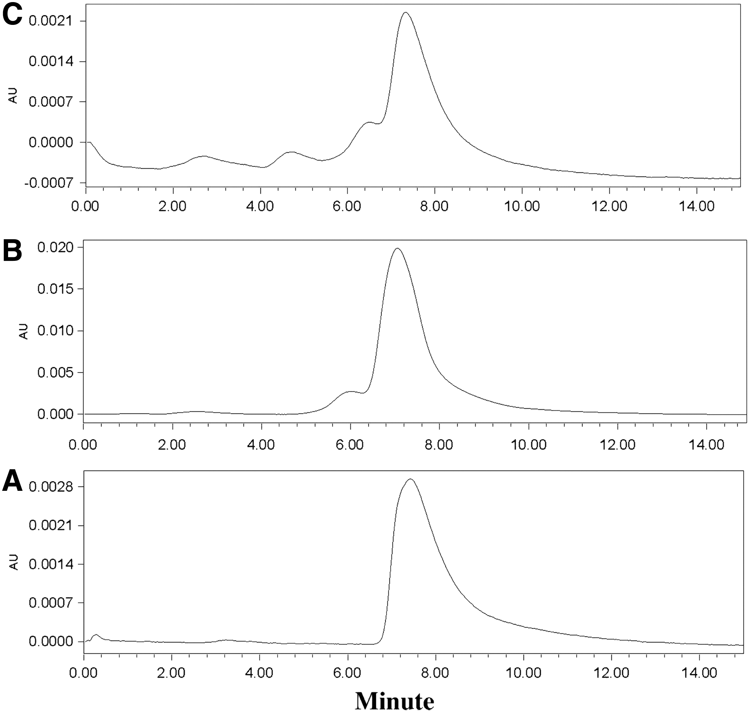
HPLC characterization of unconjugated and SATA-conjugated h8C3 antibody:
Evaluation of 188Re labeling of h8C3-SATA
Radiolabel incorporation was initially evaluated by iTLC showing 188Re incorporation from 70% to 90%; however, due to the limitation of iTLC the integrity of the antibody could not be determined and HPLC was used. HPLC results showed that despite high protein uptake, the structure of the antibody was degraded (Fig. 4A, B). To determine if the cause of this degradation was attributed to radiolysis by 188Re or by the perrhenate reduction conditions required, deprotected h8C3-SATA was incubated with sodium gluconate and SnCl2 using the same experimental conditions. It was found that the presence of 188Re was not required to cause antibody degradation (Fig. 4C). To determine if the observed degradation of the antibody was specific to h8C3, the same labeling conditions were tested on a nonspecific SATA conjugated human IgG isotype control, and a similar degradation was observed (Fig. 4D, E).

HPLC characterization of 188Re-labeled SATA-conjugated h8C3 antibody:
Discussion
It is expected that there will be 91,000 newly diagnosed cases of malignant melanoma in the United States in 2018, resulting in over 9300 fatalities. 12 Due to the difficulty of treating metastatic melanoma, new treatments are desperately needed. One of those potential treatments is radioimmunotherapy (RIT) when a radionuclide “warhead” is attached onto a targeting antibody capable of seeking out and destroying tumor deposits. Radiolabeled with 188Re first generation murine 6D2 antibody to melanin with IgM isotype showed promise in RIT of patients with metastatic melanoma. 3 Subsequently, their laboratories developed and evaluated the therapeutic potential in experimental melanoma of second-generation murine 8C3 antibody to melanin. 13 The 8C3 antibody was subsequently humanized for future use in the clinic. In this study, the authors describe their experience with radiolabeling humanized 8C3 (h8C3) with 188Re. Their preliminary imaging studies (Fig. 2) show that the h8C3 melanin-binding antibody begins to accumulate in a murine melanoma tumor 24 h postinjection and remains present up to 9 d after initial treatment. As there is a selection of effective therapeutic radionuclides to choose from, 14 –16 evaluation of this timeline allowed for the selection of one of the promising therapeutic radionuclides, 188Re which was also utilized in clinical trial of 6D2 murine antibody.
188Re is a high-energy β−-emitter with a 16.9 h half-life. This length of physical half-life gives it ample time to accumulate in and irradiate the tumor making it a good choice for radiotherapy; however, it is short enough to prevent systemic toxicity. Very importantly, Re is not a bone seeking radionuclide and is quickly excreted through kidneys without any hematologic toxicity. Re can be incorporated into small molecules or peptides at high temperatures. 17 However, incorporation of 188Re onto antibodies is more challenging as the reaction temperature cannot exceed 37°C and is typically accomplished through direct labeling. In direct labeling sulfhydryl group is generated through the reduction of disulfide bridges, exposing cysteine residues containing free sulfhydryl groups. 9 Cleavages of these disulfide bridges can result in loss of structural integrity and function rendering the antibody nonreactive toward its respective antigen. Thus, the importance of more facile labeling conditions becomes obvious. In this study the authors report their results on utilizing SATA which adds sulfhydryl groups to proteins and other amine-containing molecules in a protected form for radiolabeling the antibodies with 188Re.
According to the manufacturer's instructions, SATA, which contains an N-hydroxysuccinimide ester, can form stable covalent amide bonds with primary amines (lysine residues), and the modified molecule can be stored indefinitely when the sulfhydryl group is protected (acetylated). Exposing the SATA labeled conjugate to HA hydrochloride will deprotect (deacetylate) the conjugate exposing a labile sulfhydryl group. 18 This exposed group can then be used for cross-linking proteins or other molecules amenable to sulfide bond formation. 19 However, this exposed sulfhydryl group can also be exploited to bind 188Re(V) without the harsh conditions typically used in the direct labeling approach.
Initial results from iTLC were promising indicating good antibody binding of 188Re. However, upon further investigation through size exclusion HPLC equipped with both a UV-detector and a radio detector significant antibody degradation was observed. Examination of the UV-Vis trace shows that the antibody is no longer intact with a single sharp peak at ∼7 min, but rather multiple overlapping broad peaks eluting from 6 to 18 min. Attempts at purification using spin filtration failed to give pure radiolabeled h8C3-SATA-188Re. To determine if radiolysis was responsible for the antibody degradation, the labeling experiment was conducted without the 188Re, and it was found that the presence of SnCl2 and sodium gluconate caused the same degradation of the antibody. As only minor degradation was observed with SnCl2 alone, it can be reasoned that the sodium gluconate might be responsible for substantial portion of the SATA-conjugated humanized antibody instability.
To determine if the humanization process for the h8C3 antibody caused general instability in the antibody, a human IgG isotype control antibody was conjugated to SATA and underwent the same labeling procedure as the h8C3 antibody. Again, it was found that there was antibody degradation. In addition, despite the claims of stability, after 4 d an insoluble white precipitate formed in the acetylated h8C3-SATA and IgG-SATA isotype control antibody solution, seemingly indicating that spontaneous cross-linking is occurring during storage. This same precipitation was never observed for either CHX-A” or DOTA conjugates of the same antibody. These results show that SATA will not be a viable BCA for the incorporation of 188Re into the humanized/human antibodies.
While humanized or fully human antibodies have now become standard of care in immunooncology, 20 the experience of dealing with such antibodies in nuclear medicine is very limited, for example, the only two antibodies approved so far for RIT in the clinic—Iodine-131 Tositumomab (Bexxar) and Ibritumomab tiuxetan (Zevalin) —are both murine. Since humanized/human antibodies are significantly different from murine antibodies in their structure, qualities, and in vivo behavior, the existing radiolabeling approaches developed in the past for murine antibodies have to be adjusted to dealing with human/humanized antibodies. For example, humanized and human antibodies have much higher isoelectric points (pI) than murine antibodies, 21,22 which obviously greatly affects the way they interact with radiolabeling reagents, as well as their stability post radiolabeling. Thus, the results of this study clearly show the necessity to rethink and redesign many radiolabeling procedures for humanized/human antibodies.
Conclusions
The present study focused on the evaluation of SATA as a potential BCA for 188Re. By conjugating SATA to h8C3 melanin targeting humanized antibody the authors hoped to find an efficient simple method to coordinate 188Re and deliver a site-specific toxic dose of radiation to melanoma cancer cells. Unfortunately, it was determined that due to antibody degradation, and spontaneous precipitation of the SATA conjugated antibody upon storage, SATA will not be a suitable BCA. This study demonstrates the difficulty of finding available chelating agents for Rhenium which would be compatible with the humanized or human antibodies. As multiple radioactive isotopes of rhenium (186, 188, and 189) 23 are becoming available for use in RIT of cancer—further research on developing efficient BCAs for Rhenium is warranted.
Footnotes
Acknowledgment
This research was supported, in part, by the NCI SBIR Contract HHSN261201600017C.
Disclosure Statement
E.D. is a coinventor on the patent currently licensed to RadImmune Therapeutics by the Albert Einstein College of Medicine, NY.