
Abstract
Purpose:
Fibrin is a perfect target for specific imaging of all types of thrombotic lesions. Cyclic peptides were introduced as the best scaffolds out of the different types of probes for thrombi detection. This study was conducted to label previously synthesized peptide-targeting fibrin with [18F]FDG and its in vitro and in vivo assessments.
Materials and Methods:
CGPRPPC peptide functionalized with 6-hydrazinonicotinamide and Eei-NHS was synthesized and cyclized using air oxidation method. The cyclic sequences were labeled with [18F]FDG at 85°C within 30 min. The stability studies were performed in human plasma. Fibrin-binding and platelet aggregation tests were performed in vitro. Biodistribution and scintigraphy imaging in normal mice and carotid thrombotic rat model were considered as in vivo studies.
Results:
Radiolabeled peptides show a good stability in human plasma and also high-affinity binding for human fibrin. Platelet aggregation test confirmed specific binding of radiopeptides to fibrin. A key problem with the authors' previous research was inability to detect small-vessel thrombi. The results of positron emission tomography/computed tomography scanning show high specific uptake of [18F]FDG-labeled CGPRPPC in small-sized thrombosis.
Conclusion:
The experiment revealed that radiolabeling of cyclic peptide (CGPRPPC) with [18F]FDG enables us to detect small thrombotic lesions in small animal models with high resolution.
Introduction
Thrombosis is a mobile blood clot in the circulatory system, and it includes fibrin chains and platelets. The diseases of thrombosis with an estimated 2200 deaths each day in the United States 1 are divided into four types, such as pulmonary embolism, deep venous thrombosis, heart attack, and ischemic strokes. Computed tomography angiography, ultrasonography, and ventilation/perfusion scintigraphy were performed in patients suspected to have thrombosis to evaluate thrombi localizations. Difficulty in early detection of thrombi lesions and poor resolution are main defects of the present methods. So, recently many studies are focusing on specific molecular imaging techniques. 2,3 Labeled antibodies and their fragments, nanoparticles, and small peptides labeled with different radionuclides are the instrumental probes for selective molecular imaging of platelets and fibrin in thrombi structures. Out of these targeting agents, peptides are scaffolds preferred due to simple synthesis, low molecular weight, good penetration into thrombi, rapid clearance from blood/nontarget tissues, and nonimmunogenicity. 4 –9
Fibrin is a good target for radionuclide imaging of thrombotic lesions because of the presence of fibrin alone in pathologic conditions, both fresh and old clots, high specificity, and sensitivity for thrombi detection. 6
The GPRPP (TP850) was designed from the tripeptide (Gly-Pro-Arg) GPR, which is the first sequence of fibrinogen/thrombin inhibitor that has shown high fibrin-binding affinity. 7 –10
For diagnostic imaging, peptides can be labeled with several radioisotopes such as [99mTc], [111In], [68Ga], [64Cu], [123/124I], and [18F] to prepare single photon emission computed tomography (SPECT) or positron emission tomography (PET) tracers. A dramatic role of PET imaging in early detection of small lesions with high resolution has been revealed. 18F is the PET radionuclide of choice because of its high positron emission (97%), favorable half-life (109.8 min), high spatial resolution, low tissue radiation dose, and high yield of production in a small biomedical cyclotron. 11,12
To label peptide sequences with [18F], the direct method is not as efficient as the indirect labeling owing to lack of peptide resistance under harsh radiolabeling conditions (extreme heating, pH, and solvents) and high concentrations of peptide requirement. For indirect labeling, different [18F]-fluorinated prosthetic groups have been used with multistep and nonselective procedures that resulted in low radiolabeling yield. Another way of indirect labeling is to functionalize peptide precursors with hydrazine or aminooxy (Aoa) for hydrazone or oxime bond formation between functionalized peptides and [18F]-fluorinated aldehydes, which leads to chemoselective conjugation of peptides at some points in the sequence far from the binding site. 12,13
Based on literature review, human serum albumin protein and Arg-Gly-Asp (RGD), octreotide, and substance P as peptides were functionalized with 6-hydrazinonicotinamide (HYNIC) and labeled with a cyclic form of [18F]FDG as a common prosthetic group with high stability through hydrazone bond formation. 14 –16
In the authors' recent study, the cyclic peptide CGPRPPC was radiolabeled with [99mTc] using HYNIC conjugation. The radiopeptide showed good affinity to fibrin. The uptake of radiopeptide in rabbit thrombotic model was assessed using SPECT/CT imaging. 17 To evaluate thrombotic lesions in small animals such as rat or balb/c mice, PET/CT cameras are preferred.
In this study, the CGPRPPC peptide was synthesized and conjugated to HYNIC and aminooxy moiety (Aoa) to be labeled with [18F]FDG in one step. Moreover, the radiochemical purity of [18F]-radiolabeled peptides was assessed. Then, plasma stability, fibrin binding, and platelet aggregation tests were performed. Finally, in vivo evaluation of [18F]FDG-labeled peptides and imaging was performed using rat carotid thrombotic model.
Materials and Methods
All the required amino acids and 2-chlorotrityl chloride resin were purchased from Bachem Co. (Switzerland), and other reagents for peptide synthesis (TBTU and DIPEA) were obtained from Sigma-Aldrich. All chemicals, solvents, and reagents were of analytical reagent grade. Succinimidyl-N-Boc-HYNIC (Kimia Pajouh Dorsa, Tehran, Iran), cold FDG (ABX Advanced Biochemical Compounds, Germany), Eei-NHS (IRIS Biotech GmbH, Marktredwitz, Germany), fibrin (Sigma-Aldrich), adenosine diphosphate (ADP), arachidonic acid (AA), and collagen (Bio/Data, Corp, Germany) were obtained from the respective companies. [18F]FDG was obtained from routine in-house synthesis at the PET/CT unit (Ferdous Nuclear Medicine Center, Dr Masih Daneshvari Hospital, Shahid Beheshti University of Medical Sciences, Tehran, Iran). Thin-layer chromatography (TLC) was performed on silica gel 60 F254 precoated aluminum TLC plates from Merck. SepPak C-18 cartridge was obtained from Waters Corporation (USA). The radiochemical purity was determined using a TLC scanner (Mini-Scan MS-1000). The peptides were analyzed using mass spectra on a liquid chromatography-mass spectrometry (LC-MS) Triple Quad 6410 (Agilent) through a series 1200 high performance liquid chromatography (HPLC) column with the following specifications: C-18; 250 × 4.6 mm; 5 μm; mobile phase A: H2O + 0.1% trifluoroacetic acid, B: acetonitrile; flow rate 1 mL/min; sample volume 20 μL; and total run time 40 min. Antiplatelet aggregation activities of fibrin-targeted radiopeptides were determined using APACT-4004 aggregometer (LABiTec, Ahrensburg, Germany). A NaI well counter (Triathler Multilabel Tester, Hidex, Finland) and a dose calibrator (AtomLab 100; Biodex) were used to measure the low and high dose levels, respectively.
Synthesis of cyclic CGPRPPC-HYNIC and CGPRPPC-Aoa conjugates
Fmoc solid-phase synthesis method was used to synthesize CGPRPPC peptide based on the authors' previous study. 17 The sequences were functionalized using HYNIC or Aoa during solid-phase synthesis. Cyclization was performed using air oxidation method. Finally, LC-MS was used to confirm peptide sequences. Each step was described in detail in the authors' previous study. 17,18
Platelet aggregation test
Light transmission aggregometry was performed to show specific binding of CGPRPPC to fibrin. In brief, the citrated blood samples were centrifuged to prepare platelet-rich plasma (PRP) and platelet-poor plasma using a method described previously. 19 Then, aliquots of 200 μL (50 × 106) of PRP were transferred to four cuvettes and incubated at 37°C in APACT-4004 aggregometer (LABiTec, Ahrensburg, Germany). Then, 1 μL of cold peptide (0.05 and 0.1 mM) and 1 μL of dimethyl sulfoxide (DMSO) were added to test and control cuvettes, respectively. After 5 min of incubation, AA, collagen, and ADP were separately used to induce platelet aggregation. The changes in the optical density were recorded, and the results are expressed as percentage of inhibition by comparison with those measured for DMSO alone.
Labeling studies
HYNIC/Aoa-CGPRPPC labeling with [19F]FDG
The conjugation reaction of HYNIC/Aoa-functionalized peptides (2 mg) and [19F]FDG (2 mg) was carried out in 96% ethanol in saline (200 μL) as a solvent at 100°C for 30 min. The reaction pH was adjusted ranging from 2 to 3. The reaction mixture was diluted (1 mL) after reaching ambient temperature. Then, free [19F]FDG and [19F]FDG-peptide conjugates were separated by passing 5 mL water and 2 mL of 96% ethanol through preactivated Sep Pak® C18 cartridge, respectively. 20 The [19F]FDG-labeled cyclic peptides were identified by LC-MS.
Cyclic HYNIC/Aoa-CGPRPPC radiolabeling with [18F]FDG
A detailed protocol of radiolabeling reaction of hydrazine-functionalized peptide with [18F]FDG was made available by the authors. 18 In brief, to radiolabel peptides with [18F]FDG, a liquid kit containing 250 μg of dissolved peptide in 250 μL of 96% ethanol was prepared and then 1 mCi (200 μL) of [18F]FDG was added. The optimized radiolabeling conditions were achieved at 85°C, pH 3, after 30 min of incubation. Reaction mixtures were purified by a Sep-PakC18 cartridge. To measure radiochemical purity, TLC was performed on TLC silica gel 60 F254 and acetonitrile, using water (95:5) as a solvent. 20,21
In vitro studies
Partition coefficient (log Pp) determination
To determine the peptide distribution between the organic phase and aqueous phase, 10 μCi (0.2 μCi/μL) of radiolabeled peptides was added to 1 mL of phosphate buffered saline (PBS)–n-octanol (1:1) mixture and the mixture was completely vortexed for 10 min. After two-phase separation, 100 μL of organic phase was transferred to a mixture of PBS–n-octanol (500:400 μL), and the final mixture was vortexed and centrifuged. The aforementioned step was followed three times. The radioactivity of 100 μL organic and aqueous phases was measured using a γ counter.
Human plasma stability test
To evaluate the interaction of radiolabeled peptides with plasma proteins, 50 μL of [18F]FDG-labeled peptides was used to treat 450 μL of fresh human plasma and incubated at 37°C. At different time intervals in the range of 10–120 min, the precipitation of plasma proteins was performed by adding 500 μL cold acetonitrile. After centrifugation (10,000 g for 10 min), the radioactivity of precipitate and supernatant was counted. The supernatant was analyzed by radio TLC to confirm intact [18F]FDG-labeled peptide in the presence of plasma proteins.
Fibrin binding test
The affinity binding of radiolabeled peptides to fibrin was assessed using human fibrin as previously reported. 17 Human fibrin (1 mg) was dissolved in 400 μL of 0.05 M Na3PO4 (pH 12) and then incubated with 2–70 nM (2.5–3 Ci/mmol) of [18F]FDG-labeled peptides for 30 min at 37°C. At the end of incubation, sample solutions were passed through a Centricon molecular filtration device (Sartorius; MWCO 30000). The activity of Centricon and filtered solution was measured using a radionuclide dose calibrator.
An irrelevant peptide, [18F]FDG-labeled HYNIC-LIKKPF, was used as a negative control. To block nonspecific binding site, an excess of cold peptide (100 nM) was used.
In vivo studies
Rats were purchased from the breeding facility of the Department of Pharmacology and Toxicology of the School of Pharmacy at Shahid Beheshti University of Medical Sciences. All animal studies were followed based on the guidelines for the ethical care and use of animals as approved by the Shahid Beheshti University of Medical Sciences.
Induction of carotid thrombosis in rat
The thrombosis was induced using 10% FeCl3 saturated paper as described earlier. 22,23 Carotid artery was chosen because of its small internal diameter that allows one to assess the small lesions using a radio probe.
PET/CT and SPECT/CT imaging
In an attempt to detect thrombotic lesions in a small animal, 200 μCi of [18F]FDG-HYNIC-CGPRPPC and [99mTc]-HYNIC-CGPRPPC in saline was injected into the tail vein of normal and thrombotic rat models. After general anesthetizing, PET/CT and SPECT/CT imaging were performed on the Siemens Biograph 6 and Symbia T1 Siemens clinical scanners (Siemens AG, Erlangen, Germany), respectively. Rats were anesthetized during imaging sessions with a mixture of ketamine (80 mg/kg, IP) and xylazine (8 mg/kg, IP). Additional anesthesia was given during imaging if necessary. The rats were fixed in supine position and CT scans were performed for anatomical reference and attenuation correction (spatial resolution 1.25 mm, 80 kV, 50 mA).
PET acquisition was performed 30 min after injection. PET images were reconstructed using TrueX algorithm with attenuation correction. The reconstruction settings were 2 iterations and 21 subsets to a 336 × 336 matrix, with a postfiltering of 5 mm.
SPECT imaging was performed 30 min postinjection. The acquisition protocol was flash 3D with 256 × 256 matrix size, 2.29 zoom factor, and ninety 20-s views with both detectors in a noncircular orbit. The total counts of the thrombotic site, liver, kidneys, and bladder were measured. Reconstruction was performed using the flash 3D algorithm with 4 iterations and 20 subsets.
Transmission data were reconstructed into a matrix of equal size by means of filtered back projection, yielding a coregistered image set. The reconstructed PET and SPECT images were then fused with CT images.
Results
All experiments were done in triplicate. The data represent mean ± standard deviation unless noted otherwise. The peptides were synthesized using standard Fmoc strategy, functionalized with HYNIC and aminooxy at the N-terminal, and analyzed using LC-MS.
Cyclic HYNIC-CGPRPPC was calculated for C35H51N13O9S2: 861.3; m/z = 862.4 [M+H]+; m/z = 431.7 [M+H]+/2; analytical reverse phase-high performance liquid chromatography (RP-HPLC): Rt = 4.0 min; 85% A, 15% B. 18
Cyclic Aoa-CGPRPPC was calculated for C31H49N11O10S2: 799.3; m/z = 800.2 [M+H]+; analytical RP-HPLC: Rt = 5.13 min; 85% A, 15% B (Fig. 1).
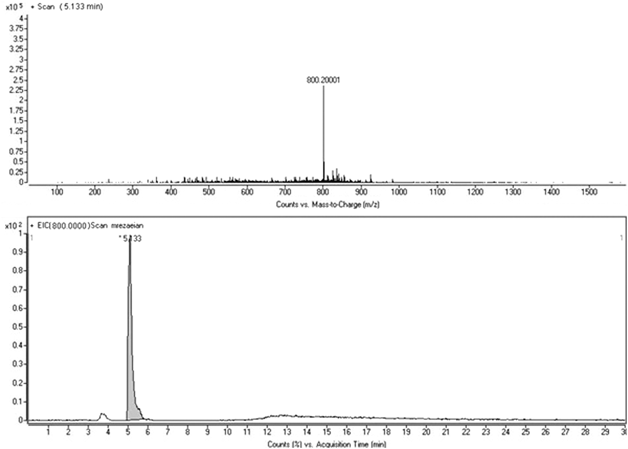
Liquid chromatography-mass spectrometry chromatogram of Aoa-CGPRPPC.
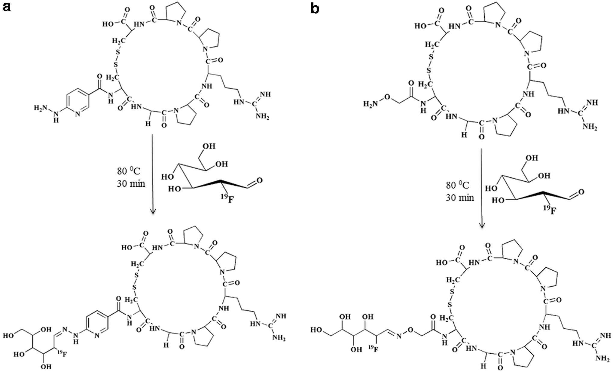
The functionalized peptides were conjugated with cold FDG (Fig. 2). The yield of labeling was 51%. LC-MS spectrometry of purified [19F]FDG-labeled peptide conjugated to HYNIC and Aoa (purification by C18SepPack cartridge) showed a single corresponding peak in m/z = 1026.4 for cyclic [19F]FDG-HYNIC-CGPRPPC and 964.4 for cyclic [19F]FDG-Aoa-CGPRPPC. Cold labeling was done to confirm the identity of hot labeling.
The functionalized peptides were radiolabeled using [18F]FDG with specific activities (SAs) of [18F]FDG-HYNIC-CGPRPPC (2.87 Ci/mmol) and [18F]FDG-Aoa-CGPRPPC (2.66 Ci/mmol).
The optimal conditions of [18F]FDG labeling were achieved in the presence of 250 μg peptide (1 mg/mL), 85°C, pH 3 for 30 min treating by 1 mCi (200 μL) [18F]FDG. The highest radiochemical purity of [18F]FDG-labeled CGPRPPC was 93% ± 1.02%.
For HPLC radiochromatograms, the outlet of LC-MS column was disconnected from mass and connected to a fraction collector. Thirty fractions (0.5 mL/min) were collected, counted, and the radiochromatograms were produced using MS Excel. The retention times of [18F]FDG-peptide and [19F]FDG-peptide were approximately the same between 4 and 5 min. Chromatograms are attached as supplementary data.
As expected, the presence of CGPRPPC had negligible effect on inhibition of platelet aggregation in all agonist-induced aggregation studies.
The log p results showed the hydrophilic nature of [18F]FDG-HYNIC-CGPRPPC and [18F]FDG-Aoa-CGPRPPC (log p = −1.3 and −1.7, respectively). The presence of coligands (Tricine and EDDA) used for labeling of cyclic peptide with [99mTc] made [99mTc]-HYNIC-CGPRPPC more hydrophilic (log p = −2.3) than [18F]FDG-labeled ones.
Treatment of [99mTc]-labeled and [18F]FDG-conjugated peptides with human plasma at 37°C resulted in only 5% release of radioactivity within 6 and 2 h, respectively.
Saturation binding experiments were performed in the presence of excess amount of cold peptide to determine the affinity of radiopeptides to fibrin (Kd) (Table 1). Concentration–response curves were fitted using GraphPad Prism software.
Biologic Characteristics of Radiolabeled Peptides
HYNIC, 6-hydrazinonicotinamide.
The in vivo study on biodistribution of [18F]FDG-labeled peptides in normal and pretreated and nontreated carotid thrombotic rat models showed a rapid renal clearance from nontarget tissues. The renal clearance of peptides confirmed the hydrophilicity of radiolabeled peptides (log p < 0). The specific uptake of radiopeptides in the carotid thrombotic region was reported as a percentage of injected dose per gram tissue (%ID/g), same as the other organs. The parallel nontreated control tests were done in a thrombosis-induced rat model. The percentage of uptake of [18F]FDG-HYNIC-CGPRPPC and [18F]FDG-Aoa-CGPRPPC in carotid region was similar in 1 h. The results after 15, 30, and 60 min postinjection are shown in Figure 3. The percentage of uptake for both [18F]FDG-labeled cyclic peptides was significantly higher in carotid thrombosis and kidneys versus the other organs. The blood clearance profile of [18F]FDG-labeled peptides as compared with [99mTc]-labeled peptide was the same within 120 min.
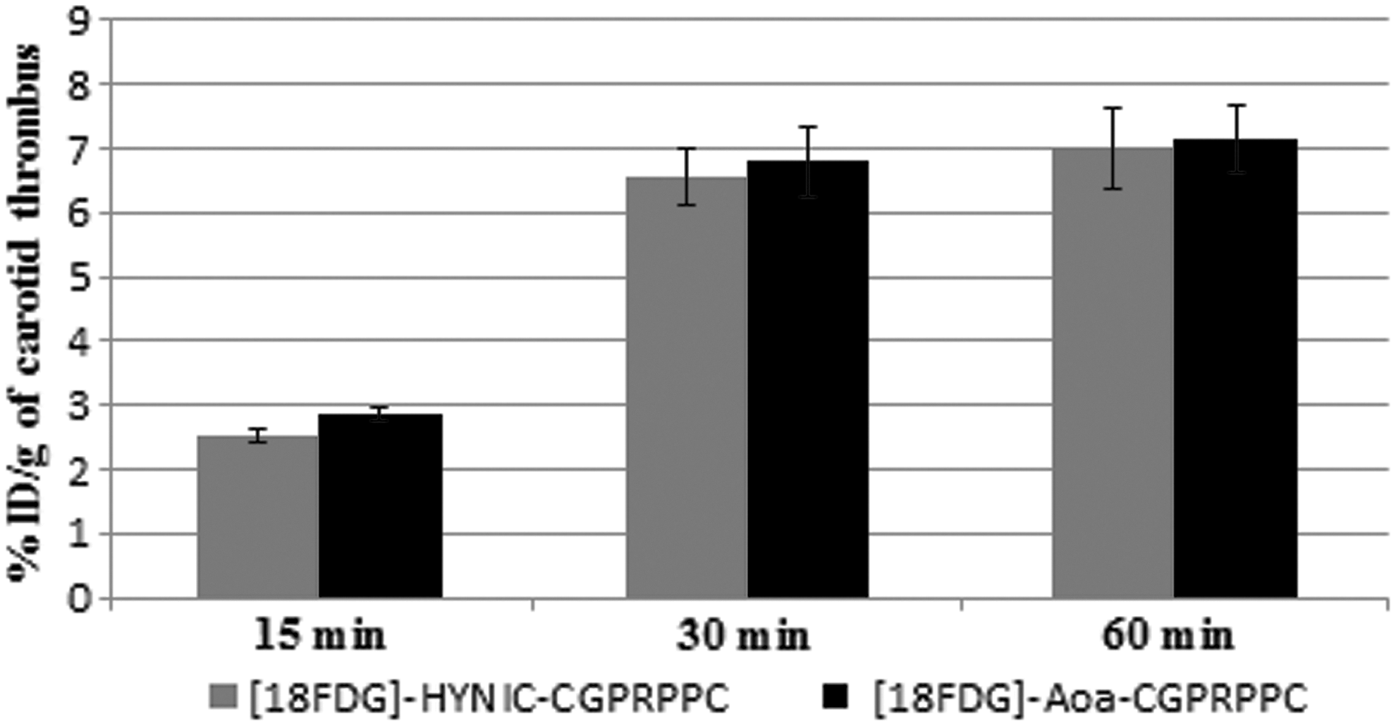
Uptake of 200 μCi [18F]FDG-Aoa-CGPRPPC and 200 μCi [18F]FDG-HYNIC-CGPRPPC in carotid thrombotic rat models at 15, 30, and 60 min postinjection (n = 3). Radioactivity is shown in terms of %ID/g carotid thrombosis.
The PET/CT and SPECT/CT images of carotid thrombotic rat models were recorded to characterize the pattern of uptake at 30 min postinjection. As shown in Figure 4, PET/CT images of thrombosed and normal rats (a, b) clearly show a high activity concentration in the kidneys and bladder as expected. However, there was a significant uptake in the carotid of the thrombosed rat compared with normal rat. Figure c and d shows the fused coronal SPECT/CT images of the carotid artery thrombotic rat model and the normal one. There was no uptake in the thrombotic site in the representative rat model.
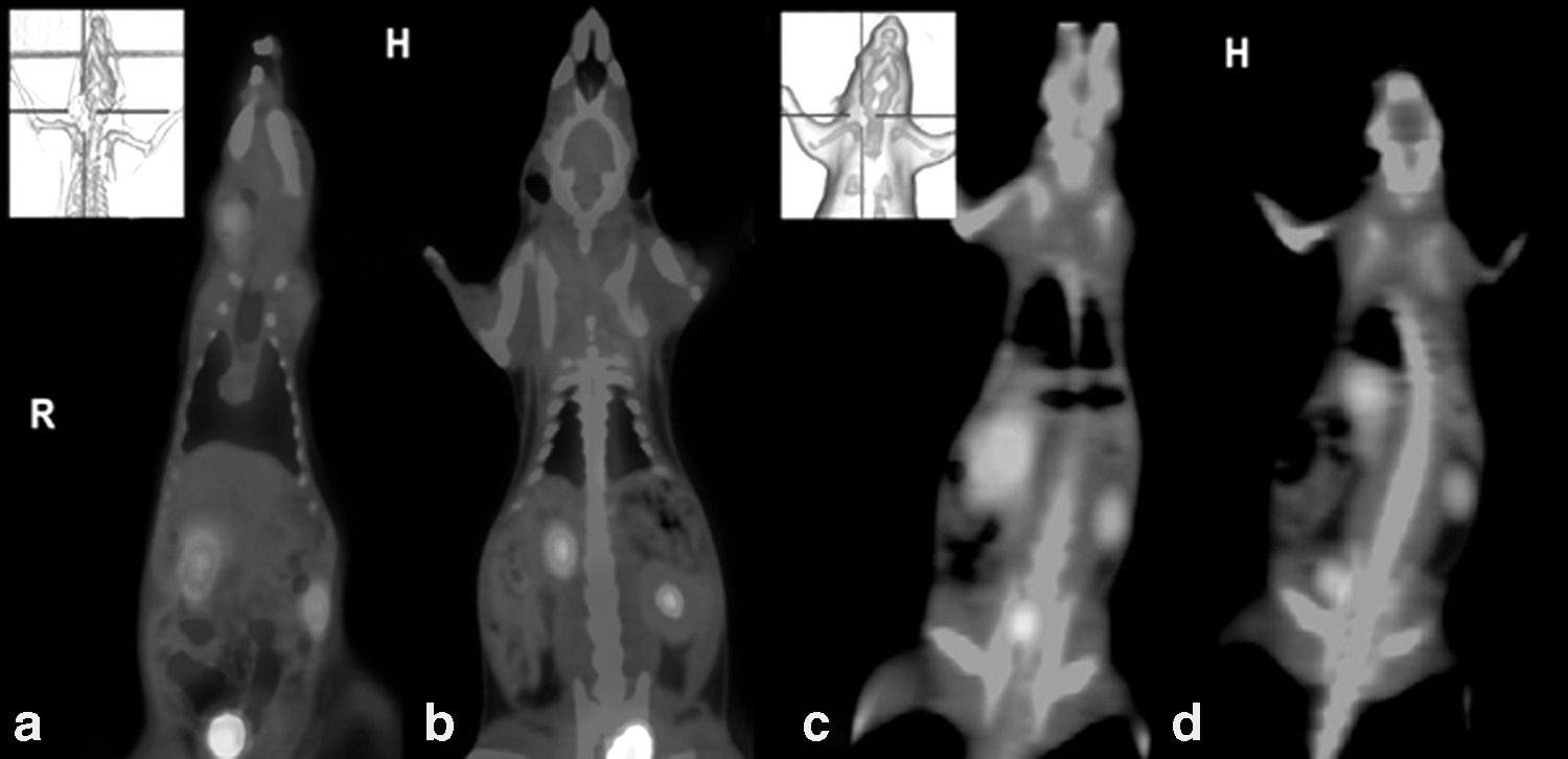
Whole-body PET/CT images of live rats 30 min postinjection of 200 μCi [18F]FDG-HYNIC-CGPRPPC in
For quantitative assessment of target uptake behavior, maximum activity concentration (Bq/cc) and maximum counts were measured in PET and SPECT imaging studies. The results show that the maximum activity concentration ratio of the thrombus was significantly higher than the background (about eight times more uptake) in PET imaging, while there was no significant difference between measured counts in the SPECT images of the thrombotic model versus normal rat.
Discussion
Fibrin is one of the main components of thrombosis, which is found in all types of thrombi but absent in the circulation. Molecular imaging of fibrin is an important step in thrombi detection. Of the different types of probes used for thrombi detection, cyclic peptides are introduced as the best scaffolds. In the authors' previous study, the cyclic peptide CGPRPPC was synthesized and radiolabeled with [99mTc] using HYNIC. After 30 min postinjection of [99mTc]-CGPRPPC to femoral thrombotic rabbit models, the thrombotic site was detected using SPECT/CT, but no visualization was found in carotid thrombotic rat model. The higher resolution and sensitivity of PET over SPECT encouraged us to label peptide with [18F]FDG.
CGPRPPC peptide was synthesized, conjugated to HYNIC and Aoa, and cyclized. Then, radiolabeling procedure was carried out with [18F] using [18F]FDG as previously reported. 18
Different radiolabeling conditions, including peptide concentration, [18F]FDG radioactivity, temperature, and incubation time, were examined. The lowest radiochemical purity was attained with a low concentration of peptide and high amount of activity. The best radiolabeling conditions were observed as and when the concentration of glucose in [18F]FDG solution was <50 μg/mL. 18 Since glucose competes with [18F]FDG for conjugation to peptide, the SAs of radiolabeled peptides as compared with those of [99mTc]-labeled peptides were very low (Table 1).
Stability of radiopeptides was determined by measuring the percentage of intact radiopeptide using HPLC. Comparison of these data with the authors' previous study showed no significant difference in plasma stability of peptide CGPRPPC labeled with [18F]FDG or [99mTc].
To assess the possibility of fibrin targeting by [18F]FDG-conjugated peptides, fibrin powder was used. The affinity (Kd) of [18F]FDG-labeled peptides was ∼50 times less than that of [99mTc]-labeled peptide. It was assumed that the lower affinity of [18F]FDG-labeled peptides for fibrin resulted from their low SAs. In other words, most of the fibrin binding sites were occupied by cold peptides.
Biodistribution studies have shown that peptide CGPRPPC has enough affinity to fibrin to be trapped in thrombotic lesions. The uptake of radiolabeled peptides with [18F]FDG and [99mTc] was higher in thrombotic lesions than in the other organs, except kidneys. The amount of [18F]FDG-labeled peptide in carotid thrombotic lesion was less than that of [99mTc]-labeled peptide because of its low specific activity. PET/CT image clearly showed the uptake of [18F]FDG-labeled peptide in the lesion; while SPECT/CT was not able to show the uptake of the same [99mTc]-labeled peptide in carotid thrombotic lesion. The [99mTc]-labeled peptide was accumulated in femoral thrombotic lesion of rabbit as visualized using SPECT/CT image. 17
Inability to detect small thrombi was an important restrictive factor in the authors' previous studies. Since there was no high-resolution small-animal SPECT scanner, peptide was radiolabel with [18F]FDG and PET/CT imaging system was used. High spatial resolution of PET/CT scanner makes it possible to accurately diagnose small lesions independent of its size and location. In this study, minimal thrombotic regions were detected in the small animal model (rat) by applying the same [18F]FDG-labeled peptide carrier.
Conclusions
As regards the essential role of early detection of small thrombotic lesions in cardiovascular disease prognosis, a previous peptide-targeted fibrin was synthesized and functionalized with HYNIC and Aoa to incorporate [18F] into peptides chemoselectively. In vitro and in vivo evaluations of [18F]FDG-HYNIC-CGPRPPC and [18F]FDG-Aoa-CGPRPPC were performed in carotid thrombotic rat model. The authors' observations support that conjugation of HYNIC and Aoa had negligible effect on either in vitro or in vivo results. Furthermore, the exact locations of thrombi were diagnosed 30 min postinjection of [18F]FDG-labeled cyclic peptides using high-resolution PET/CT scanning. Recent important findings in peptide labeling with 18 and 68Ga provide an efficient and practical way to target fibrin in small thrombotic sites.
Footnotes
Acknowledgment
This research was supported by Shahid Beheshti University of Medical Sciences, Tehran, Iran.
Disclosure Statement
No competing financial interests exist.