
Abstract
Purpose:
Overexpression of the HER2/neu (HER2) is linked to an adverse outcome in ovarian cancer. Short-interfering RNA (siRNA) is a HER2 inhibitor, which in combination with chemotherapy improves survival rate. The aim of this study was to evaluate the efficacy of adenovirus mediated HER2-siRNA in combination with cisplatin (Ad-HER2-siRNA+DDP) on treating HER2-positive ovarian cancer xenografts and explore the effectiveness of 131I-Herceptin immunoSPECT imaging for monitoring the tumor's progression.
Methods:
Mice with ovarian cancer xenografts were treated with different therapy regimens and imaged at 1, 4, 8, 12, 24, 48, 72, and 96 h postinjection with 131I-Herceptin. Concurrently, the tumor/background (T/B) ratios were calculated. In addition, HER2 protein expression levels were determined by immunohistochemistry (IHC).
Results:
The in vivo therapy experiments revealed that tumor weight and volume of Ad-HER2-siRNA+DDP group was the least of all. The IHC results further confirmed that HER2 protein level was significantly downregulated in this group. The results of SPECT imaging showed that the T/B ratios at each time point in Ad-HER2-siRNA +DDP group was the lowest (p < 0.05).
Conclusions:
The data demonstrate that the growth of xenografts of human ovarian cancer with high expression of HER2 could be inhibited effectively by Ad-HER2-siRNA+DDP. 131I-Herceptin had potential use for noninvasive imaging of HER2 expression.
Introduction
Ovarian cancer is the most lethal gynecologic malignancy. According to the National Cancer Institute 22,280 cases of ovarian cancer were diagnosed and 14,240 will die from the disease in 2016. 1 Ovarian cancer remains a serious challenge, with the highest fatality rate of gynecologic cancers. The primary treatments, including surgery, radiotherapy, chemotherapy, and endocrinotherapy, give unsatisfied therapeutic effect. Although surgical and medical treatments advanced, 5-year survival rate had not seen a definite improvement in the last decade. Thereby, it is necessary to optimize the available treatment strategies and develop a novel therapeutic agent.
HER2/neu (HER2) gene is a member of the epidermal growth factor receptor family and encodes a 185 kDa protein with tyrosine kinase activity. 2 Observed rates of HER2 overexpression have been reported to range from 8% to 66% in ovarian cancer, and it plays a major role in cell growth, proliferation, and differentiation. 3,4
Trastuzumab (Herceptin) is the first anti-HER2 monoclonal antibody applied in a clinical trial of patients with HER2-positive metastatic breast cancer. 5 For patients with HER2-positive advanced gastric cancer, trastuzumab in combination with chemotherapy significantly prolonged overall survival compared with chemotherapy alone. 6 However, intrinsic and acquired resistance to this therapy is common, which leads to disease progression. 7 For now, the clinical issue about how to precisely select patients who will surely benefit from trastuzumab has troubled doctors.
RNA interference (RNAi) is a mechanism by which double-stranded RNA acts as a signal to promote degradation of mRNA with sequence identity. 8 Short-interfering RNA (siRNA) is a class of double-stranded RNA molecules that can induce RNAi, which can be used to silence target gene expression for tumor growth. 9 The demonstration of the RNAi-mediated mechanism of target mRNA cleavage in human tumors from patients treated in Phase I clinical trials provides hope for clinical use of siRNA. 10 To establish stable gene knockdown cell lines by siRNA, several examples using plasmid vector systems have been reported. 11,12 Nevertheless, transient siRNA expression and low transfection efficiency remained the problematic for synthesized and vector-derived siRNA. Recently, viral vectors have been developed for efficient delivery of siRNA into cells. 13 Previously, the authors' group had successfully established lentivirus-mediated HER2-siRNA vector. 14 However, lentivirus system has a number of limitations in its ability to be used in clinical gene therapy. Unlike lentivirus, adenoviral vector is safe and easy to infect cells for gene therapy. 15
The occurrence of tumor is an extremely complex process, so a combined application of drugs with different anticancer mechanisms can play a better role in enhancing anticancer activity. Currently, cisplatin (DDP) is the first-line chemotherapy drugs for the treatment of ovarian cancer. It belongs to the cell cycle nonspecific drug and interferes with DNA replication, which kills the fastest proliferating tumor cells. 16 Thus, their group used an adenoviral vector to construct the adenovirus-mediated HER2-siRNA (Ad-HER2-siRNA) in this study and tested the Ad-HER2-siRNA combination with cisplatin for their ability to downregulate HER2 expression in HER2-positive ovarian cancer xenografts.
In addition, it is quite difficult to monitor the effect of RNAi in tumor gene therapy. In fact, evaluating treatment response is the key to improve the therapeutic effect. As the authors know, the molecular imaging technology is efficient, sensitive, and accurate, 17 and it may be a useful tool to solve the above problems in ovarian cancer RNAi therapy. In this study, they described the evaluation of 131I-Herceptin for the in vivo imaging and explored the effectiveness for detecting and monitoring the progression in ovarian cancer xenografts.
Experimental
Materials and methods
Recombinant adenovirus construction and identification
A HER2-siRNA with target sequence TCTGCGGTGGTTGGCATTC (2204–2222) and a negative control sequence TTCTCCGAACGTGTCACGT were previously synthesized by their group. 14 In this study, the authors, respectively, inserted them into adenovirus vector and constructed Ad-HER2-siRNA and scrambled control adenovirus vector (Ad-HER2-siRNA-NC). In brief, the double-stranded oligonucleotides were inserted into the GV119 Vector (Jikai gene technology co, Shanghai) into AgeI and EcoRI sites to construct Ad-HER2-siRNA. The Ad vector without therapeutic siRNA was prepared as a control adenovirus. Then, positive clones were selected and confirmed by PCR and DNA sequencing analysis. The resultant recombinant adenoviral DNA with HER2-siRNA was transfected into HEK293 cells growing in Dulbecco's modified Eagle's medium (DMEM; Gibco) supplemented with 10% fetal bovine serum (FBS; Gibco) with Lipofectamine 2000 (Invitrogen, Burlington, ON). At the stipulated time, the supernatant from HEK293 cell cultures were collected for adenovirus purification with Adeno-X™ Virus Purification Kit (BD Biosciences, Clontech). Next, the viral titers were determined using end-point dilution.
Cell culture
The human ovarian cancer cell line SKOV3 was obtained from Chinese Academy of Sciences Cell Center (Shanghai) and cultured in DMEM supplemented with 10% fetal bovine serum at 37°C in a humidified incubator with 5% CO2.
Animal models
All animal procedures were carried out under a protocol approved by the University of Soochow University Guidelines (Guide for the Care and Use of Laboratory Animals in Experimental Animal Center of Soochow University). Subcutaneous tumors were engrafted into 3–4-week-old female athymic nude mice that were obtained from Shanghai Experimental Animal Center (China). For implantation, 1 × 10⁸ SKOV3 cells in 100 μL Matrigel were subcutaneously injected into the right armpits of each mouse.
Xenograft therapy experiments
Xenografts were established over a 7-d period, when tumor diameter reached 3–4 mm, the mice were then randomly divided into five groups (n = 6), namely Ad-HER2-siRNA, DDP, Ad-HER2-siRNA+DDP, CON, and NC. In brief, they are as follows: Ad-HER2-siRNA group: mice in this group received Ad-HER2-siRNA 0.1 mL (1010 PFU/mL)/B.i.d for 2 d; after a week, normal saline was given 0.1 mL every other day for seven times. DDP group: mice in this group received PBS 0.1 mL/B.i.d for 2 d; after a week, DDP was given 3 mg/kg every other day for seven times. Ad-HER2-siRNA+DDP group: mice in this group received Ad-HER2-siRNA 0.1 mL (1010 PFU/mL)/B.i.d for 2 d; after a week, DDP was given 3 mg/kg every other day for seven times. CON group: mice in this group received PBS 0.1 mL/B.i.d for 2 d; after a week, normal saline was given 0.1 mL every other day for seven times. NC group: the mice received a control adenovirus vector 0.1 mL (1010 PFU/mL)/B.i.d for 2 d; after a week, normal saline was given 0.1 mL every other day for seven times.
Total time of drug treatment was 4 weeks. Noticeably, DDP and normal saline were administered intraperitoneally, while others were injected intratumorally and subcutaneously around the tumor. During the period of therapy, the xenografts size and the mice weights were observed closely by recording at least twice per week. Mice were sacrificed at last and tumors were collected and weighed, and the tumor inhibiting rate was calculated.
Immunohistochemistry for HER2 expression
After 4 weeks, three mice of each group were used to check the expression level of HER2. In brief, xenografts ovarian tumor tissues were excised, paraffin wax embedded, and sliced. Herceptin was used as the primary antibody to analyze the HER2 expression, followed by detection with a secondary antibody (Abcam, Inc., England).
131I radiolabeling
Herceptin (Roche, Inc., Germany) was labeled with Na131I (China Nuclear Power Institution, Shanghai, China) using Iodogen (Pierce, Inc.). In brief, 1 mL phosphate buffer, 50 μL Herceptin (21 mg/mL), and 10 μL Na131I (100 μci/μL) were added in the precoated Iodogen tube and reacted for 2 min at 0°C. Subsequently, the reaction solution was removed to end this reaction. Labeled antibody was purified by gel column separation. The labeling efficiency, radiochemical purity, and specific radioactivity were estimated using thin-layer chromatography.
Radioimmunoimaging study
Determination of optimal imaging dose
The authors did the preexperiment to explore the optimal imaging dose of 131I-Herceptin. When tumor diameter reached 10 mm (44 d), Ad-HER2-siRNA group (n = 9) was randomly divided into three groups (n = 3). First group of tumor-bearing mice was injected i.v. (tail vein) with 131I-Herceptin 50 μL (3.7 MBq), the second group was injected i.v. (tail vein) with 131I-Herceptin 100 μL (7.4 MBq), and the third group was injected i.v. (tail vein) with 131I-Herceptin 150 μl (11.2 MBq). SPECT/CT scans (Siemens, Munich, Germany) were conducted at 1, 4, 8, 12, 24, 48, 72, and 96 h after injection as described in the following section.
SPECT imaging
When tumor diameter reached 10 mm, the three mice left in each group were used for gamma imaging studies. Before conducting imaging studies, thyroid uptake of radioiodine was blocked by pretreatment of the mice with 0.1% KI in 5% glucose solution for 5 d. Tumor-bearing mice were intravenously (i.v.) injected with 100 μL (7.4 MBq)131I-Herceptin. SPECT/CT scans were conducted at 1, 4, 8, 12, 24, 48, 72, and 96 h after injection. The mice were placed on the SPECT/CT bed, ventral side down. A planar scintigraphic camera took images with an acquisition matrix of 128 × 128 and a preset count of 500 kcts. Regions of interest were drawn over the tumors, and then tumor/background (T/B) ratios were calculated.
Statistical analysis
Quantitative data are displayed as mean ± standard deviation (SD). The comparison of data for more than two groups was analyzed with one-way ANOVA test, and p-values <0.05 were considered statistically significant.
Results
Recombinant adenovirus construction and identification
The Ad-HER2-siRNA and Ad-HER2-siRNA-NC vectors were successfully constructed, respectively, as fragments of correct size could be amplified using PCR analysis (Fig. 1A). As shown in Figure 1B, DNA sequencing analysis was performed to confirm that the correct sequence was inserted in the vectors. The adenoviral DNA was further transfected into HEK293 cells with different dilution ratios (10−6–10−12). After 10–15 d, they found that the HEK293 cells were massively dead under a fluorescence microscope in dilution ratios of 10−6–10−9. Dilution ratio 10−10 was found to be a good condition for transfection, with a large amount of green fluorescence protein signal seen under microscope as a sign of successful transfection (Fig. 2). The recombinant adenovirus purified from the supernatant was packaged successfully with high-titer of 1 × 10¹¹ PFU/mL.

Construction of Ad-HER2-siRNA vector.

The adenovirus with different dilution ratios (10−6–10−12) transfected HEK293 cells, and the results were observed under fluorescence microscope. (a, brightfield view; b, fluorescence view with green fluorescence protein signals indicating successful transfection).
Xenograft therapy experiments
As can be seen in Figure 3A, treatment with both DDP and Ad-HER2-siRNA+DDP markedly reduced the weight growth of athymic nude mice bearing ovarian carcinoma xenografts from 2 weeks post-tumor injection when compared with CON, NC, and Ad-HER2-siRNA-treated groups (p < 0.05), but there was no significant difference among CON, NC, and Ad-HER2-siRNA-treated groups (p > 0.05). This result showed that the adenoviral vectors did not affect mice in terms of body weight, however, DDP caused some side-effects, such as weight loss and spirits atrophy.
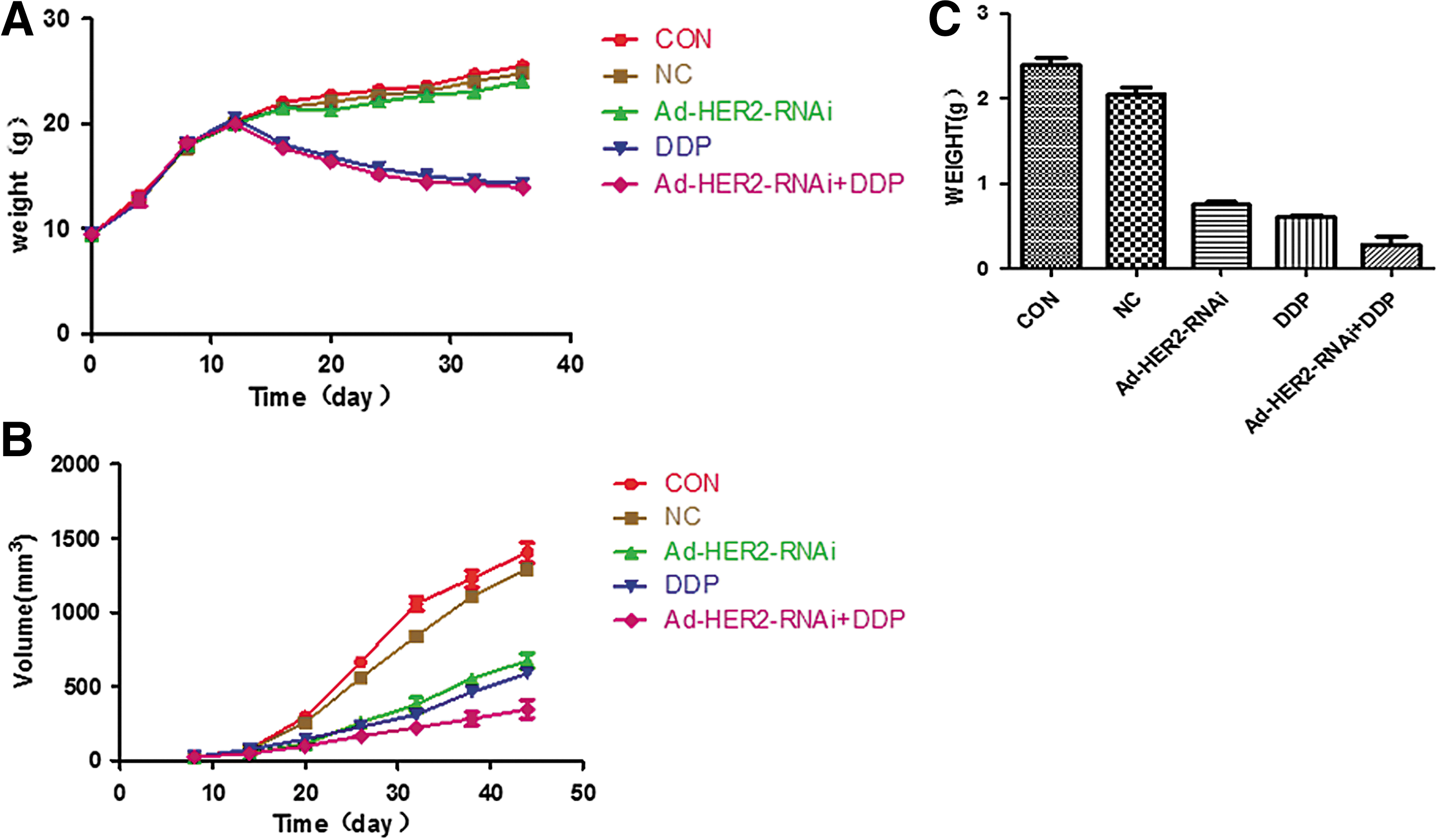
Xenograft therapy experiments.
The results of tumor volume measurement are shown in Figure 3B and Table 1. Starting from the eighth day after implanting subcutaneous tumors, the tumor volume of five groups was measured every 5 d. The results showed that the treatment with DDP, Ad-HER2-siRNA, and Ad-HER2-siRNA+DDP markedly reduced the volume of tumor from 20 d, when compared with CON and NC-treated groups (p < 0.01), and there were significant differences between Ad-HER2-siRNA+DDP group and DDP, Ad-HER2-siRNA groups, respectively (p < 0.01), but no significant difference in tumor volume was observed between Ad-HER2-siRNA and DDP groups (p > 0.05). The results of tumor weight are shown in Figure 3C. Similar with above results, compared with CON and NC groups, DDP, Ad-HER2-siRNA, and Ad-HER2-siRNA+DDP markedly decreased the weight of tumor (p < 0.01), and there were significant differences between Ad-HER2-siRNA+DDP group and DDP, Ad-HER2-siRNA groups, respectively (p < 0.01), but no significant difference in tumor weight was observed between Ad-HER2-siRNA and DDP groups (p > 0.05). Although it showed that Ad-HER2-siRNA was equally effective in reducing tumor volume and weight as the chemotherapy with cisplatin, their synergistic effect implied that Ad can decrease the tumorigenicity and increase the sensitivity of SKOV-3 cells to DDP.
Compared with CON and NC group, the values of p < 0.01 (identified by symbol *); compared with Ad-HER2-siRNA and DDP group, the values of p < 0.01 (identified by symbol #).
Immunohistochemistry for HER2 expression
Tumor sections in CON and NC groups showed higher expression of HER2, while Ad-HER2-siRNA, DDP, and Ad-HER2-siRNA+DDP groups displayed lower level, the Ad-HER2-siRNA+DDP group showed the lowest of all (Fig. 4). HER2 staining intensity correlated well with tumor uptake in all five groups, further supporting the utility of 131I-Herceptin SPECT as a noninvasive assessment of HER2 expression and interference effect in ovarian cancer.
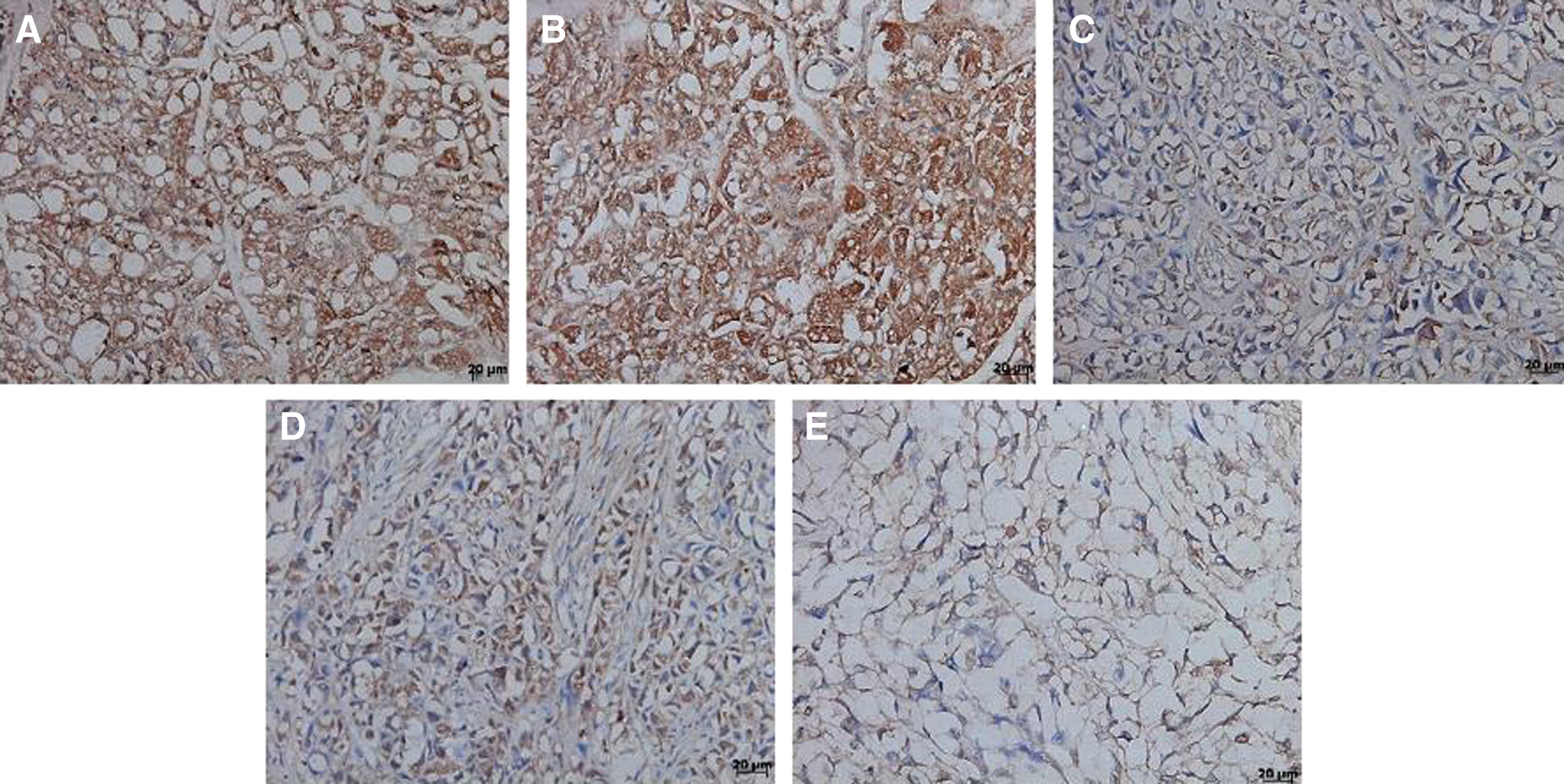
Expression of HER2 protein in tumors of ovarian cancer xenografts from different therapy groups
131I radiolabeling
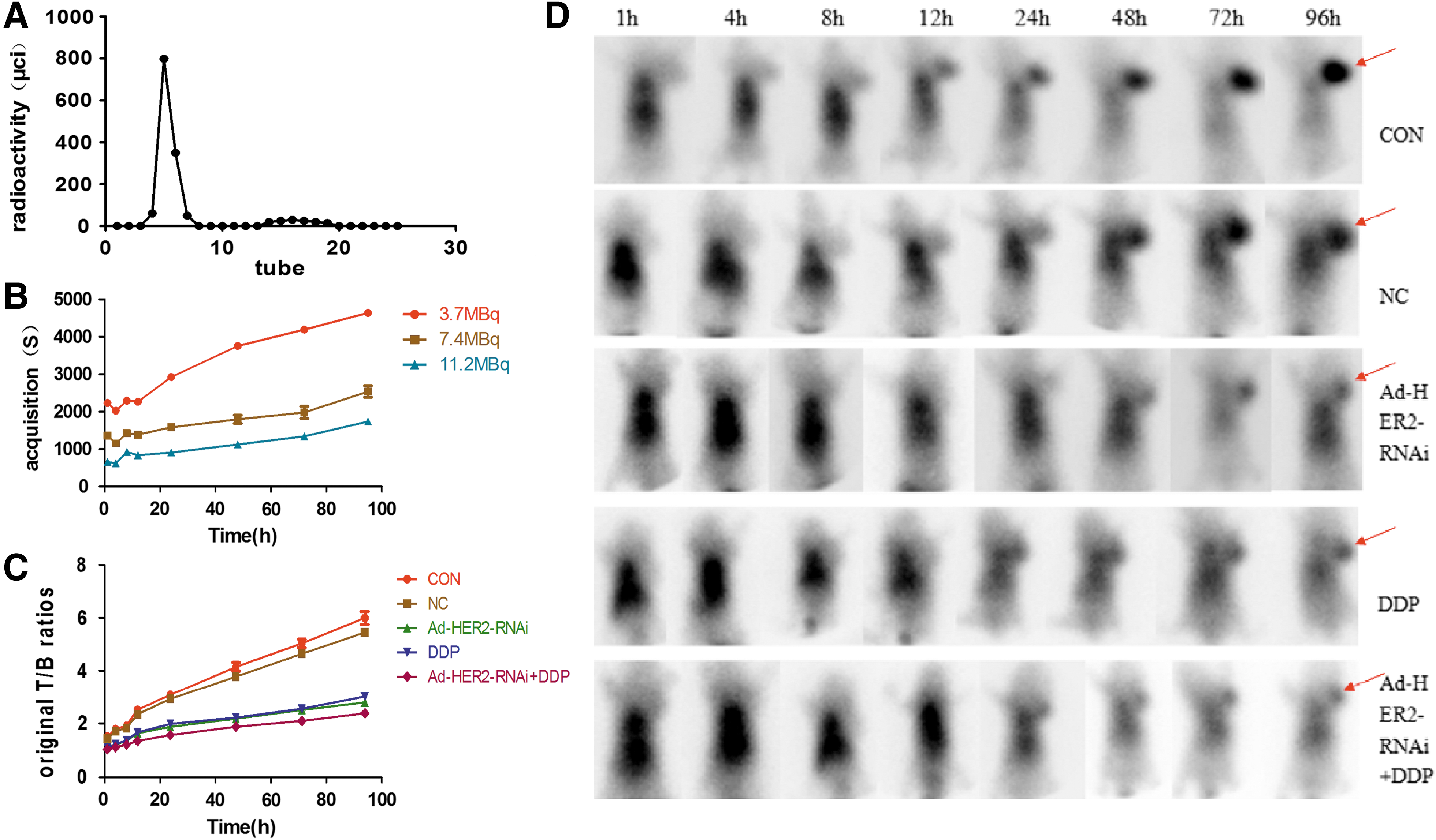
The labeling rate and the radiochemical purity of 131I-Herceptin were (90.32 ± 1.55)% and (97.29 ± 0.87)%, respectively. The specific radioactivity was 3.316 × 10⁻² MBq/μg. The elution curve of Sephadex G50 chromatographic column is shown in Figure 5A.
Radioimmunoimaging study
The Preexperiment of imaging results showed that the acquisition time (Fig. 5B) of three groups was different, the higher the dose, the shorter the acquisition time. When comparing the acquisition time of 7.4 MBq and 11.2 MBq at each time point with 3.7 MBq, the differences were significant (p < 0.01). However, for T/B ratios (Table 2), no significant difference was observed (p > 0.05) in the three groups. The determination of optimal imaging dose called for balancing two ends. On the one hand, the selected dose cannot be too low to compromise image quality; on the other hand, it should not be too high to damage important organs. Hence, the authors selected 7.4 MBq as their optimal dose for the next step of study.
Compared with 3.7 MBq group, the values of p > 0.05 (identified by symbol *)
(T/B), tumor/background.
The T/B ratios (Fig. 5C and Table 3) at each time point in the three treatment groups were lower than those of CON and NC group, respectively (p < 0.01). In addition, comparing the T/B ratios of Ad-HER2-siRNA+DDP group at each point of time with those of Ad-HER2-siRNA and DDP group, the differences were significant (p < 0.05). However, no significant difference was observed between Ad-HER2-siRNA and DDP group (p > 0.05). The tumor mass of nude mice bearing ovarian carcinoma xenografts was observed at 1 h following injection (Fig. 5D). As time went on, the contour of the transplanted tumor became gradually clear. The concentration of radioactivity was obvious compared with the surrounding nontumor tissue, while the high-contrast SPECT imaging could be obtained at 96 h postinjection, especially in CON and NC groups. For Ad-HER2-siRNA, DDP, and Ad-HER2-siRNA+DDP groups, the radioactivity uptake of tumor site was less than other groups using visual analysis, especially in Ad-HER2-siRNA+DDP group.
Compared with CON and NC group, the values of p < 0.01 (identified by symbol★ *); compared with Ad-HER2-siRNA and DDP group, the values of p < 0.05 (identified by symbol▴#).
Discussion
HER2 encodes a transmembrane protein tyrosine kinase receptor and is associated with a poor prognosis and occurrence in ovarian cancer. 18,19 It promotes cell proliferation and opposes apoptosis, and therefore must be tightly regulated to prevent uncontrolled cell growth from occurring. HER2 proteins have been shown to form clusters in cell membranes that may play a role in tumorigenesis. 20,21 The researchers have found that the surface HER2 pool is not static but instead dynamic, undergoing basal endocytosis combined with rapid, efficient recycling. They also found that trastuzumab does not downregulate surface HER2, but instead efficiently recycles the receptor after endocytosis, demonstrating that surface downregulation is not important to its mechanism of action. 22 Trials of HER2-targeted agents in patients with HER2-positive ovarian tumors have demonstrated improvements in both progression-free and overall survival in a small proportion of patients. The treatment of ovarian cancer that overexpresses HER2 remains a challenge in many therapeutic contexts in the clinic. Recently, considerable efforts have been made to find a treatment using gene therapy. For example, siRNA is a valuable therapeutic method due to its crucial role in silencing genes in cells and has attracted much attention. In contrast to targeting the HER2 receptor, HER2 expression can be directly inhibited by siRNA through the RNAi mechanism, where the antisense strand of siRNA binds the complementary sequence of target mRNA and triggers its degradation, inducing knockdown of the specific gene sequence. 23 This makes siRNA a promising therapeutic approach for cancer therapy.
In their previous work, the authors had successfully constructed lentivirus-mediated HER2-siRNA vector and did many related experiments. 14 This is in consistent with what has been reported recently from other investigators, in which siRNA-combined vector systems were demonstrated to be efficient in silencing the gene of interest. 24 However, there are a number of problems of lentivirus that limit its use in clinical gene therapy.
Adenovirus vector, the most popular system for gene delivery, does not integrate itself into the host genome at an appreciable frequency, which eliminates the possibility of insertional mutagenesis. Thus, it is safer from an oncogenic perspective. In this study, the authors successfully packaged the adenovirus-mediated HER2-siRNA vector by AdMax adenovirus package system and tested it for its ability to downregulate HER2 expression by immunohistochemistry (IHC) and xenografts experiments. As shown in Figure 4, HER2 staining intensity of Ad-HER2-siRNA+DDP was the least obvious in five groups. The tumor weight and volume of Ad-HER2-siRNA+DDP were also the least in five groups, which correlated well with the IHC results. Taken together, their results suggested that Ad-HER2-siRNA+DDP may represent a significantly more effective therapeutic tool when compared to Ad-HER2-siRNA or DDP alone. Moreover, it indicated that combined therapy showed a synergistic antitumor effect. Noticeably, to decrease the toxic effect of DDP, further studies of dose reduction are their focus.
The above results also further support the use of 131I-Herceptin SPECT as a noninvasive assessment of HER2 expression and interference effect in ovarian cancer xenografts. Herein, they conducted animal imaging studies. ImmunoSPECT imaging is a relatively new field of research that relies on the innate specificity of antibody toward its corresponding antigen. Molecular imaging of HER2 expression was first reported over 10 years using radiolabeled trastuzumab. Trastuzumab, a HER2-specific monoclonal antibody, is the most widely investigated platform for HER2-targeted imaging tracers. 25 Perik et al. reported that 111Indium-tratuzumab scintigraphy in patients with human epidermal growth factor receptor 2-positive metastatic breast cancer and discovered that radiolabeled trastuzumab scintigraphy was not valuable in predicting trastuzumab-related cardiotoxicity in metastatic breast cancer patients, but it could be used to identify HER2-positive tumors. 26 In 2009, Dijkers et al. did some preclinical studies of 89Zr-trastuzumab and revealed high uptake of radioactivity in HER2-expressing cancers in vivo. 27 Inspired by their studies, they deduced that radiolabeled trastuzumab allowed for noninvasive imaging of HER2 expression in ovarian cancer. However, the real-time monitoring and evaluation of siRNA treatment that targets HER2 in vivo has been a challenge in ovarian cancer. The present study revealed that 131I-trastuzumab is an effective SPECT tracer to specifically and selectively target HER2 expression in subcutaneous models of ovarian cancer.
Conclusion
The results indicated that the combined therapy between Ad-HER2-siRNA and DDP showed an effective treatment in athymic nude mice bearing ovarian carcinoma xenografts. Moreover, the immunoSPECT imaging was a novel way to monitor the progression of ovarian cancer xenografts. Therefore, the focus of the future investigation is to further explore the use of 131I-Herceptin as a potential tracer to evaluate the interference effect of HER2-siRNA in vivo.
Footnotes
Acknowledgment
This research was funded by the following grant: SDFEYGJ1104 from the Second Affiliated Hospital of Soochow University, Suzhou, China.
Disclosure Statement
No competing financial interests exist.