




Abstract
Background and Objective:
Doxorubicin (DOX), despite having antitumor properties, also exhibits cardiotoxicity. Resveratrol has antitumor property for breast cancer cells. 99mTc-MIBI has higher absorption rate in human breast cancer cell line MCF-7. In the present study, the authors intend to investigate the effect of DOX and resveratrol on the absorption of 99mTc-MIBI in breast cancer cell xenografts in mice.
Materials and Methods:
Sixteen xenograft models in nude mice were divided into four groups. Group I (S, control) received 2% DMSO in 0.9% saline, group II (D) 2.5 mg/kg DOX, group III (D + R) 20 mg/kg/d resveratrol with 2.5 mg/kg DOX (total dose of 15 mg/kg in six injections), and group IV (R) 20 mg/kg/d resveratrol for 2 weeks. Single-photon emission computed tomography (SPECT) images were taken for the determination of 99mTc-MIBI absorption. Mice were sacrificed, and the percentage of injected dose per gram (%ID/g) of the heart, liver, tumor, and muscle was measured using a gamma counter. Hematoxylin–eosin staining and Masson's trichrome staining were used for investigation of histopathological changes.
Results:
The %ID/g of tumor was lowest in group D + R. The severity of tumor necrosis or apoptosis was highest in group D + R, but there is no significant difference in pathological injuries and %ID/g of tumor between the group D + R and group D. In addition to the results of the %ID/g, the severity of pathological injuries to the liver and heart cells in group D + R was higher compared with group D. There is a significant difference in the %ID/g of the liver between the group D + R and group D. SPECT images showed that the lowest amount of %ID/g was observed in the tumor of group D + R.
Conclusions:
According to the results of pathology, biodistribution study, and imaging, the combination of DOX and resveratrol has shown higher antitumor effect; hence, 99mTc-MIBI can be used to evaluate their antitumor effect.
Introduction
Breast cancer is one of the prevalent cancer types common among women in recent years worldwide, about 70,000 cases of this cancer are annually diagnosed. The diagnosis and treatment of breast cancer have progressed in recent days. Chemotherapy is one of the most successful areas for cancer treatment. 1,2 Doxorubicin (DOX) is an effective anticancer drug, but a high dose of DOX can cause cardiac complications. 3 DOX is widely used as the first-line treatment for various cancers, such as breast cancer. 4 Cardiac toxicity of DOX is caused due to the heart's innate antioxidant defense. 3
Resveratrol (trans-3,4,5-trihydroxystilbene, RES) is a polyphenolic natural ingredient, which exists in a variety of food sources such as red grapes and peanuts. 5 RES has many medicinal effects, including cardioprotective, antioxidant, anti-inflammatory, and antiplatelet activities. 5,6
Previous studies have reported that RES causes resistance to oxidant injury in new born rat cells (H9c2) by maintaining the mitochondrial integrity, and therefore, it reduces the toxicity of DOX in heart cells. 3 Several in vitro studies have indicated that RES has several anticancer effects, including the ability to prevent the development of malignant tumors. 5 RES can suppress the cell cycle by preventing the expression of tumor-driven enzyme, nitric oxide synthase, which leads to apoptosis in tumor cells. RES is effective in preventing the spread of secondary malignant tumors, which are induced by the mutagenic effects of chemotherapy. 6
RES suppresses several transcription factors, such as AP-1, EGR-1, and NF-I B. Therefore, it has a powerful chemopreventive effect on different types of cancers, such as skin, head and neck, prostate, lung, lymph node, and myeloid cancers. 7 RES can prevent the growth of tumor cells when used at high concentrations alone or in combination with the chemotherapy drugs. 5 According to previous investigations conducted, RES also has antioxidant and anticancer properties that can be used as a supplement in patients treated with DOX. 3
Lipophilic cationic agent, 99mTc-hexakis-2-methoxyisobutylisonitrile or 99mTc-MIBI, is normally used in a myocardial perfusion scan. 99mTc-MIBI is stored in mitochondria and cytoplasm of cells by a simple diffusion process. Malignant breast tumors enhanced the transmembrane potential due to the metabolic demands, which caused an increase in the uptake of 99mTc-MIBI in breast tumors. In vitro studies showed that 99mTc-MIBI have higher absorption in breast cancer cells (MCF-7) compared with other tumor cell lines. 2
Research showed that in addition to antitumor properties, RES can protect the heart, and in combination with DOX, RES exhibits a higher anticancer effect than each drug alone. 8,9 In the present study, the authors intend to investigate the effect of RES and DOX on the absorption of 99mTc-MIBI in MCF-7 xenografts in mice.
Materials and Methods
Drugs and chemicals
DMSO and RES (trans-3,4,5-trihydroxystilbene) were purchased from Sigma-Aldrich (Germany). DOX HCL (Adriamycin® CS; 50 mg/25 mL) was manufactured in Australia. Na99mTcO4 was obtained from a commercial 99Mo/99mTc generator (Heart Center, Fatemeh-Zahra Hospital, Sari, Iran). 99mTc-MIBI (Cardiolite®) was prepared according to the kit instructions (Pars Isotope Company, Iran), and the radiochemical purity was above 95%.
Cell line and culture conditions
The MCF-7, human breast adenocarcinoma cell line, was provided by the National Cell Bank of Iran (NCBI, Amol, Iran). The cells were cultured in the Roswell Park Memorial Institute medium (RPMI-1640, Nuaillé, France), supplemented with 10% (v/v) fetal bovine serum (Gibco, Waltham, MA) and 100 IU/mL penicillin/100 μg/mL streptomycin (Biosera, Sussex, United Kingdom). The cells were incubated at 37°C in a humidified atmosphere containing 5% CO2.
Animals
The nu/b6 nude mice bearing human MCF-7 adenocarcinoma xenografts (Pasteur Institute, Amol, Iran) were used for histopathological, biodistribution, and imaging experiments. Mice were 6- to 8-week-old and weighing 18–20 g. For the induction of tumor xenografts, MCF-7 cells at a concentration of 7 × 10⁶ cells were injected subcutaneously in the right flank of each nude mouse. Tumors were grown for 45 d until reached to approximate diameters of 0.5–0.9 cm. The mice were housed in standard cages, and the room temperature (between 22°C and 26°C) and humidity (between 45% and 70%) were calculated. Standard diet and sterile drinking water were provided for animals.
Experimental design
The mice were divided into four groups. There were four mice in each group, which received the following treatments:
Group I (S, control): 2% DMSO in 0.9% saline was injected intraperitoneally (i.p.) for 2 weeks.
Group II (D): 2.5 mg/kg DOX (for a total dose of 15 mg/kg in six injections) was injected i.p. for 2 weeks. 3,8
Group III (D + R): 20 mg/kg/d RES with 2.5 mg/kg DOX (for a total dose of 15 mg/kg in six injections) were injected i.p. for 2 weeks.
Group IV (R): RES was injected i.p. in a dose of 20 mg/kg/d for 2 weeks. 3,5,8
After taking the drugs for 2 weeks, the mice were transferred to the nuclear medicine department in Sari (Iran). After quality control of radiochemical purity of 99mTc-MIBI, 200 μCi of 99mTc-MIBI was injected through the tail vein (0.1 mL of 99mTc-MIBI was injected intravenously into each mouse). After 1 h, the mice were anesthetized with ketamine–xylazine (0.1 mL of ketamine–xylazine i.p. into each mouse), and single-photon emission computed tomography images were taken to investigate the 99mTc-MIBI absorption in tumor cells. In the next step, the mice were sacrificed, and the heart, liver, muscle, and tumor were excised for analysis. The activity of different organs (uptake) was calculated as the percentage of injected dose per gram (%ID/g) by using a gamma counter.
Imaging of tumor-bearing mice
Imaging study was performed on MCF-7 tumor-bearing mice anesthetized with 0.1 mL of ketamine–xylazine. Images were acquired after 1 h intravenous injection of 99mTc-MIBI (100 μL, 200 μCi) using an E-CAM dual head (Siemens Medical Solutions, Hoffman Estates, IL) equipped with a low-energy high-resolution collimator. Sixty minutes after intravenous injection of 200 μCi 99mTc-MIBI in 100 μL into the tail vein, static planar images were obtained in anterior view by using Siemens E-Cam dual head gamma camera equipped with a low-energy high-resolution collimator. The duration of acquisition was 5 min, the matrix size was 128 × 128, and the pixel size was 1.8 mm. A region of interest was drawn around the tumor, and the left thigh was selected as the reference site for comparison.
Histopathological investigation
Heart, liver, and tumor tissues of mice were carefully removed and fixed in 10% formalin. Then, they were passed through a typical grade of alcohol and xylol for dehydration. Then, the cleaned samples were embedded in paraffin. In the next stage, 4 μm sections of samples were stained with hematoxylin and eosin. To evaluate the histopathological changes, the Masson's trichrome staining was performed. Cardiac fibrosis was determined by measuring the percentage of interstitial fibrosis of painted samples with trichrome by the pathologist under a light microscope. The collagen fibers were stained blue, whereas the muscle fibers were stained red. Morphological alterations, including apoptosis, necrosis, inflammation, hemorrhage, and congestion, were observed under a light microscope.
Statistical analysis
After entering data into the SPSS software, the statistical indicators such as the mean and standard deviation were used for data analysis. The obtained data from the CPM radioactivity was expressed as mean ± SD (%ID/g). Group-to-group comparisons were performed by one-way ANOVA followed by Tukey's post hoc test. The Kruskal–Wallis or Mann–Whitney U tests were also conducted for various group comparisons. The average severity (mean rank) of pathological injuries was calculated and compared using the Kruskal–Wallis test. A p-value of <0.05 was considered to be statistically significant.
Ethics statement
Animal studies were conducted according to standard ethical practice. Mouse experienced no pain during surgery and radiation. Radiation protection principles were considered. This study has been approved by the ethics committee of the Mazandaran University of Medical Sciences with ethic code: IR.MAZUMS. REC.95.2133.
Results
Biodistribution
The rate of 99mTc-MIBI absorption in different organs is shown as mean ± SD of %ID/g. The biodistribution data are presented in Table 1.
Percentage of injected dose per gram of tissue/organ (%ID/g) (mean ± SD, n = 4).
As shown in Figure 1A and Table 1, the %ID/g of breast tumor (MCF-7) was lowest in group D + R. The %ID/g showed the highest rate in group S (control). In addition, the %ID/g was lower in group D + R than group R (p = 0.882). As shown in Figure 1A, the %ID/g of tumor in group S and group R were almost at the same level (10.01 and 7.08, respectively, p = 0.949). The %ID/g of tumor in group R was higher than that in group D (p = 0.026).
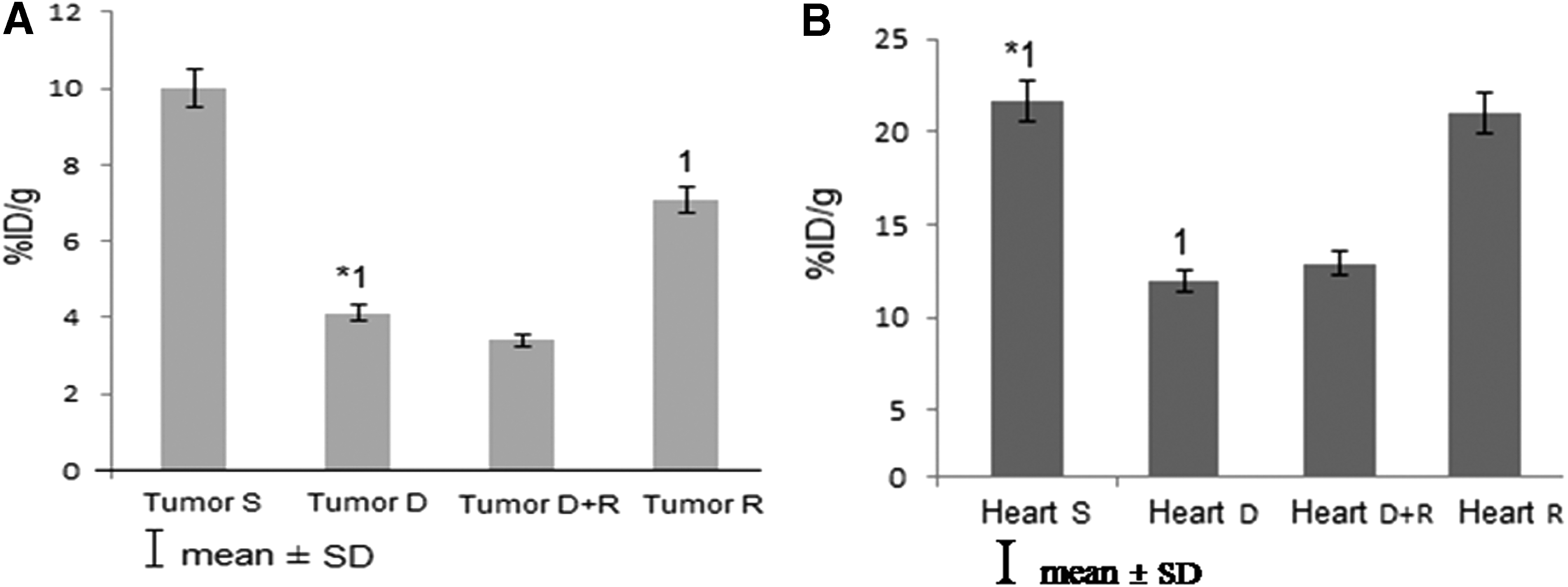
As presented in Table 1, the %ID/g of the liver was the lowest in group D (6.75), and the %ID/g obtained from groups D + R, R, and S were comparatively higher (13, 12.69, and 13.67, respectively). The differences between the two groups, D + R and D, were significant (p = 0.006). The %ID/g of the liver in group R was higher than that in group D, and there was a significant difference between these two groups (p = 0.012).
The lowest %ID/g of the heart was observed in group D, as shown in Figure 1B and Table 1. Also, the rate of %ID/g in group R was higher than that in groups D + R and D. The %ID/g in group R was similar to group S (21 and 21.64, respectively), and there was no statistically significant difference between the group R and group S (p = 0.239). As shown in Figure 1B, there was a noticeable difference between the %ID/g of group D and group S (p = 0.044). This means that the toxic effects of DOX on the heart cells were impressive and important. There was no significant difference between the group D + R and group D (p = 0.46) in the heart.
The %ID/g of muscle was calculated for each group and is shown in Table 1.
Pathology
The authors have defined the severity of pathological injuries (inflammation, congestion, hemorrhage, necrosis, and fibrosis). Thus, if the pathological injury was not observed in an organ, the severity was reported as zero. The numbers 1, 2, and 3, respectively, indicate the mild, moderate, and severe severity for the intended pathological injury. The effects of drugs showed that the severity of tumor necrosis and apoptosis was significantly different in the four groups (p < 0.05). This study showed that the severity of tumor necrosis and apoptosis increased in group D + R compared with group D, whereas the difference was not significant (p > 0.05).
As shown in Figure 2, necrosis was observed in a group treated with D or R, whereas more sever necrosis was observed in the group D + R compared with other groups. RES increased the antitumor effect of DOX in MCF-7 cells. The effects of drugs showed that the severity of liver inflammation, congestion, hemorrhage, necrosis, and fibrosis was significantly different in the four groups (p < 0.05).
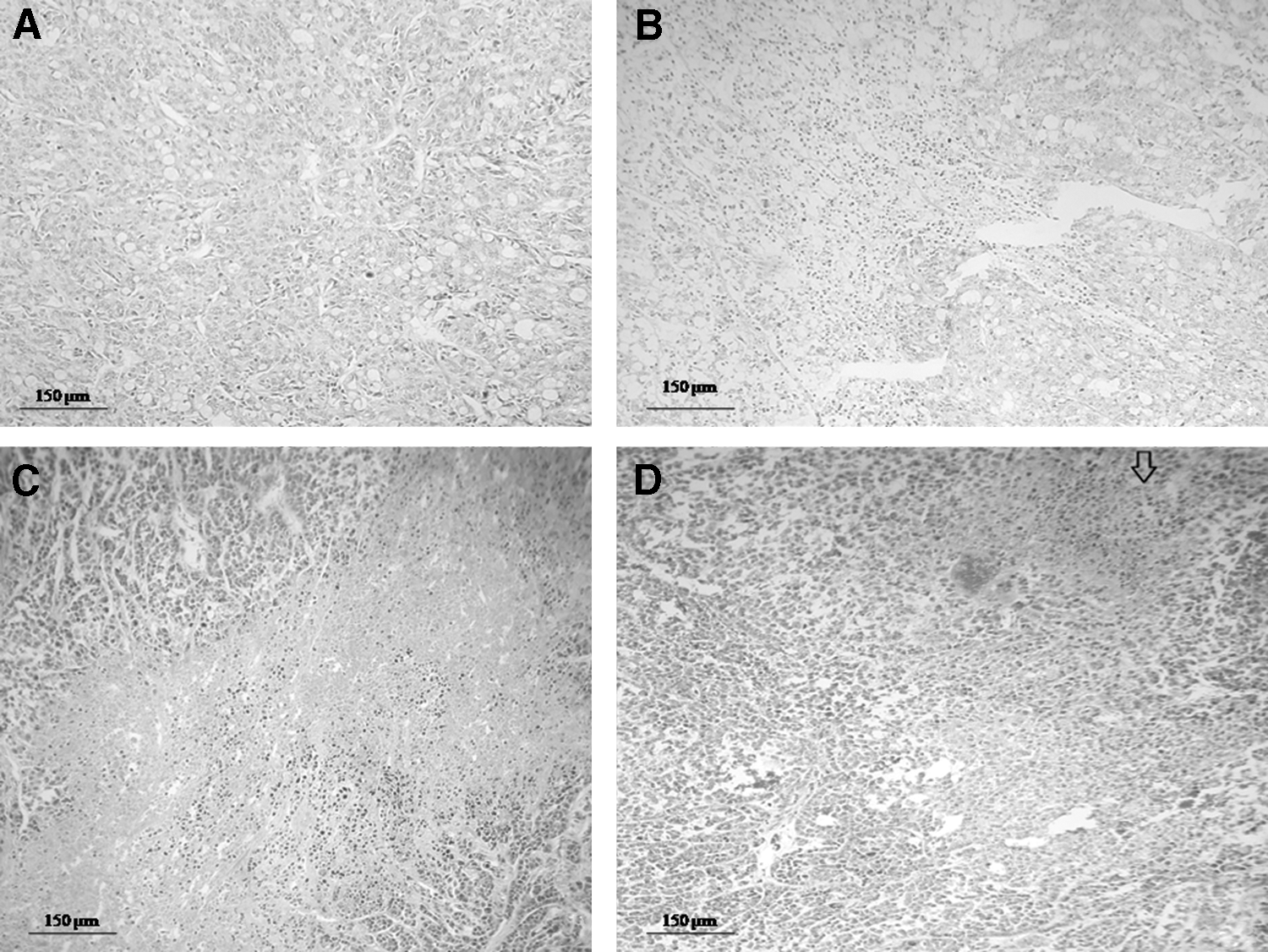
The tumor of mice treated with saline (control) was intact
As shown in Figure 3, the liver cell injury in DOX-treated mice included massive hemorrhage, necrosis, dysplasia, and perivascular fibrosis. The severity of liver congestion, hemorrhage, and necrosis in the group D + R was significantly lower than that in the group D, and the difference was significant (p < 0.05). The effects of drugs showed that the severity of heart inflammation, necrosis, and fibrosis was significantly different in the four groups (p < 0.05).
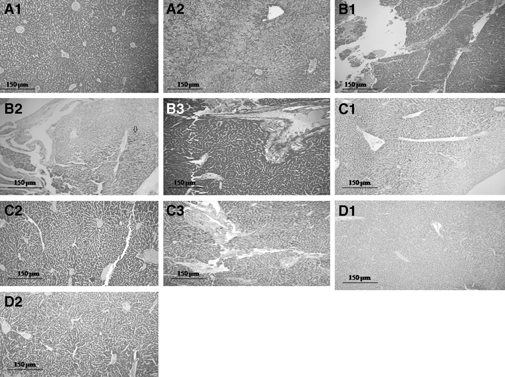
A photomicrograph of control group liver (H&E, × 40. Scale bar = 150 μm) showing normal histology
As shown in Figure 4, the cardiac cell injury in DOX-treated mice included congestion, hemorrhage, and moderate fibrosis. The severity of heart necrosis and fibrosis in the group D + R was significantly lower than that in the group D, and the difference was significant (p < 0.05).
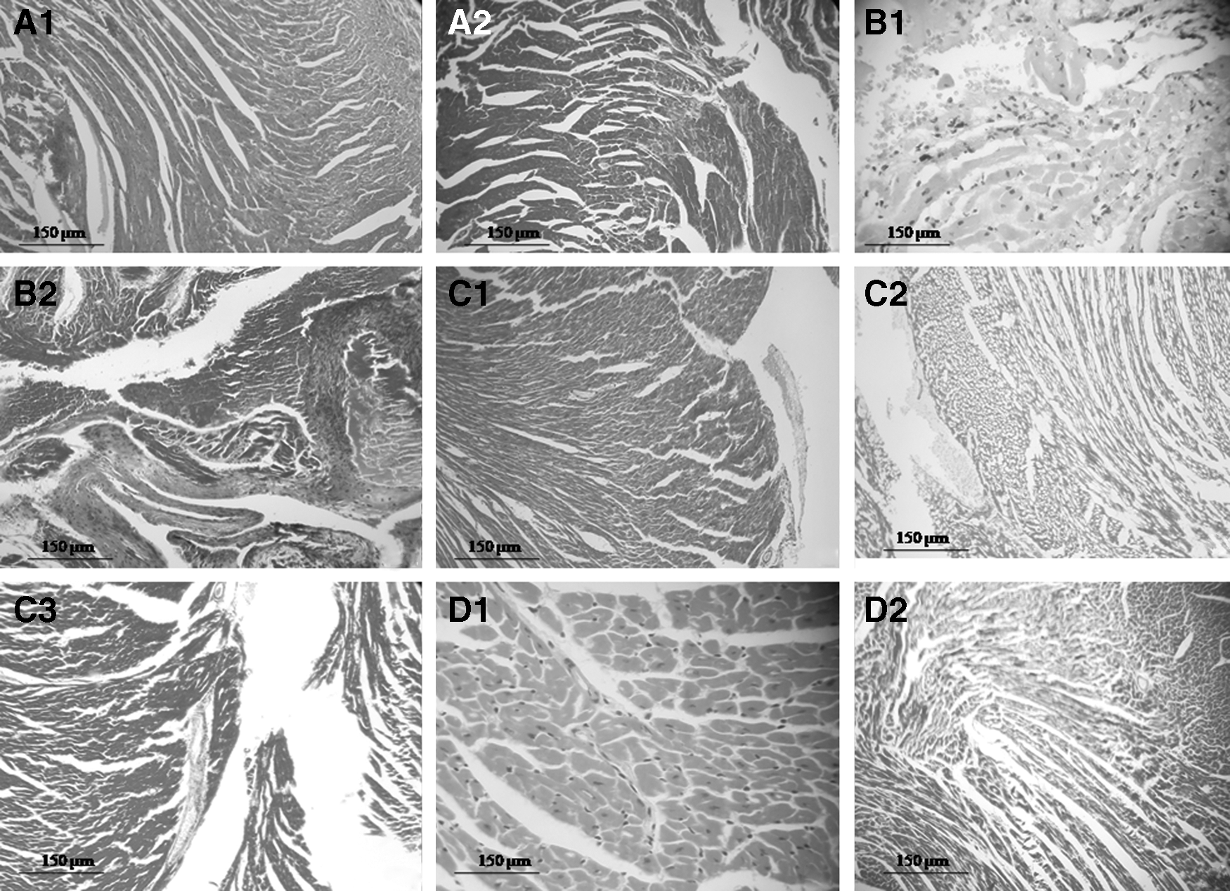
A photomicrograph of S group heart (H&E, × 40. Scale bar = 150 μm) showing normal histology
Imaging of tumor-bearing mice
Static planar images of mice are shown in Figure 5. The quantities are indicated by colors. 99mTc-MIBI is absorbed in the heart and tumor. The signals were also observed in the liver. The tumor-to-muscle ratios of S, D, D + R, and R groups were 7.89, 1.96, 1.72, and 7.66, respectively. The group S showed the highest tumor uptake, and the group D + R showed the lowest tumor uptake.

Planar images of MCF-7-xenografted nude mice 1 h after 99mTc-MIBI injection. Same size ROI was drawn around the tumor (T) and contralateral thigh muscle (M), and then the software automatically summed the amount of counts in both ROIs and calculated the tumor-to-muscle uptake ratio. This ratio only compares the uptake counts between the tumor and the contralateral muscle, and it is not correlated with the amount of the injected drug dosage. Group control
Discussion
RES is a polyphenolic compound extracted from red grape, peanut, and so on. 10 The beneficial effects of RES include anti-inflammatory, cardioprotective, neuroprotective, and antioxidant effects. 11 DOX is a chemotherapy drug with specific antitumor effects on cancer cells. However, due to drastic side-effects and cardiac disorders, high dosage of this drug is not recommended. 3
In fact, chronic cardiac toxicity caused by chemotherapy drugs is the big obstacle in the use of DOX. 12 Cardiac toxicity of DOX can be explained in many ways, such as an increase in cardiac oxidative stress; increase in reactive oxygen species leading to damage in lipid, protein, mitochondria and cardiac cells; and decrease in antioxidant protective mechanisms. In fact, these defensive mechanisms prevent damage to the heart tissue. 8 There are different approaches to decrease the negative cardiovascular effects of DOX, such as using lower dosages of DOX and application in combination with other drugs with less cardiac toxicity. 13
Arafa et al. showed that DOX induces oxidative stress and fibrosis in the heart cells, whereas RES decreases harmful effects of DOX on the heart. This effect was the result of antioxidant, antiapoptotic, anti-inflammatory, and antifibrosis properties of RES. This finding indicated the good protective effects of RES on the heart tissue. 8 RES prevents cells from lipoprotein oxidation, decreases the accumulation of platelets in blood, reduces obstruction of vessels, fights with atherosclerosis, and finally reduces the death toll. These events present the protective effects of RES on cardiac cells. 14
It was clarified that the use of RES with DOX leads to an increase in antioxidant capacity of cardiac cells. Cardiomyopathy leads to apoptosis in cardiac cells. Most medical treatments aim to reduce apoptosis of cardiac cells to prevent maximum damage caused by DOX on the cardiac cell. Research indicates that RES prevented apoptosis of cardiac cells. 8 RES prevents damage to the liver, kidneys, and brain. These tissues may be at risk of inflammatory and oxidative damages. 15
This study shows that the amount of %ID/g of the heart in group D is lower than that in group S. This may be the result of the toxic effect of DOX on the heart cells (Fig. 1B, Table 1). In the findings of this study, there was no significant difference between the group D + R and group D, but the %ID/g of the heart in group D + R is higher than that in group D (12.91 and 12, respectively). Consequently, the toxic effect of group D on the heart was similar to that of group D + R. The amount of %ID/g in group R was similar to group S, so there is no statistically significant difference between the group R and group S. The effects of group S (control) and group R on the heart cells were almost identical, and the group R showed a good cardioprotective effect.
In this study, the amount of %ID/g of the liver obtained from groups D + R, R, and S is higher than that in group D. This shows that the toxic effect of DOX is high on the liver cells, and it minimizes the amount of %ID/g (Table 1). In the group treated with DOX + RES, the level was higher than that in group D. The authors found that the use of the combination of RES and DOX prevents damage to the liver that is caused by DOX. The rate of %ID/g of the liver in the group that received RES is higher than that in the group that received DOX (p = 0.012). It shows that the toxicity of DOX is more on liver cells compared with RES. The rate of %ID/g of the liver in group R was similar to group S (12.69 and 13.67, respectively). Consequently, RES shows a good protective effect on the liver.
In addition to the results of the %ID/g, histopathological examination showed that the severity of pathological injuries on the liver and heart cells in the group D was highest in comparison with other groups, indicating a higher toxicity of DOX on the liver and heart tissues. The severity of pathological injuries on the liver and heart cells in the group R was the lowest and almost the same as in the group S. When RES was added to DOX and injected into mice, the severity of pathological injuries on the liver and heart cells in the group D + R was significantly reduced compared with the group D. This apparent change in photomicrographs was well illustrated (Figs. 3 and 4). Similar to the previous findings, 8,9 the result of pathology and the %ID/g of the liver indicate that by using the combination of DOX and RES, it is possible to prevent liver failure that is caused by sole use of DOX.
Anticancer properties of RES have been studied by various researchers. 11 In 1997, it was suggested the antitumor properties of RES. 16 Several in vitro studies suggested the anticancer effects of RES in breast cancer cell lines. RES prevents the reproduction of MCF-7 cells. It also stops cell cycle and leads to apoptosis in cancer cells. 6,10 Singh et al. showed that α-receptor of estrogen plays an important role in regulating the anti-inflammatory effect of RES. Apoptosis activity of RES was observed in stem-like cells derived from breast tumor by increasing the expression of proapoptosis genes, such as BNIP3 and DAPK2. 16 DOX is a chemotherapy drug that is widely used in breast cancer. In fact, DOX prevents the DNA from cellular cycles in G2 phase that leads to apoptosis. 17
Recent studies have shown that the DOX induces apoptosis in cancer cells. In another study, DOX showed toxic effects on tumors through generating free radicals, inhibiting topoisomerase II enzyme and destroying DNA structure. 4 In a study, it was observed that the combination of RES and DOX exerts chemosensitization effects on MCF-7 cells. 8 It was also reported that the use of DOX and RES prevents the proliferation of MCF-7 cells. 18 These two materials have synergistic antitumor effect in both in vivo and in vitro models. 9
Furthermore, RES destroys DOX-resistant breast cancer cells. 18 As shown in Figure 1A and Table 1, the %ID/g of group D + R was lower than that of group D (3.4 and 4.12, respectively, p = 0.186), suggesting that cytotoxicity of DOX + RES on tumor was similar to DOX alone. As observed in this study, the level of %ID/g of group R is higher than that in group D. The difference between these two groups is statistically significant. This means that the group D has the most damaging effect on the tumor. These results indicate that the combination of DOX + RES had a higher anticancer efficacy in MCF-7 cell line compared with any of these compounds alone. This means that the lower %ID/g of the group D + R shows more tumor necrosis in breast cancer.
Whatever tissue necrosis or tissue damage was more, the amount of radioactivity that is collected from that organ (%ID/g) was less. The %ID/g of tumor was lowest in group D + R. There is no significant difference in %ID/g of tumor between the group D + R and group D. The results of the present study showed that the severity of pathological injuries on tumor cells in the group D was significantly higher than that of the group S. Also, the severity of pathological injuries on tumor cells in the group R was higher than that of the group S. When RES was added to DOX, the antitumor effect increased.
The results of present study showed that DOX + RES has not induced a significant synergistic antitumor effect in MCF-7 cells compared with DOX (p > 0.05), which is consistent with the findings of previous studies. 8,9 In this study, the results of pathology and %ID/g demonstrate that RES can elevate DOX-induced tumor cell apoptosis and necrosis. The combination of RES and DOX could not only enhance the antitumor efficacy but also reduced the cardiotoxicity and hepatotoxicity induced by DOX. Consequently, the combination of RES and DOX can be an effective and important strategy in clinical treatment for cancer.
Due to high uptake of 99mTc-MIBI by the liver, demonstration of hepatotoxicity of a drug only by imaging is difficult, and the main signs of hepatotoxicity in 99mTc-MIBI image are decreased radiotracer uptake by the liver, increased background activity, and delayed radiotracer entrance from hepatobiliary system to the bowels. All these signs correlate with the severity of the liver damage. In this study, the liver damage was evaluated mainly by quantitative study using gamma counter. In this study, no significant difference of some data may be due to low dosages of drugs or number of MCF-7-xenografted nude mice or inaccurate biological distribution of mice tissue. These cases need more studies.
99mTc-MIBI scintigraphy is an effective approach in the detection of breast malignancies and is commonly used for cardiac perfusion scan. Initially, the radiotracer 99mTc-MIBI is trapped within the normal mitochondria on the basis of a highly negative transmembrane electrical potential. 2,19 Many studies have reported that 90% of 99mTc-MIBI is accumulated within the mitochondria of tumor cells.
The rate of uptake depends on tumor progression and angiogenesis, sectional blood flow, enhanced metabolism, and transport through the plasma membrane and mitochondrial membranes. 20 Metabolic demand in tumor cells tends to increase, and therefore, the mitochondrial activity in cancer cells is high. 2,21 Researches indicate that MCF-7 cell line is suitable in an in vitro environment compared with other breast cancer cell lines. Preparation of these cells is easy. Studies show that high absorption of 99mTc-MIBI has been observed in MCF-7 cells after 1 h. 2,22
Imaging of tumor-bearing mice with 99mTc-MIBI revealed tumor uptake at 1 h postinjection (Fig. 5). Apoptosis decreases the mitochondrial transmembrane electrical potential toward cytosol, which decreased the accumulation of 99mTc-MIBI in cells. 2 The results of this study show that 99mTc-MIBI uptake in MCF-7 cells (Fig. 5) is significantly reduced in group D + R. It represents more tumor apoptosis in group D + R compared with other groups.
Conclusions
The results of pathology and biodistribution showed that 99mTc-MIBI can be used to evaluate the antitumor effect of DOX + RES and its protective effect on the liver. The results of nuclear imaging with 99mTc-MIBI confirm the synergistic antitumor effects of DOX + RES. It can be concluded that the use of nuclear imaging with 99mTc-MIBI is helpful merely to show antitumor property of DOX and RES.
Footnotes
Acknowledgments
This work was the subject of the thesis of Fereshteh Hallajian as an MSc student of the Mazandaran University of Medical Sciences and was supported with grant number 2133. The authors also appreciate Dr. Reza Ali Mohammadpour (Biostatistician, Faculty of Health, Mazandaran University of Medical Sciences, Sari, Iran) for his sincere contribution to statistical analysis.
Disclosure Statement
No competing financial interests exist.