
Abstract
Objective:
This article describes the preparation of a 170Tm source by chemical deposition technique, its encapsulation in a titanium holder, and preliminary quality evaluation for potential utility as a brachytherapy source.
Methods:
The procedure consisted of electrodeposition of Ni on a Cu wire followed by chemical deposition of 170Tm on it. Influence of feed solution pH, carrier Tm concentration, and reaction time were studied for optimum deposition of 170Tm on substrate. After sealing the source core in a titanium capsule, quality control tests were performed. Distribution of 170Tm on substrate was evaluated by autoradiography. Inactive Tm source was characterized by scanning electron microscope (SEM) and energy-dispersive X-ray (EDS) analyses.
Results:
Under optimized conditions (pH 5, 10 μg Tm carrier, 5 h), 170Tm source core could be prepared by deposition of >95% of 170Tm radioactivity on substrate. Swipe tests and immersion tests on encapsulated sources confirmed that removable radioactivity and radioactivity leakage levels were within stipulated limits. Autoradiography of 170Tm source confirmed uniformity of radioactivity distribution. While SEM analysis confirmed good adhesion of Tm on substrate, EDS analysis confirmed elemental constituents of the Tm-deposited substrate.
Conclusion:
The objective of preparing a 170Tm source by chemical deposition for potential brachytherapy applications could be successfully achieved.
Introduction
Brachytherapy using sealed radioactive sources is one of the principal treatment modalities for patients with small, well-localized tumors. Leveraging the technological advancements in radioactive source preparation techniques, image-guided delivery systems and radiation treatment planning techniques, modern brachytherapy is able to deliver precise doses of radiation to the tumor, achieving successful outcomes. Availability of sources containing radioisotopes, such as 192Ir, 60Co, 137Cs, 125I, 106Ru, 103Pd, and 131Cs with diverse nuclear decay characteristics has expanded the scope of brachytherapy for cancer treatment. Brachytherapy treatment can be either low dose rate (LDR) or high dose rate (HDR), which differ from each other by the intensity of the radiation source and its dimensions, radiation dose delivered and the time of contact with the tumor.
By virtue of its nuclear decay characteristics and cost-effective availability, 192Ir has emerged as the radioisotope of choice for HDR brachytherapy, 1,2 whereas low-energy photon emitters, such as 125I (T ½ = 59.4 d, E γ = 35 keV), 103Pd (T ½ = 17 d, E γ = 21 keV), and 131Cs (T ½ = 9.7 d, E γ = 30 keV) remain the mainstay of LDR brachytherapy. 3 –7 Over the years, radioactive sources based on 57Co (T ½ = 271.74 d, E γ = 122.1 keV [85.6%], 136.5 keV [10.7%]), 153Gd (T ½ = 242 d, E γ = 97.4 keV [29%] and 103.2 keV [21%]), 169Yb [T ½ = 32.0 d, E γ = 63.1 keV [43.6%], 109.8 keV [17.4%], 130.5 keV [11.4%], 177.2 keV [22.3%], 198 keV [35.9%], and 307.7 keV [11.1%]), 75Se (T ½ = 119.8 d, E γ = 121.1 keV [17.2%], 136 keV [58.5%], 264.7 keV [58.9%], 279.5 keV [25%], and 400.7 keV [11.4%]), and 170Tm [T ½ = 128.6 d, E γ = 84.25 keV (2.5%)] have been explored as potential alternatives to the established ones for use in brachytherapy. 8 –18 While majority of reports on these sources address the dosimetric aspects, hardly any technical information is available on their fabrication. While the nuclear decay characteristics of 57Co are suitable for brachytherapy applications, its availability at exorbitant costs is a major impediment for its clinical use.
Among the potential radionuclides, 170Tm is promising owing to its nuclear decay characteristics. It has a half-life of 128.6 d and decays predominantly by β− emission (E βmax 968 keV) to 170Yb emitting γ photons. A number of X rays are also emitted during the decay process. 19 –21 Cost-effective production of 170Tm in adequate specific activity is feasible by the neutron activation of 169Tm target (100% abundance) owing to its appreciably high-thermal neutron capture cross-section (σ = 103 b). 22,23 Detailed literature survey has revealed that there are only few publications on the preparation of 170Tm sources. 19 –21 In one article, Levinger and Shani reported the fabrication of 170Tm seeds by the neutron irradiation of Tm wire (100% in 169Tm) in the nuclear reactor. 19 Ayoub and Shani also used the same procedure for fabricating a 170Tm source. 20 Tm-170 source was also produced by neutron irradiation of 169Tm(NO3)3·5H2O powder taken in a Ti capsule. 21 However, if the patient requires multiple sources for therapy, multiple capsules containing Tm wire/powder need to be irradiated in the reactor, which can be a limitation for large batch sizes. Moreover, any unscheduled reactor shutdown would affect the radioactivity of the source. Hence, the authors aimed to standardize a simple procedure for the batch production of LDR 170Tm sources as the required radioactivity levels are much less in comparison to that for HDR sources. Toward this aim, chemical deposition of 170Tm on a solid matrix was pursued owing to the ability to obtain homogeneous adherent deposit of the radioisotope on the substrate.
There are literature reports on the chemical deposition of rare earths on the surface of metals such as Al, Zn, Fe as well as on alloys such as stainless steel by immersing the metal in an aqueous rare earth salt solution wherein the rare earth metal forms a thin oxide/hydroxide layer on the metal surface. 24 In similar lines, Kobayashi and Fujiwara had reported the chemical deposition of cerium on the surface of a Ni-electroplated Cu substrate by immersing it in aqueous solution of cerium ions. 25 The authors had employed this technique for the preparation of 147Pm source in which Ni was electrodeposited on the surface of a copper wire followed by the chemical deposition of 147Pm from its aqueous solution. 26 Herein, they report the utility of chemical deposition technique to prepare 170Tm source, hermetic sealing of the source core in a Ti capsule and preliminary quality evaluation of the sealed source to confirm compliance with the regulatory requirements.
Experimental
Materials and instruments
Tm-170 was produced by the neutron irradiation of natural Tm2O3 target (100% abundant in 169Tm) in the Dhruva nuclear reactor at a thermal neutron flux of 5.9 × 10 13 n·cm−2·s−1 for 91 d followed by radiochemical processing at Radiopharmaceuticals Division, Bhabha Atomic Research Center (BARC). 22,23 Boric acid, nickel sulfate, sulfuric acid, and ammonium hydroxide of spectroscopic grade were procured from BDH (Mumbai, India). Thulium nitrate pentahydrate (99.9% pure) used as the carrier was purchased from Sigma-Aldrich (India). Copper wires and platinum wires of high purity with material testing certificates were procured from M/s. Hindustan Platinum Ltd. (Mumbai, India). Polystyrene granules (>95% purity) were procured from BDH.
Electrodeposition of nickel on the Cu wire was carried out using a 1–28 V, 0–2.5 A regulated power supply procured from Aplab Ltd. (India). Titanium capsules [0.8 mm optical density [OD] × 4.75 mm (l)] were fabricated at the Center for Design & Manufacture of their institute. A 50 W Nd:YAG laser welding system (M/s. Quanta Systems, Italy) was used for welding the titanium capsules containing 170Tm sources. Microstructural features of the inactive Tm-deposited substrates were analyzed using a Scanning Electron Microscope (SEM) (Model AIS-2100; SERON). Energy Dispersive X-ray (EDS) Microanalysis Technique (Model INCA E350; Oxford) was used to identify the elemental constituents of the samples. A well-type NaI(Tl) scintillation counter (ECIL, Hyderabad) was used for radioactivity counting. Industrial X-ray films were obtained from Agfa India Pvt. Ltd. (Mumbai). Optical Density (OD) measurements of the exposed X-ray films were carried out using an OPTEL Transmission densitometer Model No. 125. 3E NDT, LLC. (Pasadena, CA).
Electrodeposition of Ni on Cu substrate
The electrodeposition of Ni on the Cu wire was carried out following a method, which was standardized in our laboratory previously. 26 A Pt strip and a Cu wire (0.5 mm diameter and 5 cm length) used as the anode and cathode, respectively, were connected to a DC power supply. The surface of Cu wire was extensively cleaned so as to obtain a rough finish followed by rinsing with deionized water and air drying. It was then placed vertically in boric acid bath (30 mg·mL−1) at pH 2–3 containing 50 μg of carrier NiSO4·7H2O. Nickel was electrodeposited on the Cu wire cathode galvanostatically at a constant current density of 6 mA·cm−2 for 2 h at ambient temperature. Subsequently, the Ni-coated Cu wire was washed with deionized water followed by ethanol and was air dried. It was then cut into the required dimensions and kept sealed in vials until use.
Optimization of chemical deposition of 170Tm
To study the influence of various experimental parameters on the chemical deposition of 170Tm on Ni-coated Cu wires, a series of experiments were performed by immersing the wires in 0.2 mL of the feed solution. The feed solution comprised of HPLC-grade water containing 1.85 MBq (50 μCi) of 170TmCl3 as tracer and appropriate amount of nonradioactive 169Tm(NO3)3·5H2O as carrier. After the incubation period, the wires were removed from the solution using tweezers and washed with deionized water to remove nonadherent 170Tm.
The influence of pH of the feed solution on the uptake of 170Tm was studied by varying its pH from 1 to 7 (10 μg carrier Tm for 5 h at ambient temperature), wherein the solution pH was adjusted appropriately using dil. HCl or dil. NaOH solutions. To determine the maximum 170Tm radioactivity, which can be chemically deposited on the substrate, carrier Tm-added studies were carried out. In these experiments, carrier Tm concentration was varied from 1 to 100 μg at the optimized pH 5 for 5 h at ambient temperature and the % 170Tm deposited was determined. Finally, the influence of incubation time on the deposition of 170Tm was studied by varying the reaction time from 30 min to 6 h (at pH 5 using 10 μg Tm carrier concentration at ambient temperature). An aliquot of the feed solution (20 μL) was counted in a well-type NaI(Tl) counter before and after the experiment, from which the percentage of 170Tm deposited on the Ni-coated Cu wire was calculated using the equation
wherein A i and A f are the initial and final radioactivity counts in the feed solution. Reproducibility of the results was ascertained by performing each experiment in triplicates.
Preparation of 170Tm source
Thulium-170 source was prepared based on the optimized reaction conditions by placing one Ni-coated Cu wire [3 mm(l) and 0.5 mm(φ)] in the feed solution (0.2 mL) containing 170TmCl3 (37–60 MBq) and 10 μg of nonradioactive Tm carrier for 5 h at ambient temperature. Subsequently, the 170Tm-deposited wire was taken out of the solution with a tweezer, washed with water, and air dried. Percentage of Tm deposited on the substrate was indirectly determined by counting suitably diluted aliquots of feed solution before and after the deposition experiment as described above. Direct measurement of the radioactivity content of the sealed 170Tm sources was also carried out using a precalibrated well-type ionization chamber.
Leachability of 170Tm source core
The 170Tm source core (∼37 MBq) was immersed in a beaker containing 100 mL of saline at ambient temperature for 48 h, after which an aliquot of the saline solution was counted on a well-type NaI(Tl) scintillation counter. Based on the counts obtained, total radioactivity released in saline was estimated. 27,28
Polystyrene coating and testing for leachability
The source core was coated with a thin layer of polystyrene by immersing it into a solution of polystyrene in chloroform (150 mg/mL) for a minute, after which it was taken out. The polystyrene-coated source core was placed on a perforated tray so that the excess of the polymer solution was drained out, after which the source core was air dried. The leachability of radioactivity from the polystyrene-coated source in saline was then tested following the standard procedure described above.
Source encapsulation in a titanium holder
A cylindrical titanium capsule [0.8 mm (φ) × 4.75 mm (l) and 0.05 mm thickness] closed at one end with a cap to fit at the open end was designed to encapsulate the 170Tm source core. The cap was sealed to the main capsule by laser welding on a 50 W Nd:YAG laser welding system as per a procedure previously standardized for 125I brachytherapy sources. 27,29 Each 170Tm source was inserted individually into a capsule through its open end, which was then closed with the cap. The laser beam was focused to the junction of titanium tube and cap by an optical fiber-based delivery system. All laser parameters were computer controlled. A camera with a separate fiber-based lighting arrangement was used to focus the laser beam to the precise area and to view the welding region. The sealed sources were ultrasonically cleaned with ethanol to remove any traces of contamination.
Quality control of encapsulated 170Tm sources
Swipe test
The encapsulated 170Tm sources were tested for surface contamination by swiping their surface with a swab moistened with ethanol and the swab was counted in a well-type NaI(Tl) scintillation counter.
Leakage tests
To ascertain leak-tightness of the titanium capsule, the encapsulated 170Tm sources were subjected to the Bubble test and Immersion test, which are described below. 30,31
Bubble test
The sealed 170Tm sources were placed in nearly boiling water (10 mL) kept in a glass beaker for about 2 min. Appearance of any bubbles was observed.
Immersion test
Each encapsulated source was immersed in 25 mL of water taken in a glass beaker and heated to 50°C for 5 h in a water bath. Subsequently, the source was removed, water in the glass beaker was concentrated to 1 mL, and the radioactivity released was counted in a well-type NaI(Tl) counter.
Assessment of uniformity of radioactivity distribution
Distribution of 170Tm on the surface of the Ni-coated Cu wire was assessed by performing autoradiography studies with the encapsulated source using a custom-made cylindrically shaped brass gadget [44 mm (φ) × 14 mm (l)] with a hole at the center to hold a 2-mm (φ) glass tube containing the 170Tm source (∼37 MBq). Eight holes of 3 mm (φ) each were drilled at an angular spacing of 45° along the circumference of the gadget. 170Tm source taken in a glass tube was mounted at the center of the gadget. An industrial radiographic X-ray film was then wrapped along the circumference of the gadget. The film gets exposed to the radiations from 170Tm at eight equidistant directions through the holes. OD of the X-ray film after exposure to 170Tm source was measured at eight different points of the film for an optimized duration of 15 min using a black and white transmission optical densitometer.
SEM and EDS analyses
SEM and EDS analyses were carried out using a dummy source prepared using nonradioactive 169Tm replicating the procedure used for the fabrication of the radioactive source. SEM analysis of a Ni-coated Cu wire was also carried out for comparison. The samples were also subjected to EDS microanalysis to identify their elemental constituents.
Results
The aim of this study was to assess the utility of chemical deposition technique to fabricate 170Tm sources of acceptable quality, encapsulation of source core in a Ti capsule, and preliminary quality evaluation of the sealed sources.
Electrodeposition of Ni on Cu substrate
Under the experimental conditions, which were optimized previously in their laboratory, 26 Ni could be successfully electrodeposited on to the surface of the Cu wire.
Optimization of 170Tm deposition parameters
To deposit the required amount of 170Tm on the Ni-coated Cu wire as well as to ensure maximum deposition of Tm on the substrate, various parameters, such as pH of the feed solution, concentration of Tm, and time duration of the reaction, were optimized.
Figure 1 depicts the influence of pH of feed solution on the deposition of 170Tm on substrate. It is evident that the deposition of 170Tm is significantly influenced by the pH of the feed solution and shows an increasing trend with pH up to pH 5. Any further increase in solution pH reduced the deposition of 170Tm on the substrate, which could be attributed to the possible formation of hydroxides of Tm in solution. At pH 5, >95% of the initial 170Tm activity in the feed solution could be deposited on the Ni-coated Cu wire. Experiments carried out to determine the influence of concentration of carrier Tm in the feed on its deposition revealed that >99% of 170Tm could be deposited in the absence of Tm carrier, whereas about 95% of the initial 170Tm radioactivity in the feed solution could be deposited on the Ni-coated Cu wire when up to 10 μg of Tm carrier was added (Fig. 2). Dependence of the percentage deposition of 170Tm on reaction time is shown in Figure 3. It is evident that deposition of 170Tm on the Ni-coated Cu surface increased with increasing contact time and reached a maximum after about 5–6 h of contact. This is characteristic of chemical deposition process governed by ionic diffusion, which in turn depends on the reaction kinetics and mass transport. With increase in contact time, more and more 170Tm ions overcome the energy barrier and diffuse onto the Ni surface resulting in increase in percentage deposition. Once the surface is saturated with Tm species, percentage deposition remained constant with further increase in contact time.
Influence of pH on the deposition of 170Tm on Ni-coated Cu wire.
Influence of carrier Tm concentration on the deposition of 170Tm on Ni-coated Cu wire.
Influence of incubation time on the deposition of 170Tm on Ni-coated Cu wire.
Preparation of 170Tm source
As per the optimized procedure, 170Tm source cores were prepared by keeping the Ni-coated Cu wire in the feed solution containing 170Tm radioactivity (∼37–60 MBq) and 10 μg of Tm carrier at pH 5 for 5 h at ambient temperature. Activity content of each source was measured as per the procedures described in the Experimental section. It was observed that the efficiency of 170Tm deposition was greater than 95% under the optimized conditions.
Leachability of 170Tm source core
After the deposition of 170Tm radioactivity on the substrate, leachability of source core was tested by keeping it in 100 mL of saline for 48 h. Subsequently, an aliquot of the saline solution was counted to check leaching of radioactivity. Leaching studies conducted on four randomly selected source cores revealed that about 0.2%–0.3% of the original radioactivity of the source leached out (Table 1), which is more than the limit (<0.01% of original activity) stipulated by the Atomic Energy Regulatory Board (AERB). 29
Quality Control of 170Tm Source
Polystyrene coating and testing for leachability
The leaching of 170Tm from the source core was retarded by coating the surface with a thin layer of polymer. The polymer coating was very thin so as to keep the source core dimensions. Among the various polymers, polystyrene was selected owing to its excellent solubility in a wide range of organic solvents and amenability for coating. Thickness of the coating could be attuned by adjusting the concentration of the polymer solution. A concentration of 150 mg of polystyrene per mL of chloroform was found to be adequate to form a barrier layer sufficient to prevent the leaching of 170Tm to an acceptable level without significantly altering the dimensions of the source core. The polymer solution initially attaches to the radioactive surface of the matrix and consecutively coalesces into a continuous protective film. Table 1 shows the results of leachability tests of four source cores after polymer coating. It is evident that the polystyrene layer on the source core significantly reduced the radioactivity leached out to below 0.01% as stipulated by the regulatory agency. 29
Encapsulation of source core
The need for safety during the use of 170Tm source necessitated its encapsulation in a Ti holder. Cylindrical capsule design used for the encapsulation of 125I brachytherapy sources was chosen owing to its proven effectiveness. 27,29 The wall thickness of the Ti capsule (0.05 mm) was adequate to provide mechanical strength to the source core while concurrently delivering the required radiation output for therapy. Due to the small wall thickness and the highly oxidizing nature of titanium, laser welding technique was chosen for encapsulation as it causes minimal distortion to the capsule while giving an aesthetic weld. Laser welding parameters, such as frequency of laser pulse, its duration, and the rotational speed of sample to be welded optimized earlier for the fabrication of 125I brachytherapy sources, were used to achieve the desired degree of hermeticity. Each source was welded with strict adherence to the optimized protocol to preclude the rejection of sources during QC testing. Figure 4 is the sketch of the encapsulated 170Tm source assembly. Figure 5a is the photograph of the 170Tm source core, whereas Figure 5b is the photograph of the encapsulated 170Tm source.
Sketch of the 170Tm source assembly.

Quality control of the encapsulated sources
The encapsulated 170Tm sources were subjected to preliminary quality evaluation to ensure their compliance with the regulations.
Swipe test
Results of the swipe test carried out on four sources revealed that the removable counts were less than 185 Bq per source, which comply with the specifications laid down by the regulatory agency (Table 1).
Leakage tests
Bubble test
In the bubble test, no bubbles were observed in the near boiling water confirming that the sources were leak tight.
Immersion test
Results of the immersion test are shown in Table 1. Leakage of radioactivity from the sealed sources was less than 185 Bq, which is the upper limit for acceptance of the source for leak tightness.
Autoradiography
To establish uniformity of distribution of 170Tm radioactivity on the sources, the encapsulated sources were subjected to autoradiography examination. Figure 6a is the picture of the autoradiography gadget, whereas Figure 6b is a typical autoradiograph of a sealed 170Tm source. Table 2 depicts the OD values at eight equidistant positions of the exposed radiographic film after 15 min of exposure to the 170Tm source (37 MBq). The variation in OD values between the positions was less than 5% confirming that 170Tm radioactivity was uniformly distributed on the Ni-coated Cu wire.

Optical Density of 170Tm Source
SEM and EDS analyses
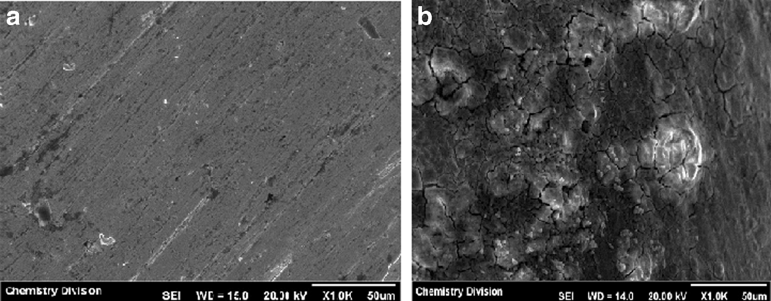
Figure 7a and b represent the morphology of Ni-electrodeposited Cu wire and Tm deposited on the Ni-coated Cu wire, respectively, as seen through the SEM. SEM micrograph of Ni-electrodeposited Cu wire indicates that a very compact layer of Ni is homogeneously distributed on the copper substrate. The SEM micrograph of Tm deposited on Ni-coated Cu wire (Fig. 7b) indicates morphological change after Tm deposition, in which aggregates are distributed throughout the surface of the substrate.
EDS spectra of Ni-electrodeposited Cu wire and Tm deposited on the Ni-coated Cu wire are shown in Figure 8a and b, respectively. Figure 8a has peaks corresponding to Cu and Ni confirming the electrodeposition of Ni on Cu. The EDS spectra of Tm adsorbed on the Ni-coated Cu wire (Fig. 8b) contains peaks corresponding to Tm in addition to that of Ni and Cu confirming the deposition of Tm on the Ni-coated Cu substrate under the optimized experimental conditions. Table 3 represents the percentage elemental composition of the Tm-deposited substrate.

Elemental Composition of Inactive Tm Source by Energy-Dispersive X-Ray
Discussion
While modern brachytherapy brought a paradigm shift in the treatment of several cancers, cost-effective availability of radioactive sources is a key determinant of its success and sustainability. Preparation of miniature radioactive sources is highly challenging owing to the necessity to deposit a required level of radioactivity on a predefined area of substrate followed by its encapsulation with highest possible degree of leak tightness to ensure patient safety during therapy. Owing to these challenging technical issues, supply of brachytherapy sources is restricted to a few commercial producers despite huge clinical demand.
The recent surge of interest in LDR brachytherapy sources containing radionuclides, which emit 40–100 keV photons having a half-life of about 100 d, were the motivating factors to prepare LDR 170Tm sources by chemical deposition. The benefits of 170Tm source for LDR brachytherapy are manyfold. The relatively longer half-life of 170Tm not only offers sufficient time for source fabrication and quality control, but it also endows longer shelf life to the source eliminating the need for frequent source replacement. Moreover, it provides logistical advantages for shipment to hospitals distant from the source production facility as the decay loss is minimal. As 170Tm emits photons having longer range compared with those emitted by 125I source, 170Tm sources may be useful for treating relatively large prostate glands using fewer number of sources. Due to the emission of β− particles, their use as β ray sources for the treatment of tumors can also be envisaged. 19
Chemical deposition of cerium on a Ni-electroplated Cu substrate reported previously by other researchers formed the scientific basis of this work. 25 Although they have not studied the mechanism of deposition of 170Tm, the mechanism of chemical deposition of rare earths, such as cerium on metal surfaces has been reported previously. 24 An electrochemical mechanism is proposed wherein the metal surface is said to be consisting of a large number of electrochemical cells, wherein cathodic reactions and anodic reactions (metal dissolution) take place. The cathodic reactions (O2 + 2H2O + 4e− ⇒ 4OH−) generate hydroxide ions resulting in alkaline conditions in the vicinity leading to the deposition of rare earth ions (which are present in the aqueous solution in contact with the metal surface) as hydroxide/oxide on the metal surface. In their work on the deposition of cerium on a Ni-coated Cu wire, Kobayashi and Fujiwara gave conclusive evidence for the presence of Ce(IV) oxide as the principal species by XPS analysis. 25 The deposition of Tm on the Ni-coated Cu substrate is also expected to follow the same mechanism.
While the chemical deposition of 170Tm on electrodeposited Ni surface is an extension of their previous work on 147Pm sources, influence of various experimental parameters on the deposition of Tm on the Ni-coated Cu substrate was thoroughly investigated to arrive at the optimum conditions for source preparation. Polymer coating of the source core followed by its encapsulation prevented the leaching out of radioactivity. For the encapsulation of the source, Titanium capsule design and laser welding technique standardized in their laboratory previously for 125I brachytherapy source were chosen given their demonstrated effectiveness. The authors have not yet carried out any dosimetric studies to determine the extent of β attenuation by the titanium capsule. Enger et al. have studied the influence of encapsulating materials, such as stainless steel, titanium, and platinum on the energy spectrum of the photons and electrons emitted from the 170Tm source. 15 Based on the thickness of the encapsulating material, dimensions and radioactivity of the source, the energy spectrum of the photons, and electrons coming out of the encapsulated source would differ.
This study is a preliminary feasibility study on the preparation of 170Tm sources by chemical deposition for possible applications in brachytherapy. Further work, including extensive dosimetric studies, bioevaluation studies in animals to assess any release of 170Tm radioactivity, and migration of source during therapy, technology development to undertake large-scale production of sources with high radioactivity content, and extensive quality evaluation as per the established national and international protocols for brachytherapy sources need to be addressed for actual clinical applications.
Conclusions
As part of the authors' efforts to develop novel radioactive sources for medical applications, potential utility of chemical deposition to prepare a 170Tm source core, its encapsulation in tiny titanium capsules, and its preliminary quality evaluation could be successfully demonstrated. Further detailed studies, including dosimetric and bioevaluation studies, would unravel the potential of the 170Tm sources for future brachytherapy applications.
Footnotes
Acknowledgments
Research at the BARC is part of the ongoing activities of the Department of Atomic Energy, India, and is fully supported by government funding. The authors are thankful to Dr. P.K. Pujari, Associate Director, Radiochemistry & Isotope Group, BARC for his support to the program. Staff of Radiochemicals Section, Radiopharmaceuticals Division, BARC are hereby acknowledged for providing the 170Tm used in the study. Thanks are also due to Mr. A.S. Tapase of Isotope and Radiation Applications Division, BARC for help in the autoradiography studies and to Mr. Pritam Bansode, Engineering Services Section, Radiopharmaceuticals Division for preparing the required drawings.
Disclosure Statement
There are no existing financial conflicts.