
Abstract
Objective:
The incidence of breast cancer in females is gradually increasing. Expression of HER-2 gene and protein is critical for predicting the prognosis of breast cancer. This study examined amplification of HER-2 gene and protein expression in breast cancer patients to analyze their correlation with clinical and pathological features.
Materials and Methods:
Specimens of breast gland tissues were collected from breast cancer patients for measuring HER-2 protein expression by immunohistochemistry (IHC) method. Fluorescent in situ hybridization (FISH) measured HER-2 gene amplification. The consistency of HER-2 protein and gene expression was analyzed in addition to their correlation with clinical and pathological features of patients.
Results:
Thirty-six percent patients showed negative expression of HER-2 protein, and 9%, 32%, and 23% of them had positive expression to different levels (+, ++, and +++). Forty percent patients were positive for HER-2 gene amplification, including 3, 21, and 14 cases of (+), (++), and (+++) patients. Expression of HER-2 protein was highly correlated with HER-2 gene amplification (r = 1.262; p < 0.05). Both parameters were correlated with tumor size, differentiation grade, lymph node metastasis, and TNM stage (p < 0.05).
Conclusions:
Combined assay of IHC and FISH for detecting HER-2 protein or gene amplification in breast cancer tissues showed their correlation with tumor size, differentiation grade, lymph node metastasis, and TNM stage.
Introduction
Molecular biology has become a new focus in medical research, as both clinicians and scientists pay efforts to identify factors that may affect the expression of oncogenes and tumor suppressor genes for malignant tumor growth, proliferation, as well as cytokines for gene expression regulation. 1 Human epidermal growth factor receptor-2 (Her-2/neu) has become a focus in current study. HER-2 protein is one transmembrane growth factor receptor with tyrosine kinase activity. The gene locates on chromosome 17q12-21.32. The incidence of breast cancer is gradually increasing in recent years and has become the second popular malignant tumor in women. The treatment strategy thus has become a major challenge in clinics. Chemo- or radiotherapy has major adverse effects on patients and compromises their life quality. Molecular-targeted treatment thus has drawn research interests. 2,3 About 25%–30% breast cancer patients had positive expression of HER-2 protein and gene amplification. 4 HER family member participates in the biological processes of growth, differentiation, invasion, and metastasis of malignant tumors. For breast cancer, HER-2 is a critical cytokine regulating breast gland growth and development, as its overexpression frequently leads to the occurrence of breast cancer. Various studies showed the overexpression of serum/tissue HER in breast cancer patients. Moreover, HER-2 also participates in the proliferation and apoptosis of breast cancer cells. 5,6 As early as 1987, Slamon proposed the oncogenic role of HER-2 as its gene amplification and multiple copies lead to tumor recurrence and unfavorable prognosis. 7 Since the application of targeted drugs in clinics, such as Herceptin and Lapatinib, patients have interests in targeted treatment. This requests larger-scale molecular pathology diagnosis of HER-2 gene in clinics. Currently the most common test measures include immunohistochemistry (IHC) and fluorescent in situ hybridization (FISH). Abundant data show frequently occurring false-positive results of IHC, thus requiring FISH assay to determine HER-2 gene amplification for HER-2 (++) patients. Herceptin treatment can only be performed with dual-positive HER-2 protein and gene amplification. 8 However, whether consistency of HER-2 protein expression plus gene amplification, whether these parameters can work as reference for drug selection, as well as any correlation with clinical or pathological features of breast cancer require more quantitative evidences. This study recruited breast cancer patients from the Central Hospital of Wuhan and measured HER-2 protein expression and gene amplification in breast cancer tissues to analyze any consistency between the two factors for a further correlation analysis between these factors and clinical or pathological features of breast cancer.
Materials and Methods
General information
A total of 100 breast cancer patients admitted to the Central Hospital of Wuhan from January 2015 to June 2016 were recruited. Patients were aged between 35 and 70 years, with an average age of 43.14 ± 10.51 years. There were 75 cases of invasive ductal carcinoma, 15 patients having invasive lobular carcinoma, and 10 cases of medullary carcinoma. TNM staging showed 32, 40, and 28 cases at phase I, II, and III, respectively.
Inclusive criteria
All patients were diagnosed by pathologic examination. No auxiliary chemotherapy was performed before surgery. No one was complicated with mesenchymal tissue disease or immune disorder. No patients presented with chronic or acute disorder of vital organs, including heart, liver, and lung. No acute or chronic infection occurred.
The study protocol was approved by the Research Ethics Committee of the Central Hospital of Wuhan, and all patients gave their informed consent before study commencement.
Experimental reagent and equipment
HER-2 blocking reagent plus primary antibody, rabbit anti-mouse IHC secondary antibody test kit, DAB substrate kit, 0.01% citric acid buffer, xylene, and absolute ethanol and paraffin were purchased from Shanfeng Chem. Other equipment included HER-2 DNA probe kit (Vysis), Ultrapure workstation (Formal), inverted and fluorescent microscopes (Olympus), tissue embedding workstation (SAKURA), ultra-thin microtome (Leica), and computer-assisted imaging analysis system (HP).
IHC for measuring HER-2 protein expression in breast cancer tissues
Tissues were fixed, dehydrated, and immersed in wax. After embedding in paraffin, tissues were sliced and processed by heating antigen retrieval. Primary antibody was added for 1 h incubation after blocking, followed by 10 min incubation in secondary antibody. DAB substrate was added for development, followed by counterstaining and differentiation. Mounted slides were observed and recorded under a light-field microscope.
FISH for HER-2 gene amplification
Embedded tissues were prepared for paraffin-based sections, which were dewaxed in xylene and rehydrated in 70%, 85%, and 100% ethanol. Tissues were then immersed in 30% sulfite for 30 min and rinsed in SSC thrice. Proteinase K was used to digest for 5 min, followed by SSC rinsing. Tissues were immersed in 0.1 mol/L HCl for 5 min, followed by dehydration in 70%, 85%, and 100% ethanol, and was incubated in acetone for 3 min. After baking, 10 μL denatured DNA probe was added for hybridization at 42°C overnight, followed by 50% formamide rinsing for 5 min. After SSC rinsing thrice and drying, 15 μL DAPI was added for 10 min counterstaining. Tissues were observed under a fluorescent microscope.
Judging criteria
Positive IHC was determined as 9 dark brown on the membrane. Negative (0) was identified when <10% cell staining was observed. Weak positive (+) means >10% of cells had incomplete membrane staining. Positive (++) means >10% of cell membrane had mild to moderate staining. Strong positive (+++) was identified with >10% of strong membrane staining.
In FISH, green signal in the nucleus identified diploid of chromosome 17. Red nuclear signal showed HER-2 gene. Red/green ratio >2, or >4 red HER-2 fluorescent genes indicated positive amplification. 10
Data processing
SPSS17.0 software was used for data processing. Enumeration data were analyzed by chi-squared test, while measurement data were presented as mean ± standard deviation and analyzed by analysis of variance. Multivariate analysis used a logistic regression model. A statistical significance was identified as p < 0.05.
Results
HER-2 protein expression in breast cancer tissues
We tested HER-2 protein expression in breast cancer tissues and showed 36 cases (36%) with HER-2 (−), 9 cases (9%) with HER-2 (+), 32 cases (32%) with HER-2 (++), and 23 cases (23%) with HER-2 (+++), as shown in Table 1 and Figure 1.
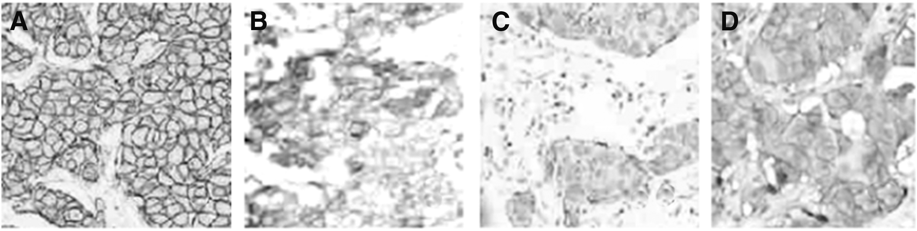
HER-2 protein expression in breast cancer tissues ( × 400).
HER-2 Protein Expression in Breast Cancer Tissues
Amplification of HER-2 gene in breast cancer tissues
In tumor cells, samples with 4–6 red/green signals for HER-2 but having <1.8 ratio were identified as no gene amplification. Clustered red signal ratio >2.2 indicated HER-2 gene amplification. In all 100 recruited patients, 40 (40%) of them had HER-2 gene amplification, and 60 without amplification (Figure 2).
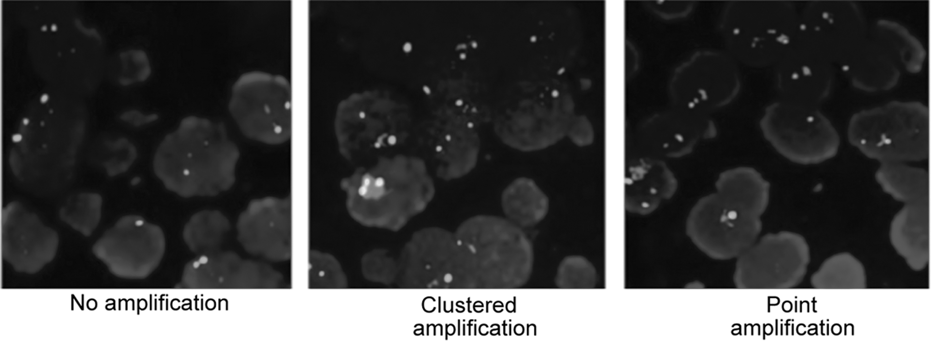
Amplification of HER-2 gene in breast cancer tissues.
Consistency between IHC and FISH results for HER-2 protein/gene amplification
In the breast cancer tissues we analyzed, 64% were positive for HER-2 protein by IHC, and 40% were positive for HER-2 gene amplification by FISH. Among all 64 tissue samples with positive HER-2 protein expression, 38 of them had positive HER-2 gene amplification. Among these there were 3 cases with HER-2 (+) for gene amplification, plus 21 and 14 samples with (++) or (+++) for HER-2 gene amplification, respectively. In 40 samples with positive HER-2 gene amplification, only 2 were negative for HER-2 protein expression. HER-2 protein expression and HER-2 gene amplification had correlation (r = 1.262; p < 0.05; Table 2).
Consistency Between IHC for HER-2 Protein Expression and FISH for Gene Amplification
FISH, fluorescent in situ hybridization; IHC, immunohistochemistry.
Correlation between HER-2 protein expression/gene amplification
We analyzed the correlation between HER-2 protein expression or gene amplification and clinical/pathological features of breast cancer. Results showed a correlation between HER-2 protein expression/HER-2 gene amplification and tumor size, differentiation grade, lymph node metastasis, and TNM stage (p < 0.05) but not with age, estrogen receptor (ER), or progesterone receptor (PR; p > 0.05). For those breast cancer patients with tumor size >3.5 cm, lower differentiation grade, lymph node metastasis, and advanced TNM stage, positive HER-2 protein rate and gene amplification rate were higher (Table 3).
Correlation Between HER-2 Protein/Gene Amplification and Clinical/Pathological Features of Breast Cancer
ER, estrogen receptor; PR, progesterone receptor.
Discussion
A previous study has indicated the involvement of HER-2 in growth, proliferation, invasion, and migration of breast cancer. The precise assay for HER-2 gene amplification can work as the reference index for Herceptin compliance or auxiliary chemotherapy and can predict patient prognosis, thus having major implications. 11,12 Currently IHC assay for HER-2 protein expression has become a common approach. Despite its lower cost and easy manipulation, IHC efficiency and accuracy are compromised by differential antibody origin, antigen retrieval approach, tissue fixation, tissue preparation, and subjective judgement, all of which lead to the deviation of HER-2 protein expression. 13,14 FISH, therefore, is proposed to further elucidate HER-2 gene amplification in HER-2 (++) individuals to better guide clinical treatment. 15 This study recruited breast cancer patients admitted to the Central Hospital of Wuhan and tested HER-2 protein expression and gene amplification to analyze their correlations with clinical/pathological features of breast cancer.
In this study, breast cancer tissues were collected for measuring HER-2 protein expression. Results showed 36% with HER-2 (−), 9% with HER-2 (+), 32% with HER-2 (++), and 23% with HER-2 (+++). FISH assay showed 40% of included patients with HER-2 gene amplification, while 60% without amplification. Further analysis about the consistency between HER-2 protein expression and HER-2 gene expression showed 64% of positive HER-2 protein expression and 40% of positive HER-2 gene amplification. In those tissues with positive HER-2 protein expression, there were 3, 21, and 14 cases for HER-2 (+), HER-2 (++), and HER-2 (+++) of gene amplification. In all 40 breast cancer tissues with positive HER-2 gene amplification, only 2 had not shown positive expression for HER-2 protein. HER-2 protein expression was correlated with HER-2 gene amplification. A research targeting breast cancer has found higher HER-2 gene amplification rate in patients with a higher positive expression rate. 16 Another study has found a higher positive HER-2 gene amplification rate in patients showing HER-2 (+++) by IHC assay than those with HER-2 protein (++) expression, indicating inconsistency between HER-2 (++) patients and HER-2 gene amplification. 17 For breast cancer patients with compliance, FISH assay can better reflect HER-2 gene amplification than IHC. 18 In addition, apart from the detection of HER-2 gene expression by FISH, a recent study has also demonstrated that the closed-system real-time quantitative polymerase chain reaction (RT-qPCR) assay showed >90% concordance with the ASCO/CAP HER-2 IHC/FISH scoring. 19 Moreover, the RT-qPCR assay was highly concordant (94%) with the continuous-variable HER-2 quantitative immunofluorescence (QIF) assay and might better reflect the true continuum of HER-2 receptor status in invasive breast cancer. This study further analyzed the correlation between HER-2 protein expression or HER-2 gene amplification and clinical/pathological features of breast cancer. HER-2 protein or gene amplification was correlated with tumor size, differentiation grade, lymph node metastasis, and TNM stage. For tumors of size >3.5 cm, lower differentiation grade, lymph node metastasis, and advanced TNM stage, positive HER-2 protein rate and gene amplification rate were higher. In a study on primary breast cancer patients, those with HER-2 upregulation had 3-year median survival time, while those with negative HER-2 protein expression had 7-year median survival period, indicating that those breast cancer patients with HER-2 overexpression usually had unfavorable prognosis. 20 Azizun-Nisa suggested that HER-2 protein participated in proliferation and apoptosis of breast cancer cells and regulated division and proliferation of cancer cells, leading to uncontrollable tumor growth. 21 A previous study showed a positive correlation between HER-2 gene amplification of breast cancer tissues and tumor size, pathological grade, lymph node metastasis, and Ki-67 expression, 22 consistent with our study.
Conclusions
HER-2 participates in the whole process of breast cancer occurrence, progression, invasion, and metastasis. IHC combined with FISH may be a better way to test HER-2 protein expression and gene amplification with higher accuracy. HER-2 protein expression is correlated with gene amplification. Both HER-2 protein expression and gene amplification are correlated with tumor size, differentiation grade, lymph node metastasis, and TNM stage of breast cancer. When the HER-2 gene shows an overexpression, it may indicate unfavorable prognosis of patients. In clinical practice, testing of HER-2 protein expression plus gene amplification in breast cancer patients can help assist clinicians to stipulate more efficient chemotherapy plans, choose optimal target drugs, make judgement of endocrine therapy and of disease progression, as well as predict patient prognosis. 23,24 Various cytokines are involved in the progression of breast cancer, with interconnection among signal transduction pathways especially with disease occurrence and progression or interaction with targeted genes through a complex mechanism. As a future perspective, a precise assay for HER-2 protein expression and gene amplification might be critical for efficient treatment of breast cancer, thus providing novel insights and strategy for breast cancer treatment with promising values, although further studies are required to elucidate detailed mechanisms.
Footnotes
Acknowledgments
This work was supported by Nanjing Science and Technology Plan project (No. 201715011) and Project of Jiangsu Provincial Six Talent Peaks (No. WSN-057).
Disclosure Statement
There are no existing financial conflicts.