
Abstract
Objective:
To investigate the expression levels of matrix metalloproteinases-9 (MMP-9) and tissue inhibitor of metalloproteinase-1 (TIMP-1) in pituitary adenomas (PAs), and to analyze the relationship of the expressions of the two with the prognosis of patients.
Methods:
A total of 108 patients with PAs diagnosed in our hospital from May 2010 to May 2012 were selected and divided into the invasive PA (IPA) group (n = 58) and the non-IPA group (n = 50) according to the invasiveness of PAs. Hematoxylin and eosin (H&E) staining was used to observe the pathological state of patients. The expression levels of MMP-9 and TIMP-1 were measured by immunohistochemistry and western blotting at protein level and reverse transcription–polymerase chain reaction at gene level, respectively. The expression levels of MMP-9 and TIMP-1 in serum of patients before operation were tested using enzyme-linked immunosorbent assay, and patients with PAs after operation were followed up.
Result:
The positive expression rate of MMP-9 in IPAs was significantly higher than that in non-IPAs, whereas that of TIMP-1 was relatively high in non-IPAs, and the differences were statistically significant (p < 0.05). At both protein and gene levels, MMP-9 was highly expressed in IPAs, whereas TIMP-1 was highly expressed in non-IPAs, and the differences were statistically significant (p < 0.05 in all comparisons). Before operation, the expression level of MMP-9 in serum of patients with IPAs was relatively high, whereas that of TIMP-1 in serum of patients with non-IPAs was relatively high, and the differences were statistically significant (p < 0.05 in all comparisons).
Conclusion:
The postoperative survival rate of patients with highly expressed MMP-9 was relatively low, whereas that of patients with highly expressed TIMP-1 was relatively high. The abnormal expressions of MMP-9 and TIMP-1 play important roles in the invasion process of PAs. The prognoses of patients with low expression MMP-9 and high expression TIMP-1 are more positive.
Introduction
Pituitary adenoma (PA) is a relatively common neuroendocrine tumor with a low intracranial growth rate. 1 Its incidence rate is second only to glioma and meningioma, accounting for >10% of intracranial tumors. 2 However, the incidence rate of PAs in recent years shows an increasing trend. 3 PA is a benign tumor in the sellar region, but some PAs metastasize and invade many peripheral tissues. This type of tumor is called invasive PA (IPA). 4,5 Although there are a large number of in-depth explorations on the pathogenesis of PAs, the specific mechanism of its invasion has not been accurately elucidated. At present, the mechanism of its pathogenesis is mainly illuminated from the following three aspects: protooncogenes (homogentisate solanesyltransferase [Hst], Ras, pituitary tumor transforming gene [PTTG], etc.), 6 tumor suppressor genes (p53, retinoblastoma [Rb], gene of phosphatase and tensin homolog deleted on chromsome ten [PTEN], etc.), 7 and various types of invasion factors (proliferating cell nuclear antigen [PCNA], matrix metalloproteinases [MMPs], fibroblast growth factors [FGFs], etc.). 8 Members of the MMP family are highly conserved and have their own characteristics based on similar functions. 9 MMPs can almost degrade many components in the extracellular matrix (ECM), destroy all kinds of histological barriers, and remove the obstacles in tumor cell invasion. 10 Matrix metalloproteinase-9 (MMP-9) has been long recognized as a key enzyme for the proteolytic degradation of ECM during tumor invasion and metastasis. 11 Its expanding roles include regulating cancer progression, activating angiogenesis, and recruiting macrophages or other bone marrow-derived myeloid cells to the preexisting metastatic niche. 11,12 Through forming a complex with MMP-9 and inhibiting its enzymatic activity, 13,14 tissue inhibitor of metalloproteinase-1 (TIMP-1) plays a negative role in the invasion and metastasis of tumor cells. Imbalance between MMP-9 and TIMP-1 is closely associated with invasion and metastasis of several tumors. Considering the critical role of MMP-9 and TIMP-1 in the pathogenesis of tumors, their role in the invasion and prognosis of PA remains poorly understood. In this study, we aimed to investigate the expressions of MMP-9 and TIMP-1 in PAs as well as the relationship of these two molecules with the prognosis of patients.
Data and Methods
Data
A total of 108 patients with PAs who underwent surgical resection in our hospital from May 2010 to May 2012 were selected, including 61 men and 47 women, aged 25–57 years old. Magnetic resonance imaging (MRI) scan was performed for all patients before operation, and they were classified into the IPA group (n = 58) and the non-IPA group (n = 50) according to Hardy–Wilson 15 and Knosp classification 16 methods. All clinical symptoms were consistent with the diagnostic criteria for PA, and confirmed by clinical imaging, surgery, and pathology diagnosis. All patients underwent surgical resection of PAs and PA tissues were obtained at surgery and adherent blood was removed followed by being frozen in liquid nitrogen immediately and stored at −80°C for extraction of protein and RNA.
This study was approved by ethics committee in the Harrison International Peace Hospital affiliated to Hebei Medical University and all the enrolled objects had signed informed consent.
Main regents
Rabbit antihuman MMP-9 monoclonal antibodies (Beijing Donglinchangsheng Biotechnology Co., Ltd.), rabbit antihuman TIMP-1 (BD), goat anti-rabbit secondary antibodies (American BD Company), β-actin (the internal reference) antibodies (BD), 3,3-diaminobenzidine (DAB) color development reagent and powder preparation of citrate buffer (Shanghai Xin Yu Biotech Co., Ltd.); immunohistochemistry (Nanjing DASF Biological Technology Co., Ltd.), reverse transcription (RT) kit (Shanghai Bo Yi Biological Technology Co., Ltd.), real-time fluorescence quantitative polymerase chain reaction (PCR) kits (Guangzhou Vipotion Biotechnology Co., Ltd.). TRIGene reagents (Beijing Kangrun Chengye Biotechnology Co., Ltd.), RT kits (Shanghai Yuduo Biological Technology Co., Ltd.), extraction kits for the total protein of cells (Jiangsu Keygen Biotech Co., Ltd.), bicinchoninic acid (BCA) protein quantification kits (Nanjing SenBeiJia Biological Technology Co., Ltd.), MMP-9 and TIMP-1 enzyme-linked immunosorbent assay (ELISA) kits (Shanghai Enzyme-linked Biotechnology Co., Ltd.).
Methods
Hematoxylin and eosin staining
Paraffin-embedded tissue sections were dewaxed, followed by washing with alcohol. After thorough hydration, the sections were stained with hematoxylin for 10 min and then washed with water. Afterward, the sections were differentiated with 1% hydrochloric acid alcohol for 3 sec, washed with water, and blued with saturated lithium carbonate for 3 sec. Then they were rinsed with running water for 20 min and stained using 1% alcohol-soluble eosin for 10 sec, and finally conventional dehydration, transparency, and mounting were conducted.
Immumohistochemical staining method
The paraffin sections were dewaxed, hydrated, and then washed with phosphate buffer. To reduce the background nonspecific staining caused by endogenous peroxidase, blocking was further conducted in the blocking solution for 20 min, and 10% serum was used for blocking for 10 min. After that, primary antibody solution (MMP-9 and TIMP-1 monoclonal antibodies; diluted at 1:50) was added for incubation at 4°C overnight. Phosphate buffer was applied for washing, followed by the addition of secondary antibody solution (diluted at 1:50) for incubation at room temperature for 30 min. The sections were washed with phosphate buffer again. Then streptavidin–peroxidase solution was used for incubation at room temperature for 30 min, phosphate buffer solution was used for washing, color development was conducted using DAB, followed by rinsing with distilled water, restaining, and mounting.
Assessment of immumohistochemistry results
A total of 100 cells on the field of view needing to be observed were randomly selected, and the average number of positive cells in the field of view was calculated as the number of positive cells expressing the protein in tissues. Intensity points: 0–3 points represented no coloring, light yellow, pale brown, and dark brown, respectively. Points of positive rate of stained cells: 0–4 points represented the percentage of positive cells of (1, 10), (10, 25), (25, 50), (50, 75), and (75, 100), respectively. The product of the points of the above two groups are as follows: 0 point for negative, 1–4 points for (+), 5–8 points for (++), and 9–12 points for (+++).
Determination of the expressions of MMP-9 and TIMP-1 messenger RNAs by real-time PCR
Primers were synthesized by Huamei Ruikang (Beijing) International Biotechnology Research Institute Co., Ltd. Primer sequences are given in Table 1. The total RNA was extracted from tissues according to the instructions of total RNA extraction kit, and complementary DNA (cDNA) was synthesized using the RT kit.
Primer Sequences
MMP-9, matrix metalloproteinases-9; TIMP-1, tissue inhibitor of metalloproteinase-1.
Ten microliters reaction solution is as follows: 2 μL 5 × genomic DNA (gDNA) eraser buffer, 1 μL gDNA eraser, 1 μg total RNA, ribonuclease (RNase)-free distilled H2O (dH2O).
Twenty microliters RT system is as follows: 4 μL 5 × PrimeScript Buffer, 1 μL PrimeScipt RT Enzyme Mix, 1 μL PrimeScript RT Enzyme Mix, 10 μL of the above reaction solution, and 4 μL RNase-free dH2O. RT reaction conditions are 37°C for 15 min and 85°C for 5 sec.
Twenty-five microliters PCR reaction system is as follows: 12.5 μL SYBR Premix Ex Taq™ II, 1 μL forward primer, 1 μL reverse primer, 2 μL cDNA, and 8.5 μL dH2O. Reaction conditions are denaturation at 94°C for 3 min, denaturation at 94°C for 20 sec, annealing at 58°C for 20 sec, and extension at 72°C for 30 sec; a total of 40 cycles. With β-actin as an internal reference, RT-PCR instrument automatically calculated and produced the relative expression levels of MMP-9 and TIMP-1 messenger RNAs (mRNAs).
Detection of proteins by western blotting
The total protein of tissues was extracted according to the instructions of cell total protein extraction kit, the concentration of the extracted protein was determined by BCA protein detection assay, and the total protein was stored at −70°C for standby application.
Gels at different concentrations were prepared for sodium dodecyl sulfate–polyacrylamide gel electrophoresis. Gel locations of the two kinds of proteins were generally identified according to the marker bands. The membrane was transferred for 35 min and then blocked with 5% nonfat dry milk for 90 min at 37°C. The primary antibody (diluted at 1:1000) was used for incubation at 4°C overnight. Then these proteins were added with Tris-buffered saline with Tween-20 (TBST) solution, and then placed and shaken on a shaker for washing three times with 15 min each time. Subsequently, the secondary antibody (diluted at 1:1000) was used for incubation at 37°C for 1 h. The proteins were added with TBST solution, and then placed and shaken on a shaker for washing three times lasting 15 min each time. In a dark room, electrochemiluminescence (ECL) solution was added for coloring, followed by exposure, development, and fixation. Finally, image analysis was conducted, and the optical density value was calculated with β-actin as an internal reference for control through the scanning of ChemiDocTMMP imaging system using the professional image analysis software, ImageJ.
Detection of the expressions of serum MMP-9 and TIMP-1 by ELISA
Five milliliters peripheral blood was extracted from all the patients and centrifuged at 2000 rpm for 5 min. The serum was collected with a half being preserved at −80°C and another half being tested. The levels of serum MMP-9 and TIMP-1 were measured in strict accordance with the procedures in the instructions of the ELISA kit.
Statistical analysis
In this experiment, the professional statistical software, Statistical Product and Service Solutions (SPSS) 17.0 (provided by Beijing Xinmei Jiahong Science and Technology Co., Ltd.) was used for data analysis. Measurement data were expressed as mean ± standard deviation. One-way analysis of variance was used for intergroup comparisons. The survival analysis was performed using GraphPad Prism5, and α = 0.05 was taken as the statistical standard.
Results
Hematoxylin and eosin staining graphs
Under the microscope, it was shown that non-IPA cells were flake with round nuclei and uniform cytoplasm, whereas PA cells were daisy-like with small nuclei and more abundant cytoplasm (Fig. 1).

H&E staining results of PAs ( × 400).
Immumohistochemical staining results
The positive rates of MMP-9 and TIMP-1 in IPAs were 86.21% (50 of 58) and 18.97% (11 of 58), respectively, and 50.00% (25 of 50) and 34.00% (17 of 50), respectively in non-IPAs. The positive expression rate of MMP-9 in IPAs was significantly higher than that in non-IPAs, and the difference was statistically significant (p < 0.05). Difference in the positive expression rate of TIMP-1 between the two types of PAs was also statistically significant (p < 0.05) (Table 2).
Expressions of Matrix Metalloproteinases-9 and Tissue Inhibitor of Metalloproteinase-1 in Different Types of Pituitary Adenomas
IPA, invasive pituitary adenoma.
Detection results of the relative expression levels of MMP-9 and TIPM-1 mRNAs by RT-PCR
The relative expression level of mRNA was detected using RT-PCR. The results showed that the expression level of MMP-9 in IPAs was significantly higher than that in non-IPAs, and the difference was statistically significant (p < 0.05). However, the expression level of TIMP-1 in IPAs was significantly lower than that in non-IPAs, and the difference was statistically significant (p < 0.05) (Fig. 2).

Detection results of the relative expression levels of MMP-9 and TIPM-1 mRNAs by RT-PCR. Compared with IPAs, *p < 0.05. MMP-9, matrix metalloproteinases-9; mRNAs, messenger RNAs; RT-PCR, reverse transcription–polymerase chain reaction.
Detection of the expression levels of MMP-9 and TIMP-1 proteins by western blotting
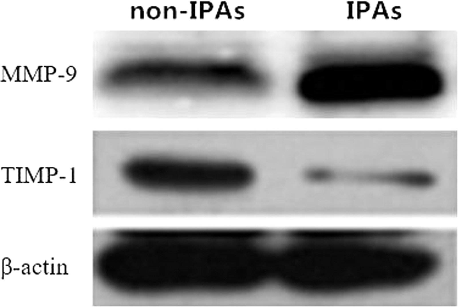
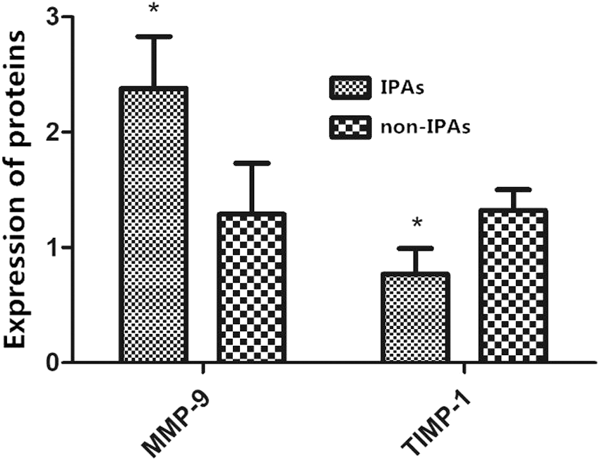
Western blotting was used to detect the relative expression of proteins, which revealed that the expression level of MMP-9 protein in IPAs was obviously higher than that in non-IPAs, and the difference was statistically significant (p < 0.05). However, the expression level of TIMP-1 protein in IPAs was obviously lower than that in non-IPAs, and the difference was statistically significant (p < 0.05) (Figs. 3 and 4).
Bar graphs of the expressions of MMP-9 and TIMP-1 proteins detected by western blotting. TIMP-1, tissue inhibitor of metalloproteinase-1.
Detection results of the expressions of MMP-9 and TIMP-1 proteins by western blotting. Compared with non-IPAs, *p < 0.05.
Detection results of serum ELISA
The results of ELISA indicated that the expression level of MMP-9 in serum of patients with IPAs was remarkably higher than that in serum of patients with non-IPAs, whereas that of TIMP-1 was significantly lower than that in serum of patients with non-IPAs, and the differences were statistically significant (p < 0.05 in all comparisons) (Fig. 5).

Expressions of serum MMP-9 and TIMP-1. Compared with non-IPAs, *p < 0.05.
Prognosis analysis
The median relative expression level of MMP-9 and TIMP-1 was taken as the standard. The level above this standard represented high expression, whereas the level below this standard represented low expression. The postoperative survival rate of patients with highly expressed MMP-9 was significantly lower than that of patients with lowly expressed MMP-9, and the difference was statistically significant (p < 0.05). On the contrary, the postoperative survival rate of patients with highly expressed TIMP-1 was significantly higher than that of patients with lowly expressed TIMP-1, and the difference was statistically significant (p < 0.05) (Fig. 6).
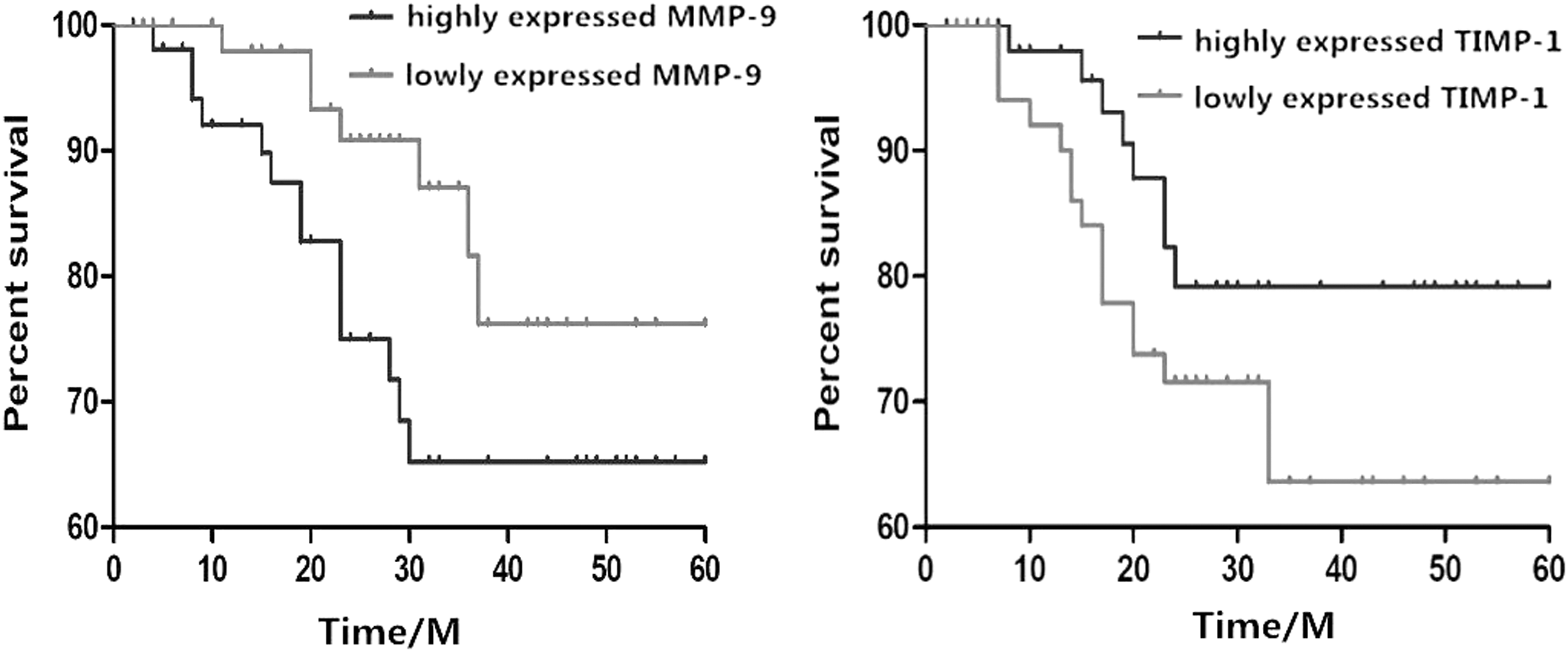
Five-year survival rates of MMP-9 and TIMP-1.
Discussion
PAs originate from the anterior pituitary gland, and the appearance of tumors in the pituitary is usually accompanied by some clinical symptoms. 17 PAs are benign in histological morphology, but some also show characteristics of malignant tumors, which are called IPAs. Surgery has become the most important treatment method for IPAs, 18 and in normal cases, the removal of non-IPAs is relatively simple. Once these non-IPAs develop into IPAs, the total resection is hard to be achieved. Some scholars speculated that this may be because of difficulties in separating fibrous tissues in IPAs from tissues in the saddle area. 19
With the deepening of the research, more and more evidence show that the invasion of PAs is a dynamic and continuous process. First, tumor cells shed off and produce adhesion in situ, and then they degrade the ECM components around. Tumor cells begin to metastasize with blood vessels and lymphatic vessels, proliferate at the secondary site, and eventually become metastases. 20,21 Therefore, the degradation of the ECM is crucial for tumor cells to complete their invasion. MMPs are a collective term for a family of proteases that can well degrade the basement membrane and ECM, and MMP-9 plays a key role in this degradation process. 22,23 TIMPs are specific inhibitors of MMPs, and they are negative regulators corresponding to MMPs in the regulation of ECM metabolism. 24 A study has shown that TIMP-1 is a specific inhibitor of MMP-9 that can inhibit the invasion and metastasis of tumor cells by reducing the activity of MMP-9. 25
The results of this study revealed that the pathological manifestations of IPA and non-IPA cells were markedly different. Non-IPA cells were flake with round nuclei and uniform cytoplasm, whereas PA cells were daisy-like with small nuclei and more abundant cytoplasm. The expression level of MMP-9 in IPAs was significantly upregulated, whereas that of TIMP-1 was relatively low. Differences in the positive expression rates of MMP-9 and TIMP-1 in both types of tissues were statistically significant (p < 0.05 in all comparisons). At the gene and protein levels, it was also found that the expression level of MMP-9 in IPAs was higher than that in non-IPAs, whereas that of TIMP-1 in IPAs was lower than that in non-IPAs. Serological test showed that the preoperative expression level of MMP-9 in serum of patients with IPAs was also relatively high (p < 0.05), suggesting that MMP-9 gene is activated in the invasion process of PAs and is closely related to the invasion.
The upregulated MMP-9 expression in patients with IPAs increased the secretion of MMP-9 in the blood, inhibited the expression of TIMP-1, and decreased the secretion of TIMP-1, thus reducing the inhibition of MMP-9 and further deteriorating tumors. Through postoperative prognosis analysis for patients, it was found that the postoperative survival rate of patients with highly expressed MMP-9 was significantly lower than that of patients with lowly expressed MMP-9, and the difference was statistically significant (p < 0.05). In contrast, the survival rate of patients with highly expressed TIMP-1 was obviously higher than that of patients with lowly expressed TIMP-1, and the difference was statistically significant (p < 0.05), suggesting that when the expression level of MMP-9 is relatively high and that of TIMP-1 is relatively low, the invasion of PAs is relatively strong, causing poor prognosis of patients, which is consistent with the research findings of Sun et al. 26
In conclusion, the abnormal expressions of MMP-9 and TIMP-1 play important roles in the invasion process of PAs, and the prognoses of patients with downregulated expression of MMP-9 and upregulated expression of TIMP-1 are better, which can be used as one of the indicators for prognosis.
Footnotes
Acknowledgment
Research supported by the Hebei Province Hengshui City Science and Technology Plan item (No. 14003A).
Disclosure Statement
All authors declare that they have no conflict of interest.