
Abstract
Purpose:
68Ga-BPAMD has recently emerged as one of the preferred radiopharmaceuticals for imaging of bone lesions due to its ability to produce high-resolution images and uncomplicated availability of 68Ga, a positron emission tomography (PET) radionuclide, from commercial 68Ge/68Ga generators. The primary objective of this work is to develop freeze-dried BPAMD kit, for the easy and convenient formulation of 68Ga-BPAMD patient dose at the hospital radiopharmacy. In addition, the kit should be compatible with 68Ga, eluted using HCl of various molarities from the 68Ge/68Ga generators sourced from different suppliers.
Procedures:
Freeze-dried BPAMD kit, comprising 50 μg of BPAMD and 150 mg of HEPES, was prepared and evaluated using 68Ga eluted from three different 68Ge/68Ga generators. Radiochemical purity (RCP) of 68Ga-BPAMD was determined by both thin-layer chromatography and high-performance liquid chromatography studies. The maximum volume of 68Ga, which can be added in the kit, was determined. The biological behavior of 68Ga-BPAMD, prepared using the freeze-dried kit, was evaluated by both in vitro and in vivo studies. Clinical studies were also performed in limited number of patients suffering from metastatic bone cancer.
Results:
68Ga-BPAMD could be prepared with >95% RCP using the freeze-dried BPAMD kit and 68Ga eluted from 68Ge/68Ga generators obtained from three different suppliers. 68Ga-BPAMD, prepared using the freeze-dried kit, exhibited adequate serum stability and ∼91% binding with the hydroxyapatite particles. Biodistribution studies in normal Wistar rats exhibited selective uptake of the agent in skeleton and fast clearance of the nonaccumulated activity through urinary route. Clinical studies in cancer patients showed excellent accumulation of the agent in bone lesions.
Conclusion:
The preliminary studies exhibited the potential of the developed BPAMD kit toward its utilization for the PET scanning of skeletal metastases.
Introduction
Skeletal metastasis is one of the common problems encountered by a large majority of cancer patients at the advanced stage of their disease. 1,2 It is reported that 80% of patients suffering from breast or prostate cancer develop bone metastases and suffer from associated complications, predominantly pain. 1 –3 In many instances, although bone metastases occur at an early stage of the disease, their symptoms are recognized quite late. 4,5 Therefore, early and accurate noninvasive diagnosis of skeletal metastases has profound implications in the proper management of cancers.
Bisphosphonates with two germinal phosphonic acid groups have found extensive usage in addressing various bone maladies, including metastatic bone lesions. 1,6 Unlike pyrophosphates with a P-O-P unit, bisphosphonates with a P-C-P unit have been found to be biologically stable and are excreted unaltered from the human body. 7 Affinity of the phosphonate groups toward the hydroxyapatite [HA, Ca10(PO4)6(OH2)2], which is the main component of the inorganic bone matrix, is considered to be the reason of preferential accumulation of the bisphosphonates in the skeleton, and this property has been extensively exploited to prepare a wide variety of radiolabeled bisphosphonates for targeting bone lesions. 1,3,6,8
99mTc-MDP (methylene diphosphonate) is undoubtedly the most widely utilized SPECT (single photon emission computed tomography) agent used for carrying out imaging of metastatic bone lesions. 1 However, due to the higher spatial resolution and superior image quality of PET (positron emission tomography) compared to that obtained with SPECT, use of bone-seeking agents radiolabeled with suitable positron-emitting radionuclides viz. 18 F [T1/2 = 110 min, β+ max (97%) = 0.633 MeV] and 68Ga [T1/2 = 68 min, β+ max (89%) = 1.89 MeV] are preferred. 5,8,9 The utility of [ 18 F]NaF as a PET agent for bone scintigraphy is well documented in the contemporary literature and the use of this agent has gained momentum in the recent past. 5,10 –12 However, availability of 18 F is limited to only those medical centers that are either equipped with a medical cyclotron or having access to the agent from some source of nearby proximity. Therefore, development of bone-seeking agents labeled with 68Ga, a PET radionuclide, which can be conveniently obtained from the 68Ge/68Ga radionuclide generator, has become popular in the recent time. 10
BPAMD [1,4,7,10-Tetraazacyclododecane-1,4,7-triacetic acid, 10-[2-[(diphosphonomethyl)amino]-2-oxoethyl]] is the outcome of one of such research interests and has recently emerged as the bisphosphonate of choice for radiolabeling with 68Ga. 5,13 BPAMD contains a macrocyclic DOTA moiety that is capable of complexation with 68Ga with a high stability constant value (log K = 26.1) 14,15 and the agent showed excellent skeletal accumulation and favorable pharmacokinetics in the animal model. 5,16,17 Clinical studies with 68Ga-BPAMD have also been reported in the contemporary literature, documenting the excellent potential of the agent in bone scintigraphy. 10,13 Therefore, an attempt has been made to develop a freeze-dried BPAMD kit, which will help easy, single-step, and convenient formulation of 68Ga-BPAMD patient dose at the hospital radiopharmacy.
Germanium-68/Gallium-68 radionuclide generators are commercially available from various reputed suppliers and depending upon the matrix used for the generator column, 68Ga activity is eluted from the generators using HCl of various molarities, which usually vary from 0.05 M (Isotope Technologies Garching, Germany) to 0.6 M (iThemba Lab, South Africa). 18 –22 Therefore, it is necessary to develop the kit in such a way that it can be utilized for the preparation of a 68Ga-BPAMD patient dose using the 68Ga eluted from any of these commercial 68Ge/68Ga radionuclide generators. It will also free the nuclear medicine centers to procure any commercial 68Ge/68Ga radionuclide generator without wasting their resources toward reoptimizing the formulation protocol of the agent. During the course of this work, focus was also given toward this direction so that the developed freeze-dried BPAMD kit becomes compatible for using with any of these commercially available 68Ge/68Ga radionuclide generators.
Herein, the authors report the details of formulation of the freeze-dried BPAMD kit and preparation of 68Ga-BPAMD with the developed kit using 68Ga eluted from three different 68Ge/68Ga radionuclide generators. They also report the pharmacokinetics and biological distribution exhibited by the radiolabeled agent in normal animal model. The authors' experience of carrying out limited clinical studies, employing the 68Ga-BPAMD, prepared using the freeze-dried kit, is also described in this article.
Experimental
Materials and Methods
BPAMD was obtained from Otto Chemie (Germany). HEPES (4-(1-hydroxyethyl)-1-piperazine-ethanesulfonic acid) was procured from Sigma-Aldrich. HA particles were synthesized following the procedure reported in the literature. 23 Sodium citrate, citric acid, and all other chemicals used in this study were purchased from reputed local manufacturers and were of analytical reagent grade.
Lyophilization was performed using the Labocene Coolsafe™ 55-4 freeze-drier (Denmark). Pharmaceutical borosilicate glass vials (10 mL capacity, crimp neck) were procured from a reputed local manufacturer and thoroughly cleaned and autoclaved following the usual procedure before use. Three different 68Ge/68Ga radionuclide generators, used for this study, were obtained from different commercial manufacturers [SiO2-based generator from ITG (Germany), SnO2-based generator from iThemba (South Africa), and TiO2-based generator from Eckert & Ziegler Isotope Products (Germany)]. All radioactive counting was carried out by using a well-type NaI(Tl) scintillation counter, obtained from Electronic Corporation of India Limited (India), after adjusting the baseline and window at 450 and 100 keV, respectively, to utilize the 511-keV positron annihilation photopeak of 68Ga.
Thin-layer chromatography (TLC) was performed using silica gel 60 F254-coated aluminum sheets, procured from Merck (India). The high-performance liquid chromatography (HPLC) system (PU 1580) was obtained from Jasco (Japan). Elution profiles were monitored by detecting the radioactivity signal using a well-type NaI(Tl) detector (Jasco, Japan) coupled with the HPLC system. All the solvents used for HPLC studies were degassed and filtered before use and were of HPLC grade.
Animal experiments were carried out using normal healthy Wistar rats, which were bred and reared in the institutional animal house facility following the standard management practice. Radioactive countings associated with the animal studies were performed using a flat-type NaI(Tl) scintillation detector, procured from Electronic Corporation of India Limited (India), employing the same counting setup mentioned earlier. Animal studies reported in this article were approved by Institutional Animal Ethics Committee (IAEC) of Bhabha Atomic Research Centre (Mumbai, India) and all the animal experiments were carried out in strict compliance with the relevant national laws relating to the conduct of animal experimentation.
Clinical studies were performed in eight male patients (46–78 years). Patients referred for a routine bone scan with a diagnosis of carcinoma prostate or for inflammatory arthropathy were included in the study. A whole-body PET-CT (computed tomography) in 6–7 bed positions was acquired 30–60 min after injection on a Siemens Biograph 6 PET-CT scanner. The protocol for clinical evaluation was approved by Institutional Ethics Committee (IEC) of Kovai Medical Center and Hospital (Coimbatore, India). Written informed consent was obtained from all the patients before carrying out the imaging studies.
Preparation of freeze-dried BPAMD kits
A stock solution of 1.25 M HEPES buffer (pH 5.0) was prepared by dissolving HEPES (23.8 g) in autoclaved double-distilled water (80 mL). BPAMD (5 mg) was also dissolved in double-distilled water (5 mL) to prepare a stock solution having BPAMD concentration of 1 μg/μL. The stock solution containing BPAMD (5 mL) was thoroughly mixed with HEPES buffer (50 mL) and the pH of the resulting mixture was checked. Subsequently, the solution was aliquoted equally into 100 glass vials. All the glass vials were incubated at 0°C for 4 h and afterward overnight at −80°C. The vials were then lyophilized for 6 h using a table top lyophilizer resulting into the formation of white pellets inside the glass vials. The kits, thus prepared, were stored at 2–8°C immediately after lyophilization. Two such batches were prepared during the course of this study.
Elution of 68GaCl3 from 68Ge/68Ga radionuclide generators
Gallium-68(III) chloride was eluted from 68Ge/68Ga radionuclide generators using different molarities of hydrochloric acid. While the 68Ge/68Ga generator obtained from ITG was eluted with 0.05 M HCl, 68Ge/68Ga generators obtained from Eckert & Ziegler and iThemba Laboratory were eluted with 0.1 and 0.6 M HCl, respectively. 68GaCl3, eluted with HCl of different molarities, was directly used for further studies without any postelution purification.
Preparation of 68Ga-BPAMD using freeze-dried kit
For the preparation of 68Ga-BPAMD, the kit was allowed to attain ambient temperature. Freshly eluted 68GaCl3 was added in the kit vial and the reaction mixture was incubated at 100°C for 20 min. The maximum volume of 68GaCl3 added in the vial was restricted to 2 mL when ITG- or Eckert & Ziegler-make 68Ge/68Ga radionuclide generator was used, while for iThemba-make 68Ge/68Ga generator, the maximum volume of 68GaCl3 added in the vial was 500 μL.
Quality control of 68Ga-BPAMD prepared using freeze-dried kit
Percentage complexation yield of 68Ga-BPAMD was determined using two chromatographic techniques, namely TLC and HPLC. TLC was performed using 0.25 M sodium citrate buffer (pH 4.0) as the eluting solvent. On the other hand, HPLC was carried out using SAX (strongly anion exchange) column employing gradient elution technique using a mixture of 1 M phosphate buffer (pH 3.0) (solvent A) and 1 M citrate solution (solvent B) as the mobile phase. Each HPLC run was continued for 20 min with the flow rate maintained at 1.5 mL/min, and the following gradient system was employed for the HPLC studies: 0–5 min 100% A, 5–7 min 100% A to 70% A, 7–12 min 70% A, 12–14 min 70% A to 100% A, and 14–20 min 100% A.
Determination of kit capacity with respect to the addition of maximum volume of 68GaCl3
Commercially available 68Ge/68Ga radionuclide generators are required to be eluted with HCl of different normalities, 22 and therefore, the maximum volume of 68GaCl3, which can be added to the kit vial, will depend on the type of 68Ge/68Ga generator from which 68GaCl3 is sourced. To find out the maximum volume of 68GaCl3, which can be added in the kit without compromising the radiochemical purity (RCP) of 68Ga-BPAMD, gradually increasing volumes of 68GaCl3 eluate were added in the freeze-dried kits and radiolabeling was performed following the protocol mentioned above. As the ideal pH for carrying out the radiolabeling of 68Ga with BPAMD lies between 3 and 4, pH of the resulting solutions was determined after the addition of increasing volume of 68GaCl3, eluted from different 68Ge/68Ga generators, in the kit vial. 68GaCl3 eluate obtained in three different molarities of HCl viz. 0.05, 0.1, and 0.6 M was used for this purpose. Addition of increasing volume of 68GaCl3 was continued until the pH of the resulting solutions reached below 3 and the corresponding radiolabeling yields were determined.
Stability of 68Ga-BPAMD
In vitro stability of 68Ga-BPAMD was determined both in HEPES buffer as well as in human blood serum. For determination of stability of 68Ga-BPAMD in the buffer medium, the preparation was incubated at 37°C for 2 h. An aliquot of the incubated 68Ga-BPAMD was spotted on a TLC strip and percentage radiochemical yield was determined by following the same procedure mentioned above. For determination of serum stability, 68Ga-BPAMD (200 μL) was mixed with human blood serum (600 μL) and the mixture was incubated at 37°C for 2 h. Acetonitrile (500 μL) was added into the reaction mixture subsequent to incubation, which led to the precipitation of serum proteins. The contents of the reaction mixture were centrifuged at 15,000 rpm for 15 min for enabling the separation of precipitate and supernatant. An aliquot of the supernatant was spotted on the TLC strip and the resultant radiolabeling yield was determined by the procedure mentioned above.
Binding studies with hydroxyapatite
To study the binding affinity of 68Ga-BPAMD, prepared using the freeze-dried kit, toward bone matrix, simulation studies involving binding of 68Ga-BPAMD with in-house synthesized HA particles were performed following the protocol mentioned below. Normal saline (1 mL) was added in three glass vials containing HA particles (∼20 mg in each vial) and the suspensions were kept on stirring at room temperature for 24 h. Tracer amounts of 68Ga-BPAMD (50 μL) were added in each glass vial. The vials were subsequently vortexed for 30 s and incubated for 10 min at ambient temperature, after which the vials were centrifuged at 10,000 rpm for 5 min. The supernatant was separated and the precipitate was washed with normal saline (1 mL). Percentage binding was determined by measuring the counts bound with precipitated HA particles and supernatant using a well-type NaI(Tl) counter.
Determination of the shelf-life of freeze-dried BPAMD kits
Lyophilized kits were randomly chosen from both the batches and stored at 2–8°C for a period up to 6 months. The kits were evaluated after 1, 3, and 6 months of storage. Toward this, 68Ga-BPAMD complexes were prepared using the 68Ga obtained from different 68Ge/68Ga radionuclide generators and quality control studies were performed following the protocol mentioned earlier.
Biodistribution studies
Biological distribution of 68Ga-BPAMD was studied by carrying out biodistribution studies in normal healthy Wistar rats. Each animal, weighing 200–250 g, was intravenously injected with ∼100 μL of the radiolabeled preparation (∼50 μCi, 1.85 MBq) through one of the lateral tail veins. The animals were maintained in the normal laboratory atmosphere till the predesignated postadministration time points and sacrificed through CO2 asphyxia at 30 min, 1 h, and 2 h postadministration of the radiolabeled agent. All the vital organs/tissues of the animals were excised, washed with normal saline, dried, weighed in a weighing balance, and radioactivity associated with each organ/tissue was measured using a flat-type NaI(Tl) counter. 24,25 Activity accumulated in various organs/tissues was calculated from these data and expressed as percentage injected activity per gram of organ/tissue (% IA/g). For each time point, three animals were used.
Clinical studies
Six patients with confirmed histopathologic cancer of the prostate, with proven or suspected bone metastases, and two patients with suspected arthritis were injected intravenously with 68Ga-BPAMD, prepared using the freeze-dried BPAMD kit, with activities in the range of 185–370 MBq (5–10 mCi). All the patients were male with an age range of 46–78 (mean age = 62) years. All patients underwent routine 99mTc-MDP SPECT scan a day before or after the 68Ga-BPAMD PET scan. Patients were asked to maintain good oral hydration and were asked to void before recording the scans. Standard protocol of acquisition, as mentioned earlier, was used for both the scans in all the patients with a waiting time of 30–60 min postinjection for the PET-CT and 180–240 min for the SPECT scan. A visual reporting system based on concurrence of two experienced nuclear medicine physicians was used to classify both the PET and the SPECT scans as normal or abnormal.
Results
Preparation of freeze-dried kits of BPAMD
Two batches of freeze-dried BPAMD kits, comprising 100 kits per batch, were prepared. Each kit vial contains 50 μg of BPAMD and ∼150 mg of HEPES. The kit vials were stored at 2–8°C immediately after lyophilization and kept at that temperature until used. The kits were allowed to attain room temperature before use for the preparation of 68Ga-BPAMD.
Quality control of 68Ga-BPAMD prepared using freeze-dried kit
Quality control studies for 68Ga-BPAMD were carried out by TLC using 0.25 M sodium citrate buffer (pH 4.0) as the eluting solvent, where the 68Ga-BPAMD complex was found to exhibit a retention factor (Rf) of 0.4–0.7, while uncomplexed 68Ga moved toward the solvent front (Rf = 0.9–1.0) under identical conditions. TLC studies showed that 68Ga-BPAMD complex could be prepared with >95% radiolabeling yield using the freeze-dried BPAMD kit. Typical TLC patterns of 68GaCl3 and 68Ga-BPAMD are shown in Figure 1. The RCP of 68Ga-BPAMD preparation was further confirmed by HPLC studies where 68Ga-BPAMD complex exhibited a retention time of 7.0 min, while free 68Ga eluted from the column at 14.5 min. HPLC studies also showed that the 68Ga-BPAMD complex could be prepared with >95% RCP using the freeze-dried kit. Typical HPLC profiles of 68GaCl3 and 68Ga-BPAMD are shown in Figure 2. The results of the quality control studies performed on the 68Ga-BPAMD complex are summarized in Table 1.
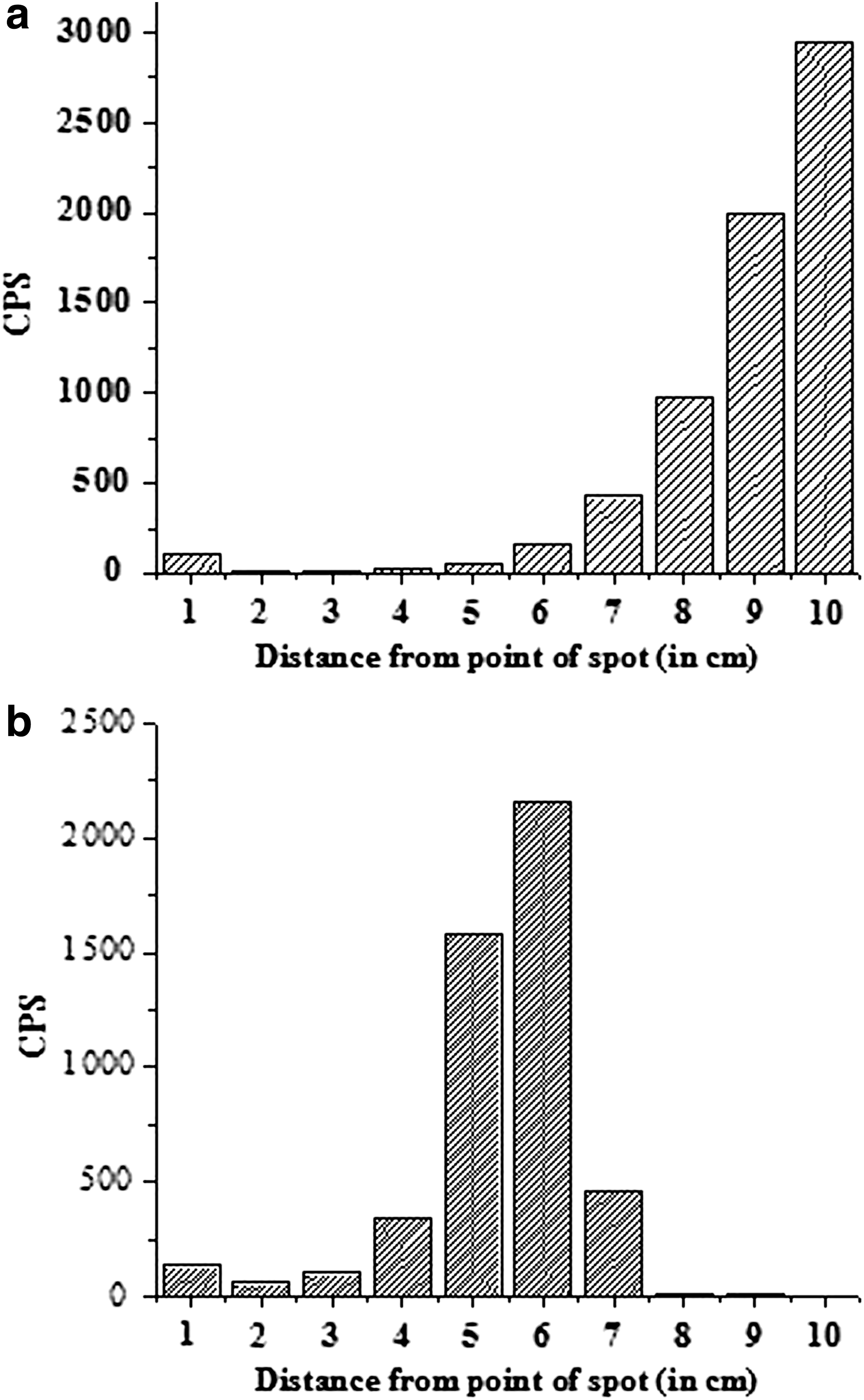
Typical thin-layer chromatography patterns of
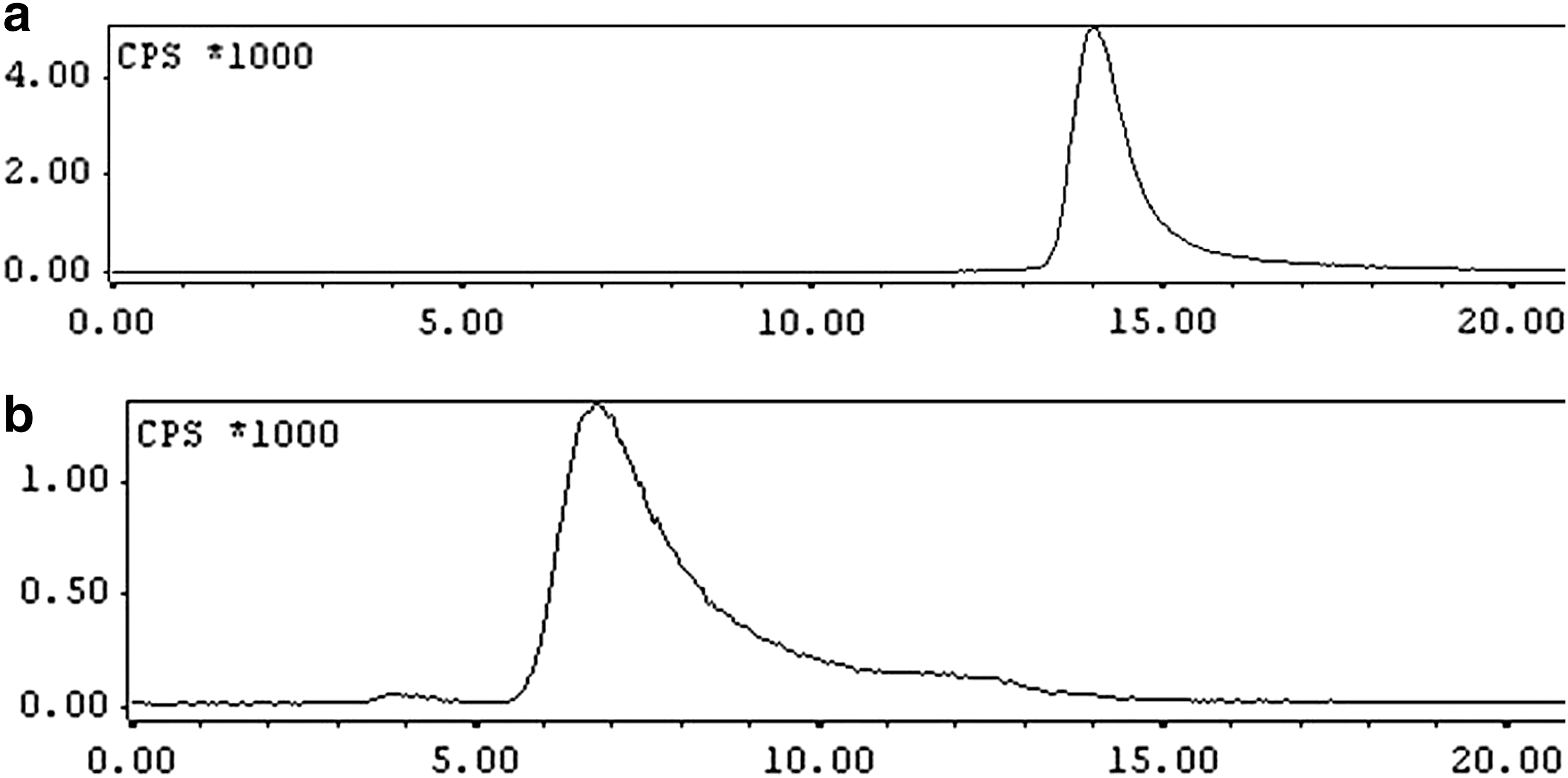
Typical HPLC profiles of
Summary of Quality Control Results for 68Ga-BPAMD Prepared Using Freeze-Dried BPAMD Kit
As per Ph. Eur. Monograph.
RCP, radiochemical purity.
Determination of kit capacity with respect to the addition of maximum volume of 68GaCl3
As the various commercially available 68Ge/68Ga radionuclide generators need to be eluted with HCl of different molarities, it is essential to know the maximum volume of 68GaCl3 that can be added in the kit vial for the preparation of 68Ga-BPAMD without compromising its RCP. Therefore, gradually increasing volumes of 68GaCl3, eluted from different 68Ge/68Ga radionuclide generators, were added in the kit vial and the corresponding complexation yields were determined. It is well reported in the literature that radiolabeling yield of 68Ga-BPAMD remains optimum for clinical usage when the pH of the reaction mixture remains in between 3 and 4. 5 Therefore, maximum volume of 68GaCl3 eluate that can be added in the kit is also limited by the fact that pH of the resulting solution after the addition of 68GaCl3 should remain within this range. Pattern of variation in pH of the reaction mixture and percentage radiolabeling yield of 68Ga-BPAMD complex upon addition of different volumes of 68GaCl3 obtained from three different commercial 68Ge/68Ga generators is shown in Table 2. It is evident from this table that a maximum volume of 2.0 mL of 68GaCl3 can be added in the kit vial if 68Ga is sourced from the ITG- and Eckert & Ziegler-make 68Ge/68Ga generators, while maximum volume of 68GaCl3 that can be added in the kit vial should be restricted to 0.5 mL when 68Ga is obtained from iThemba-make 68Ge/68Ga generator.
Pattern of Variation in pH and Percentage Radiolabeling Yields Upon Addition of Different Volumes of 68GaCl3 Eluate Obtained from Three Different Commercial 68Ge/68Ga Generators Viz. ITG, Eckert-Ziegler, and iThemba
Stability of 68Ga-BPAMD
68Ga-BPAMD was found to be stable both in HEPES buffer as well as in human blood serum as revealed by TLC studies. A percentage radiochemical yield >95 was found to be maintained even after 2 h postpreparation, up to which the study was continued.
Binding studies with hydroxyapatite
Binding studies of 68Ga-BPAMD with HA particles revealed a binding of 91% ± 0.28%. This shows that the affinity of 68Ga-BPAMD toward HA particles remained unaltered due to formulation of freeze-dried BPAMD kit.
Shelf-life of freeze-dried BPAMD kits
Shelf-life determination of freeze-dried BPAMD kits was carried out to ensure the efficacy of the kits after storage for longer periods at 2–8°C. The kits were found to exhibit good radiolabeling yields (>95%) when radiolabeled with 68Ga eluted from 68Ge/68Ga generators, obtained from different suppliers after an incubation period of 6 months, up to which the studies were continued.
Biodistribution studies
Biodistribution studies were carried out in normal healthy Wistar rats to validate the pharmacokinetics as well as skeletal affinity of 68Ga-BPAMD prepared using the freeze-dried BPAMD kit. The results of biodistribution studies are tabulated in Table 3. The biodistribution studies revealed that at all the three postadministration time points viz. 30 min, 1 h, and 2 h, majority of the injected activity (IA) is localized in the skeleton (2.22% ± 0.43%, 2.10% ± 0.75%, and 2.50% ± 0.28% IA/g at 30 min, 1 h, and 2 h, respectively) with insignificant accumulation in the nontarget organs/tissues such as, blood, muscle, lungs, liver, and kidneys. The agent exhibited fast blood clearance (retention of only 0.05% ± 0.01% IA/g at 1 h and complete clearance by 2 h postadministration time point) with majority of the nonaccumulated activity excreted through the urinary pathway. The biodistribution pattern of 68Ga-BPAMD recorded in normal Wistar rats was found to be akin to that reported in the contemporary literature. 5,16,17
Biodistribution Pattern of 68Ga-BPAMD in Normal Wistar Rats (n = 3)
Clinical studies
68Ga-BPAMD, prepared using the freeze-dried kit, was administered for whole-body scanning of patients who were referred for evaluation of skeletal metastases or arthritis. Toward this, 185–370 MBq (5–10 mCi) of the radiolabeled preparation was administered in each patient and whole-body scintigraphy was performed at 30–60 min postadministration time point. Figure 3a shows the whole-body PET scan of a prostate cancer patient recorded after 60 min of 68Ga-BPAMD administration, while Figure 3b shows the whole-body scans of the same patient recorded at 4 h postadministration of 99mTc-MDP. The PET scan clearly demonstrates focal lesions with increased uptake involving the skull, sternum, lower thoracic vertebrae, lower left ribs, and pelvic bones. The MDP images also demonstrate some of these lesions. However, the lower thoracic, sacral, skull, and rib lesions are not well appreciated in the SPECT scan due to the lower spatial resolution of the gamma camera, suggesting much higher sensitivity of the PET scans to pick up tiny lesions. SPECT (99mTc-MDP) and PET-CT (68Ga-BPAMD) scans of another prostate cancer patient are shown in Figure 4a and b, respectively. The PET-CT scans demonstrate multiple additional lesions in the ribs, pelvis, and vertebrae of the patient. The lesions were found to exhibit SUV (standardized uptake value) in the range of 39–56 compared to normal bones, where SUV was observed to be in the range of 4–6, thereby clearly demonstrating the preferential accumulation of the radiotracer in the bone lesions.
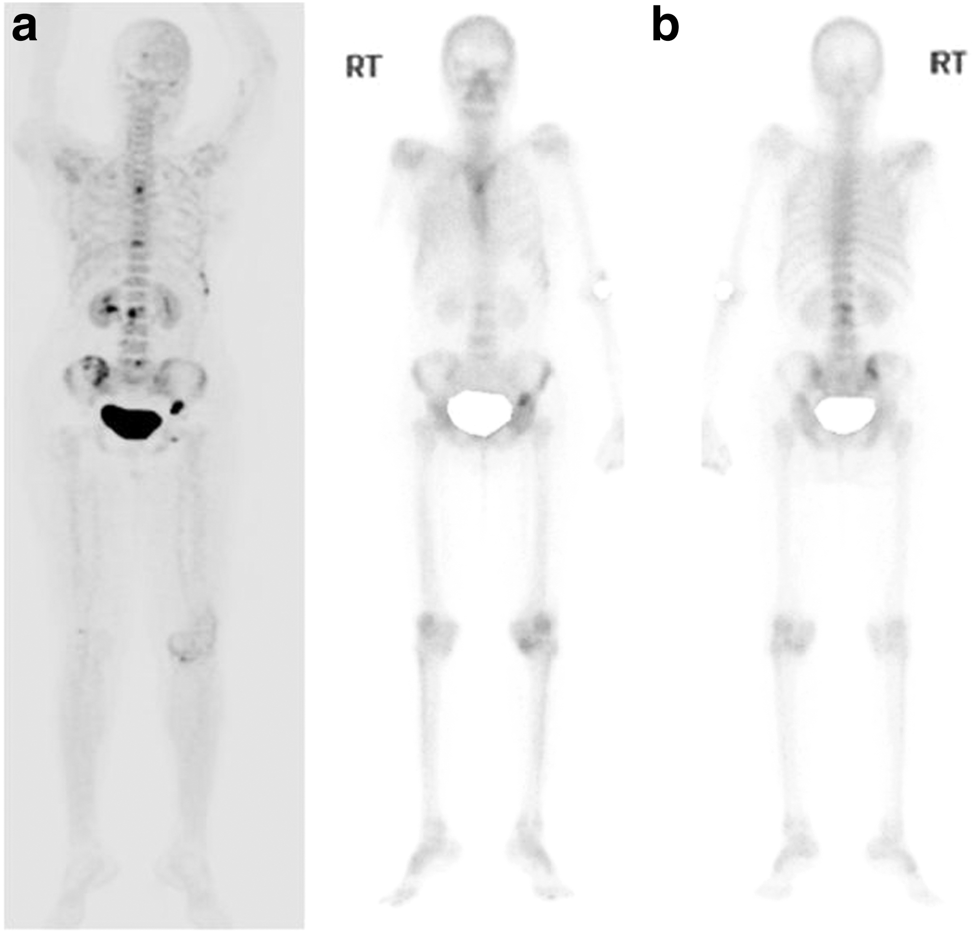
Whole-body scans recorded in the same patient after administration of

Whole-body scans (SPECT and PET-CT) of the same patient after administration of
Discussion
Skeletal scintigraphy is one of the most commonly used imaging procedures performed in the nuclear medicine centers across the globe to diagnose and assess the severity of a variety of bone diseases and conditions, specially metastatic spread of cancer, which is frequently experienced by patients suffering from breast, prostate, or lung carcinoma at the advanced stage of their diseases. 26,27 This makes 99mTc-MDP as one of the most used SPECT radiopharmaceuticals in the nuclear medicine practice. However, with the advancement of PET, which provides higher spatial resolution and thus better quality images compared to those obtained with SPECT, the use of [ 18 F]NaF is gaining momentum. 26,27 It has been reported in the contemporary literature that bone scan with [ 18 F]NaF provides improved sensitivity and specificity over conventional planar and SPECT bone scans. 28,29 However, to ensure the availability of [ 18 F]NaF at the hospital radiopharmacy, the presence of an on-site medical cyclotron facility or accessibility of the agent from the nearby region is absolutely essential. 18,30
This problem can be circumvented by using suitable 68Ga-based agents, as this PET radionuclide can readily be obtained from a commercially available 68Ge/68Ga radionuclide generator, which can be housed in the hospital radiopharmacy. 19 The possibility of multiple elutions in a single day along with the long shelf-life of 68Ge/68Ga radionuclide generator ensures regular and continuous supply of 68Ga, making it an attractive radionuclide for developing PET radiopharmaceuticals. 19 Although the development of quite a few 68Ga-based skeletal imaging agents is documented in the literature, 68Ga-BPAMD has received maximum attention owing to the very high target-to-soft tissue ratio exhibited by this agent along with fast renal clearance and comparable SUVs with those of [ 18 F]NaF. 10
The primary objective of this study was to develop a freeze-dried kit of BPAMD, which will enable convenient and single-step formulation of 68Ga-BPAMD at the hospital radiopharmacy for clinical utilization. In addition, efforts were also directed to develop the kit in such a manner so that it can be suitably used for the preparation of 68Ga-BPAMD using the 68Ga obtained from various commercial 68Ge/68Ga radionuclide generators. It is worthwhile to mention that the most widely used 68Ge/68Ga radionuclide generators are commercially available from reputed manufacturers such as Isotope Technologies Garching (ITG), Cyclotron Company (Obninsk, Russia), Eckert & Ziegler Isotope Products, and iThemba Labs, and are eluted with HCl of different molarities. 22 Availability of such a kit will help the nuclear medicine centers use any commercially available 68Ge/68Ga radionuclide generator for the formulation of 68Ga-BPAMD patient dose without the need of reoptimizing the radiolabeling protocol.
Working in this direction, the authors have developed a HEPES-based freeze-dried BPAMD kit, comprising 50 μg of BPAMD and ∼150 mg HEPES, and evaluated the performance of the kit using 68Ga eluted from three different 68Ge/68Ga radionuclide generators viz. ITG, Eckert & Ziegler (Germany), and iThemba Labs (South Africa), which were eluted with three different molarities of HCl (0.05, 0.1, and 0.6 M, respectively). Although no attempt had been made to evaluate the performance of the kit with 68Ga obtainable from 68Ge/68Ga generator supplied by Cyclotron Company (Obninsk, Russia), it is logical to believe that the kit can be successfully used with this generator also, as 68Ga is eluted from this generator using 0.1 M HCl. This study also showed that a maximum of 2 mL of 68GaCl3, obtained from either ITG- or Eckert & Ziegler-make 68Ge/68Ga generator, could be added in the kit vial without compromising the RCP of 68Ga-BPAMD. However, when the 68Ga is obtained from iThemba-supplied 68Ge/68Ga generator, maximum volume of 68GaCl3 that could be added in the kit should be restricted to 0.5 mL. This limitation of using lesser volume of 68GaCl3 is imposed due to the requirement of using HCl of higher molarity while eluting 68Ga from this type of generator.
One of the basic drawbacks of the developed kit arises from the fact that there exists a volume restriction on 68GaCl3, which can be added in the kit vial for the preparation of 68Ga-BPAMD with adequate RCP. This may become an impediment toward the usage of this kit at the later stage of generator life, particularly with iThemba-make 68Ge/68Ga radionuclide generator. To circumvent this problem, another freeze-dried BPAMD kit was formulated using higher amount of HEPES buffer (1.25 M, ∼300 mg), following the same protocol mentioned in the Experimental section. More volume of 68GaCl3 could be added in this kit for the preparation of 68Ga-BPAMD without comprising the RCP of the agent (Table 4). However, as the physiological effect of using higher amount of HEPES is not well reported in the literature, the authors have restrained from further evaluation of this kit in animal model and human patients.
Pattern of Variation in pH and Percentage Radiolabeling Yields Upon Addition of Different Volumes of 68GaCl3 Eluate Obtained from iThemba-Make 68Ge/68Ga Generator in Kits Having Higher Amount of HEPES
RCP of 68Ga-BPAMD, prepared using the developed freeze-dried BPAMD kit, was determined by TLC as well as HPLC studies and found to be >95% under optimized conditions. The validation of the efficacy of the agent was carried out by in vitro binding studies with artificial bone matrix constituent, that is, with HA particles as well as by biodistribution studies in normal healthy Wistar rats. Strong binding of 68Ga-BPAMD with HA particles (∼91%) indicated the affinity of the radioactive preparation toward bone matrix. Biodistribution studies showed preferential localization of the radiolabeled agent in the skeleton along with insignificant accumulation in the nontarget organs/tissues. The nonaccumulated activity exhibited fast and major clearance through the renal route. The pharmacokinetic behavior and biodistribution pattern exhibited by 68Ga-BPAMD, prepared using the freeze-dried kit, are akin to the biological distribution of the agent reported in the literature, 5,16,17 which proves that lyophilization and use of HEPES buffer have no adverse effect on the biological activity of the agent.
Gallium-68-BPAMD, formulated using the developed kit, has also been evaluated clinically by administering the preparation in human patients. Comparative scans recorded with 68Ga-BPAMD and 99mTc-MDP in the same patient showed picking up of the identical/additional lesions with superior image quality when 68Ga-BPAMD was used for imaging. This indicates that the developed kit can be successfully used for the easy and convenient preparation of 68Ga-BPAMD for clinical exploitation.
Conclusions
A freeze-dried formulation of BPAMD in the form of a ready-to-use lyophilized kit was prepared. The performance of the freeze-dried kit was evaluated with 68GaCl3 eluted from three different commercial 68Ge/68Ga radionuclide generators. Efficacy of 68Ga-BPAMD prepared using the freeze-dried kit was confirmed by carrying out different studies such as in vitro binding with hydroxyapatite particles, biodistribution studies in healthy animal model, and PET imaging in cancer patients. All these studies revealed that the developed kit can be suitably used with 68Ga eluted from different 68Ge/68Ga radionuclide generators for the preparation of 68Ga-BPAMD patient dose. The methodology standardized during the course of this work can be deployed for the preparation of freeze-dried kits suitable for the formulation of 68Ga-based radiopharmaceuticals using 68Ga obtainable from any commercial 68Ge/68Ga radionuclide generator.
Footnotes
Acknowledgments
The authors thankfully acknowledge Dr. P.K. Pujari, Associate Director, Radiochemistry and Isotope Group, Bhabha Atomic Research Centre (BARC), for his constant encouragement and support. The authors are grateful to Dr. V.R. Lele, Chief, Department of Nuclear Medicine and PET-CT, Jaslok Hospital (Mumbai), and Dr. V. Rangarajan, Professor and Head, Department of Nuclear Medicine and Molecular Imaging, Tata Memorial Hospital (Mumbai), for kindly providing the 68Ge/68Ga radionuclide generators used in this study. The authors thank the staff members of the Animal House Facility, Radiation Biology and Health Sciences Division, BARC, and Department of Nuclear Medicine & PET, Kovai Medical Center and Hospital, for the help received during animal experimentations and clinical studies, respectively.
Disclosure Statement
No competing financial interests exist.