
Abstract
Objective:
To investigate the correlations of miR-31 expression with cell proliferation, invasion, and prognosis of patients with head and neck squamous cell carcinoma (HNSCC).
Methods:
The expression of miR-31 in human laryngeal cancer TU686 cells, human nasopharyngeal carcinoma CNE-2 cells, and normal human oral keratinocyte (NHOK) epithelial cells was detected via quantitative real-time polymerase chain reaction (qRT-PCR). The effects of miR-31 on the proliferation and invasion of HNSCC cells were explored through transfecting miR-31 analogs (miR-31 mimics) and miR-31 inhibitors (anti-miR-31). qRT-PCR was applied to detect the expressions of miR-31 in 56 cases of HNSCC tumor tissues and tumor-adjacent normal tissues. The correlation of miR-31 expression with pathological parameters and survival prognosis of HNSCC patients was also analyzed.
Results:
The expressions of miR-31 in TU686 and CNE-2 cell lines were significantly higher than that in NHOK cells (p < 0.01). Compared with those in the negative control group, the proliferation and invasion abilities of cells transfected with miR-31 mimics were notably enhanced (p < 0.01), and those of cells transfected with anti-miR-31 were significantly reduced (p < 0.01). In addition, miR-31 mimics significantly reduced ARID1A expression (p < 0.01) and anti-miR-31 increased its expression (p < 0.05). The expression of miR-31 in tumor tissues of HNSCC patients was remarkably higher than that in tumor-adjacent normal tissues (p < 0.01). This, together with clinical data analysis, revealed that the expression of miR-31 was associated with tumor differentiation, metastasis, and staging of patients, and the survival period of patients with lowly expressed miR-31 was longer.
Conclusions:
The highly expressed miR-31 can stimulate the proliferation and invasion of HNSCC cells, closely correlated with tumor differentiation, metastasis, and staging of patients. Patients with lowly expressed miR-31 have a longer survival period. Therefore, miR-31 expression can be taken as a crucial reference indicator for the prognosis of HNSCC patients.
Introduction
Head and neck tumor is classified based on its location in the body. Statistics has manifested that the incidence rate of the tumor in China is as high as 15/100,000 per year, accounting for about 4.45% of the total proportion of malignant tumors. Among the tissue subtypes of the head and neck tumor, the squamous cell carcinoma takes up 94.6%. 1,2 According to the statistics of the World Health Organization, there are 500,000 new cases of head and neck squamous cell carcinoma (HNSCC) each year, whose most tissues have been in the middle and advanced stages when definitely diagnosed. The complicated anatomy and dense organ distribution of the head and neck lead to the high risk of surgery and a great difficulty in treatments. 3 Studies have demonstrated that the pathogenesis and progression of HNSCC are subjected to joint actions by multiple factors and mechanisms, including mutations of oncogenes and tumor suppressor genes as well as incorrect pairing of DNA during replication. 4 –10
MicroRNAs (miRNAs) are small RNAs consisting of 21–26 nucleotides, which play various biological roles in the human body. With the increase of studies in recent years, miRNAs have been found to have specific action targets and pair with the target noncoding regions, thus further regulating the translation level of messenger RNAs (mRNAs). 11,12 Under physiological conditions, miRNAs participate in normal life activities such as cell proliferation, differentiation, migration, and apoptosis. According to the existing reports, the occurrence and development of malignant tumors are often accompanied by the abnormal expression of miRNAs. 13
In some tumors, targeted detection of miRNA molecules will be of vital guiding significance for the early diagnosis and treatment of lesions. Researchers discovered miR-31 for the first time in the study on HeLa cells, 14 and studies have revealed that miR-31 expressions are upregulated in lung cancer and colorectal cancer. 15,16 miR-31 not only speeds up the growth of colon cancer cells but also stimulates the invasion and metastasis of these cells, as well as the progression of pathological staging, thereby resulting in the aggravation of poor prognosis. 17
The correlations of the expression level of miR-31 in HNSCC cell lines and the patient's tumor tissues with cell proliferation, invasion, and prognosis have not been reported. In this study, the quantitative real-time polymerase chain reaction (qRT-PCR) testing technique was adopted to detect the expression of miR-31 in HNSCC cell lines and clinically resected tissues. After transfection with miR-31 analogs (miR-31 mimics) and miR-31 inhibitors (anti-miR-31), the effects of miR-31 on the proliferation and invasion abilities of HNSCC cells were explored, and the effect of miR-31 on the prognosis of HNSCC patients was investigated in combination with clinical data.
Materials and Methods
Materials
Human laryngeal cancer TU686 cells, human nasopharyngeal carcinoma CNE-2 cells, and normal human oral keratinocyte epithelial normal human oral keratinocyte (NHOK) cells (Cell Bank of Chinese Academy of Sciences, Shanghai, China); Roswell Park Memorial Institute (RPMI)-1640 medium and fetal bovine serum (FBS; Gibco, New York); RNA extraction kit (Invitrogen, Carlsbad, CA); qRT-PCR kit (TaKaRa, Dalian, China); Transwell chamber (Corning, New York); Lipofectamine 2000 and primers (Invitrogen, Carlsbad, CA); and anti-miR-31 and negative control (NC) sequences of small interfering RNA (Biomics Biotechnology Co., Ltd., Nantong, China).
In clinical research, HNSCC tumor tissues and the corresponding cancer-adjacent normal tissues of a total of 56 cases undergoing surgical treatment in hospital from January 2013 to December 2015 were collected. The relevant specimens were taken out and quickly stored in liquid nitrogen. Among them, there were 32 males and 24 females with an average age of 63.2 ± 16.8 years, including 21 cases of hypopharyngeal squamous cell carcinoma and 25 cases of laryngocarcinoma. All the patients were definitely diagnosed with HNSCC, before which they received no radiochemotherapy, and this was the first operation for them. All the involved patients or their family members signed the informed consent, and the study was approved by the Ethics Committee of The First Affiliated Hospital of Bengbu Medical College.
Cell culture
In the experiment, SAS, CAL27, and NHOK cells were cultured using 1640 medium containing 10% FBS for cell growth. The cells were cultured at 37°C and 5% CO2 and subjected to passage for 48 h, and those in the logarithmic growth phase were taken and paved on the plate for application in subsequent experiments.
Detection of the expression of miR-31 mRNA in cells and tissue specimens of patients via qRT-PCR
Total RNAs in each group of cells and tissues were extracted using the TRIzol method, and 1 μg of total RNA was taken from each group and subjected to reverse transcription according to the kit instructions to obtain complementary DNA. The expressions of miR-375 and miR-221 mRNAs were detected according to the instructions of the fluorescence qRT-PCR kit, with U6 RNA as an internal reference control. The miR-375, miR-221, and U6 primer sequences are shown in Table 1. Reaction conditions were as follows: at 95°C for 10 min, 95°C for 15 s, and 60°C for 1 min, and 40 cycles of amplification. The Ct value was output from the instrument software, and the relative expression level was calculated using the 2−ΔCt method according to the following formula: ΔCt (target gene) = Ct (target gene) − Ct (control gene). The relative expression was presented as a fold change relative to U6.
Polymerase Chain Reaction Primer Sequences
Transfection methods of miR-31 mimics and anti-miR-31
The miR-31 mimics and anti-miR-31 sequences as well as the NC sequences are shown in Table 2. The operating procedures are as follows. The cells in the logarithmic growth phase were paved on six-well culture plates, and each well was added with 2 μg of transfection reagents together with miR-31 mimics and anti-miR-31 (final concentration: 15 nmol/L). Transfection was carried out with Lipofectamine 2000. Patients in the experiment were divided into NC group, miR-31 mimics group, and anti-miR-31 group.
Transfection Sequences of miR-31 Mimics and Anti-miR-31
NC, negative control.
Effects of miR-31 mimics and anti-miR-31 on the cell proliferation ability
After the cells were transfected for 48 h, they were digested into single cell suspensions and inoculated in 96-well culture plates at a density of 10 4 cells/well. After standing in 24 h of culture, 20 μL of 3-(4,5-dimethylthiazol-2-yl)-2,5-diphenyltetrazolium (MTT) solution at a concentration of 5 μg/μL was added. After culturing for 4 h in a dark place, 150 μL of dimethyl sulfoxide solution was added, shaken, and evenly mixed. Then, the optical density value was measured at a wavelength of 490 nm, and the cell activity was calculated.
Effects of miR-31 mimics and anti-miR-31 on the cell invasion ability
The Transwell chamber was applied to examine the invasion ability of cells. After the chambers were processed based on the operating procedures of the instructions, the transfected cells were prepared in the single cell suspension at a density of 4 × 10 5 /mL. Subsequently, to the upper chamber were added 100 μL of serum-free medium and 100 μL of cell suspension, which were evenly mixed. Then, to the lower chamber was added 500 μL of medium with FBS. Forty-eight hours later, the cells were fixed, stained, observed under a microscope, and analyzed through photographing.
Western blot analysis of ARID1A expression
After transfection, protein was isolated followed by separation on 10% sodium dodecyl sulfate/polyacrylamide gel electrophoresis and subsequent transferring to polyvinylidene fluoride membrane. Then, the membrane was blocked with 5% defatted milk powder for a 2-h room temperature incubation. After being rinsed in phosphate-buffered saline, the membrane was incubated with a primary antibody (ARID1A at 1:2000 or β-actin at 1:2000) at 4°C overnight. On the next day, the membrane was rinsed in PBST and incubated with the horseradish peroxidase-conjugated goat anti-rabbit secondary antibody at room temperature for 30 min. The membrane was developed using ECL reagent for 1 min, followed by X-ray exposure. Protein imaging processing software and Quantity One system were used to scan X-ray film and to measure band densities. All experiments were repeated for four times (n = 3) for statistical analysis. The expression of ARID1A was presented as a ratio relative to β-actin.
Correlations of the expression of miR-31 in tissues of HNSCC patients with pathological parameters and survival prognosis
Based on the median expression level of miR-31 in HNSCC tissues, the patients were divided into the miR-31 low-expression group and miR-31 high-expression group. Tumor volume was measured by magnetic resonance imaging or computerized tomography scan and presented as cm 3 . The correlation between the expression of miR-31 and the patient's pathological progression was analyzed on the basis of relevant clinical data. All the 56 patients were followed up after operation for 5 years, with the follow-up rate of 100%. The survival period was from the first day after the operation to the last date of the follow-up or death of patients. Statistical analysis was conducted monthly.
Statistical processing
Data of this study were processed using Statistical Product and Service Solutions (SPSS) 17.0 software (International Business Machines Corporation) and expressed as mean ± standard deviation. The t-test was adopted for comparison between groups. The univariate survival analysis was performed using the Kaplan–Meier method, and log-rank was applied to examine the differences in survival curves. p ≤ 0.05 represented that the difference was statistically significant.
Results
Detection of the expression of miR-31 in cell lines via qRT-PCR
The results revealed that compared with that in normal human oral keratinocyte epithelial NHOK cells, the relative expression levels of miR-31 in SAS and CAL27 cells were notably increased, displaying statistically significant differences (p < 0.01) (Fig. 1).
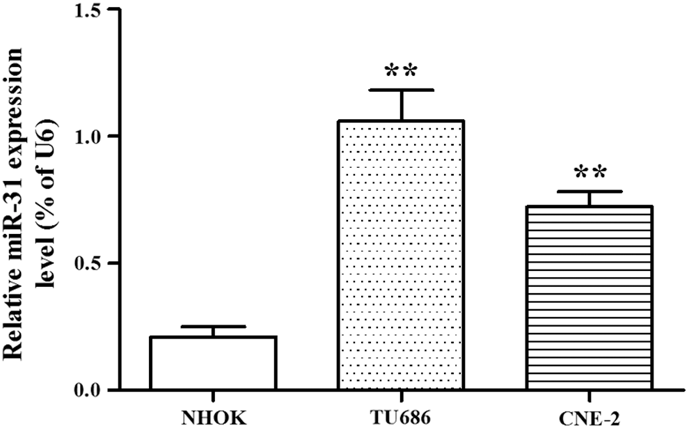
Detection of the expressions of miR-31 in NHOK, SAS, and CAL27 cells via quantitative real-time polymerase chain reaction. **p < 0.01 versus NHOK cells. NHOK, normal human oral keratinocyte.
Effects of transfection with miR-31 mimics and anti-miR-31 on the cell proliferation ability
The transfection efficacy was 68% for miR-31 mimics and 72% for anti-miR-31. The miR-31 expression was significantly increased (p < 0.01) after transfection of miR-31 mimics and decreased after transfection of anti-miR-31 (p < 0.05) compared with NC transfection (Fig. 2A, B). The effects of miR-31 mimics and anti-miR-31 on the proliferation abilities of SAS and CAL27 cells were detected by MTT assay, which manifested that the proliferation rates of SAS and CAL27 cells were markedly increased after transfection with miR-31 mimics (p < 0.01) but evidently decreased after transfection with anti-miR-3 (p < 0.01) (Fig. 2C, D).
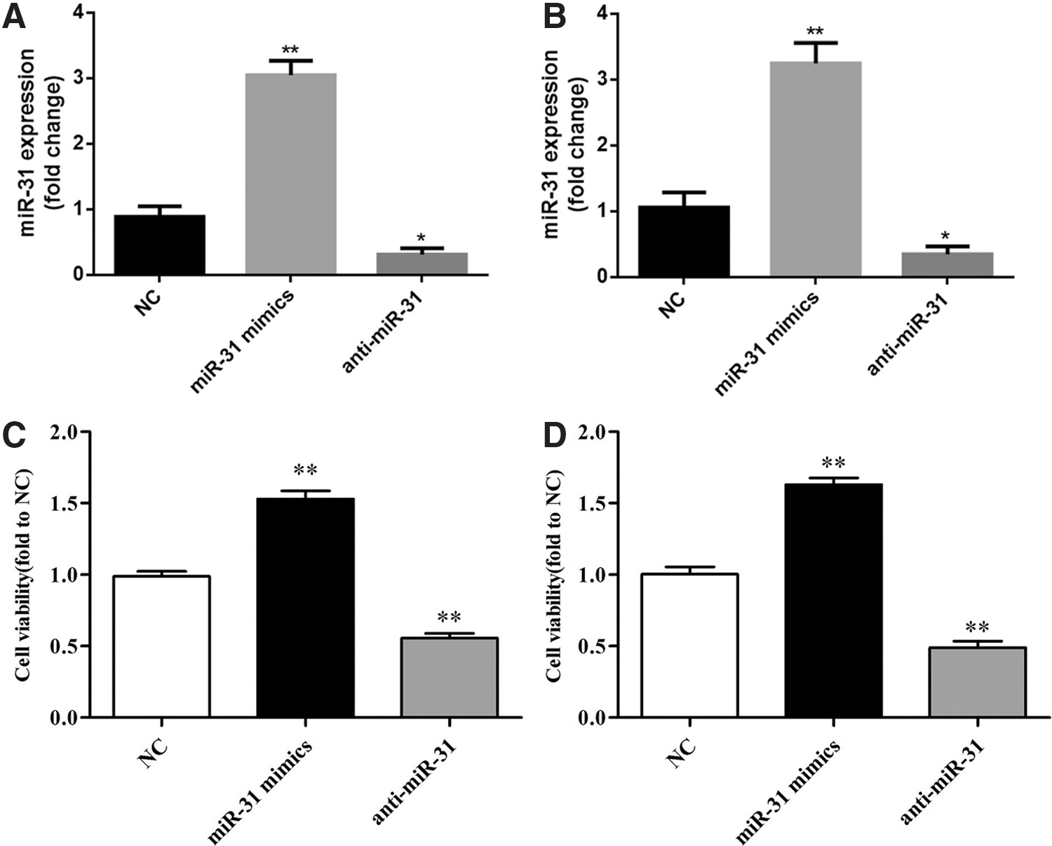
miR-31 expression
Effects of transfection with miR-31 mimics and anti-miR-31 on the cell invasion ability
Transwell cell invasion assay was adopted to detect the effect of miR-31 on the invasion of HNSCC cells. The results demonstrated that after miR-31 mimics were transfected, the number of SAS and CAL27 cells passing through the membrane was remarkably increased (p < 0.01), but after transfection with anti-miR-31, the number significantly declined (p < 0.01) (Fig. 3).
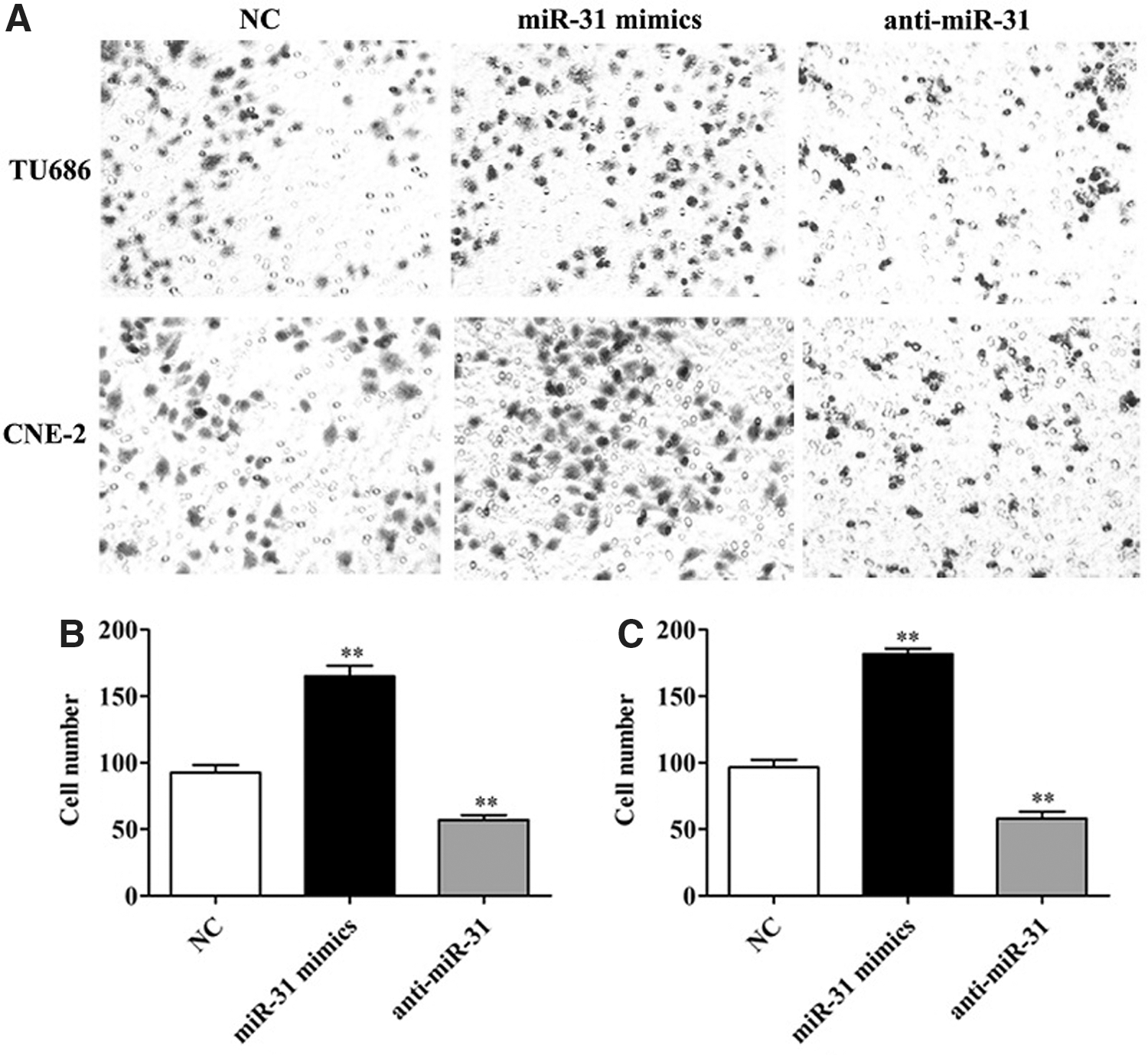
Cell invasion ability of SAS and CAL27 cells after transfection.
Effect of miR-31 on ARID1A expression
As ARID1A is a tumor suppressor gene, the authors next evaluated whether miR-31 affects the expression of ARID1A expression. As seen in Figure 4, compared with NC, miR-31 mimics' treatment significantly reduced ARID1A expression (p < 0.01). However, anti-miR-31 treatment significantly increased ARID1A expression compared with NC (p < 0.05).
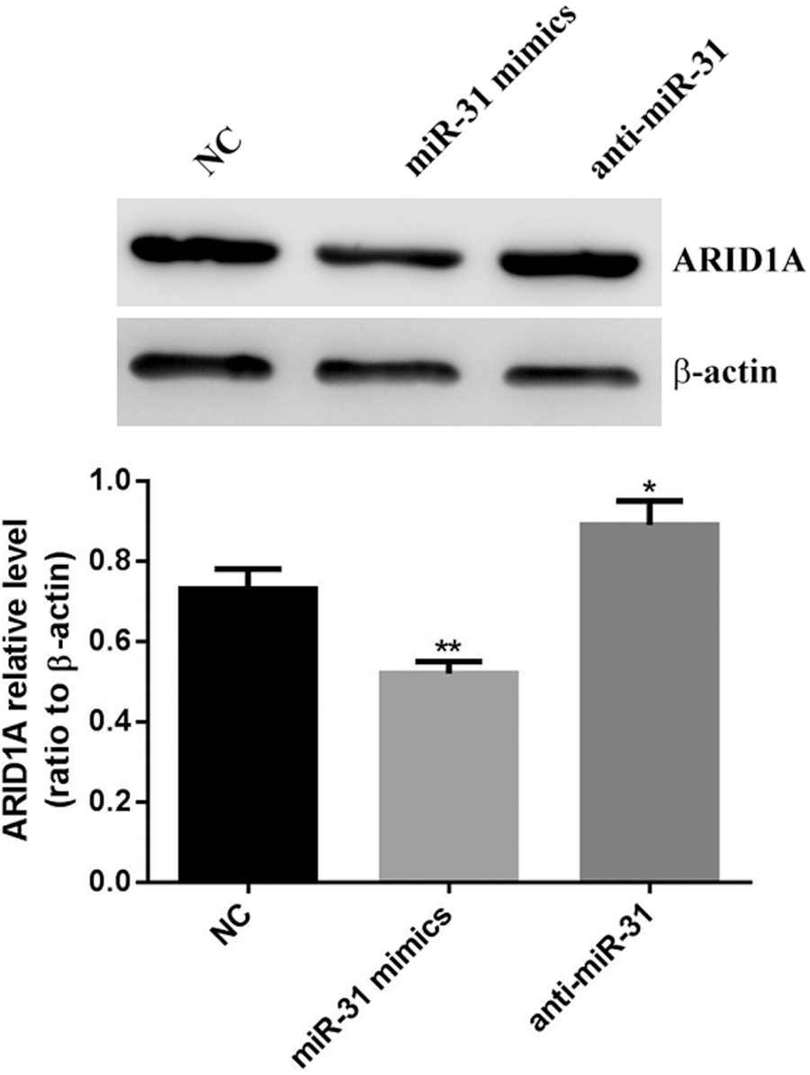
Expression of ARID1A after miR-31 transfection. **p < 0.01 and *p < 0.05 versus NC group.
Detection of the expression of miR-31 in specimens via qRT-PCR
The results manifested that compared with that in cancer-adjacent normal tissues, the expression level of miR-31 in HNSCC tissues was obviously raised, and the difference was statistically significant (p < 0.01) (Fig. 5).
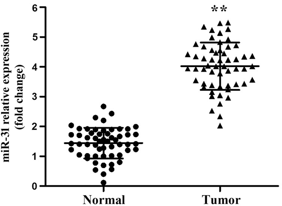
Expressions of miR-31 in tumor tissues and cancer-adjacent normal tissues of HNSCC patients. **p < 0.01 versus cancer-adjacent normal tissues. HNSCC, head and neck squamous cell carcinoma.
Correlations of the expression of miR-31 with HNSCC clinicopathological parameters
The analysis results of the correlations of the expression of miR-31 with HNSCC clinicopathological parameters illustrated that the expression of miR-31 was related to the degree of tumor differentiation, tumor metastasis, and clinical staging (p < 0.01), but not associated with the patient's gender, age, and tumor size (p > 0.05) (Table 3).
Correlations of the Expression of miR-31 with Clinicopathological Parameters of Head and Neck Squamous Cell Carcinoma Patients
Relationship between the expression of miR-31 and the survival prognosis of HNSCC patients
Analysis of Kaplan–Meier survival curves in 56 patients with HNSCC revealed that the survival prognosis of patients with low expression of miR-31 was better, and differences in overall survival curves were analyzed using the log-rank test. The effect of miR-31 on the overall survival rate of HNSCC patients was statistically significant (p < 0.05) (Fig. 6). In addition, multivariable Cox proportional hazards analysis was performed to identify whether miR-31 was an independent prognostic covariate for HNSCC patients and showed that high miR-31 expression was associated with poor prognosis in terms of overall survival (p = 0.036), independent of other clinical covariates (Table 4).

Kaplan–Meier survival curves of the expression of miR-31 and HNSCC patients.
Multivariate Analysis of Various Prognostic Factors Using Cox Regression Analysis
CI, confidence interval.
Discussion
Reports have revealed that miRNAs, on the one hand, can regulate the expression of tumor-associated genes, and, on the other hand, have their own functional characteristics of tumor suppressor genes or oncogenes. 18 According to this study, miR-31 expression levels in different tumor cells are not the same. Besides, Valastyan 19 discovered that miR-31 can make the formed tumors smaller after lung metastasis in breast tumors, and the inhibition of miR-31 function will significantly enhance the invasion and metastasis abilities of breast cancer cells. As such, it can be concluded that miR-31 acts as a tumor suppressor gene in the occurrence and development of breast cancer. On the contrary, the highly expressed miR-31 in colon cancer has sped up the invasion and metastasis of tumors, 20 so miR-31 also possesses the characteristics of oncogenes.
Numerous studies have manifested that the expression and function of miR-31 vary in different tumor cells. Specifically speaking, the expression of miR-31 is remarkably upregulated in colorectal cancer, 21 lung cancer, 22 and oral cancer, 23 but significantly downregulated in gastric cancer, 24 breast cancer, 25 and ovarian cancer. 26 Using next-generation sequencing as an agnostic discovery platform, Leidner et al. 27 found that miR-31 showed frequent downregulation only in patients with high-grade dysplasia and esophageal adenocarcinoma, suggesting it might be associated with transition from Barrett's metaplasia to high-grade dysplasia, implying miR-31 plays a role in the early-stage malignant progression.
It was found in this experiment that compared with that in normal tissues, the expression of miR-31 in esophageal squamous cell carcinoma obviously went up. Hence, the expression level of miR-31 differed in different tumor tissues, and the miR-31 expression was increased in squamous tumors but tended to decline in the reproductive system cancer cells. The above studies have manifested that the expression of miR-31 may be tissue specific. 28 It is speculated that the reason for this may be that miRNAs can be secreted and identified by different cells. In different specific cancer cells, if miR-31 mainly regulates tumor suppressor factors, the specific biological function of miR-31 is manifested as a tumor suppressing tendency. Conversely, if miR-31 mainly exerts an effect on regulating oncogenes, the specific biological function of miR-31 shows a tendency to promote cancer. Some miRNAs do not express proteins themselves, but the nature of target genes determines their tissue specificity.
In the present study, the authors showed a significantly higher miR-31 expression in patients with HNSCC, consistent with a previous study revealing a significantly higher level of miR-31 expression in patients with HNSCC, 29 supporting a role of miR-31 in the development and pathogenesis of HNSCC. In addition, miR-31 was upregulated in 77.8% of the ESCC tissues together with significantly increased serum miR-31 levels. 30 Furthermore, following in vitro studies showed that miR-31 promoted ESCC colony formation, migration, and invasion. Consistent with this, the present study also demonstrated that miR-31 promotes HNSCC cell proliferation and invasion.
Conclusions
qRT-PCR was applied in this study to detect the expression levels of miR-31 in human oral squamous cell carcinoma SAS cells, human tongue squamous cell carcinoma CAL27 cells, and normal human oral keratinocyte epithelial NHOK cells, which indicated that the expressions of miR-31 in SAS and CAL27 cell lines were significantly higher than that in NHOK cells. Through transfection with miR-31mimics and anti-miR-31, the effects of miR-31 on the proliferation and invasion abilities of HNSCC cells were investigated. Compared with those in NC group, the proliferation and invasion abilities of cells transfected with miR-31 mimics were obviously increased, while those of cells transfected with anti-miR-31 were evidently reduced. These results indicate that miR-31 has the function of promoting tumor cell proliferation and invasion in HNSCC cells. The expression of miR-31 in tumor tissues of 56 patients with HNSCC was markedly higher than that in cancer-adjacent normal tissues. This, in combination with clinical data analysis, demonstrated that the expression of miR-31 was related to tumor differentiation, metastasis, and staging of patients, and the survival period of patients with lowly expressed miR-31 was longer.
In summary, highly expressed miR-31 can stimulate the proliferation and invasion of HNSCC cells, closely related to tumor differentiation, metastasis, and staging of patients. The survival period of patients with lowly expressed miR-31 is longer, and miR-31 expression can be regarded as a crucial reference indicator for the prognosis of patients with HNSCC.
Footnotes
Disclosure Statement
No competing financial interests exist.