
Abstract
Cancer Biotherapy and Radiopharmaceuticals
officially retracts the paper entitled, “miR-424-5p Regulates Hepatoma Cell Proliferation and Apoptosis,” by Lianshu Piao, Fei Wang, Yanyan Wang, Zirong Yang, Qianwei Li, Lifeng Cui, and Qinggong Yu (Cancer Biother Radiopharm 2019;34(3):196–202. doi: 10.1089/cbr.2018.2625) due to the discovery that the paper was purchased from a paper mill by the lead author, Lianshu Piao, and that the conducted experiments were not those of the team of authors:
“I am very sorry to inform you that the paper was purchased. This paper was written and published by others. I did not do the experimental results myself, so I can't guarantee the authenticity of the experimental results. And [o]ther members of the paper did not know anything about it, including the corresponding author, Qinggong Yu.
[sic]”
The Editor and Publisher of Cancer Biotherapy and Radiopharmaceuticals are committed to preserving the scientific literature and the community it serves and does not tolerate any violations of scientific misconduct.
Introduction
Hepatocellular carcinoma (HCC) is a common malignant tumor in clinics. Its morbidity and mortality rates are relative high around the world.
1
–4
Yes-associated protein 1 (YAP1), a major effector and target protein of the classical Hippo-YAP signaling pathway, regulates the expression of target genes by entering the nucleus at the transcriptional coactivation form. Its expression level was significantly increased in various tumor tissues and cells.
5
–7
It was shown that YAP1 expression and function were obviously enhanced in HCC tumor tissues, suggesting that YAP1 plays an oncogenic role in the development of HCC.
8
–10
MicroRNA (miRNA) is a newly discovered noncoding, single-stranded small RNA 19–25 nucleotides in length. It is an important gene regulatory substance. miRNA mainly complementary binds with the 3′ untranslated region (3′-UTR) of the target gene mRNA to degrade the target mRNA or inhibit post-transcriptional translation, thereby participating in regulation of biological processes, such as cell growth, differentiation, apoptosis, and migration.
11,12
It was found that miR-424-5p expression was significantly reduced in tumor tissues of HCC patients, suggesting that miR-424-5p may play a role as a tumor suppressor gene in HCC.
13,14
Materials and Methods
Main reagents and materials
Hepatoma cells, HCCLM3 and MHCC97-L, and human normal liver cells, HL-7702, were purchased from Shanghai Cell Bank of Chinese Academy of Sciences. DMEM was purchased from Hyclone. Fetal bovine serum (FBS) was purchased from Gibco. Lip 2000 and Opti-MEM were purchased from Invitrogen. The ReverTra Ace qPCR RT Kit and SYBR dye were purchased from Toyobo (Japan). miR-NC, miR-424-5p mimic, and miR-424-5p inhibitor were purchased from Ribobio (Guangzhou, China). Rabbit anti-human YAP1 (Clone ID: EP1674Y) and β-actin antibodies were purchased from Abcam. Horseradish peroxidase-conjugated goat anti-rabbit secondary antibody was purchased from Jackson ImmunoResearch. RIPA protein extraction fluid was purchased from Beyotime (China). Annexin V/propidium iodide (PI) apoptosis detection reagent was purchased from BioLegend. The dual-luciferase reporter assay system and pMIR plasmid were purchased from Promega. The EdU cell proliferation assay kit was purchased from Molecular Probes, and this clinical trial was approved by the Affiliated Zhongshan Hospital of Dalian University.
Clinical information
HCC patients who underwent resection in our hospital from January 2017 to September 2017 were enrolled, including 39 males and 21 females with mean age of 54.8 (35–68) years. All patients did not have hepatitis and did not receive anti-inflammatory treatment during sample collection. No patients received antitumor treatment before surgery. In each case, the HCC diagnosis was confirmed by postoperative pathological examination. In brief, 5-μm-thick, formalin-fixed paraffin-embedded slides for further hematoxylin and eosin staining were formulated in accordance with the protocol of the Department of Pathology of our hospital. The tumor tissues were obtained from the operation.
Another 20 cases of normal liver tissues were collected as normal controls (Supplementary Fig. S1). Determination of tumor stage was based on the classification system of the American Joint Committee on Cancer/International Union Against Cancer (tumor–node–metastasis) 2002. Tumor differentiation was based on the Edmondson grading system. There were 11 cases in stage I, 12 cases in stage II, 20 cases in stage III, and 17 cases in stage IV. All patients had signed an informed consent, and this study was approved by the hospital ethics committee.
Cell culture
HCCLM3, MHCC97-L, and HL-7702 cells were cultured in DMEM containing 10% FBS and 1% penicillin–streptomycin at 5% CO2 and 37°C. The cells were passaged at a ratio of 1:4 and used for experiments in the logarithmic phase.
Dual-luciferase reporter gene assay
The HEK293T cell genomic mRNA was used as a template to amplify the fragment containing the targeted binding site in the 3′-UTR region of the YAP1 gene or its mutant fragment using the QuikChange Site-Directed Mutagenesis kit (Stratagene; Agilent Technologies, Inc., Santa Clara, CA). The PCR product was digested and connected into pMIR vector to transform DH5α. The positive clone was screened by PCR. The wild type (WT) or MT of YAP1 3′-UTR was cloned into the downstream of the firefly luciferase-coding region of pMIR-GLO™ luciferase vector (Promega Corporation, Madison, WI). The cloning procedure was performed by Yearthbio. The plasmid with a correct sequence was selected and named as pMIR-YAP1-WT or pMIR-YAP1-MUT.
pMIR-YAP1-WT (or pMIR-YAP1-MUT), miR-424-5p mimic (or miR-NC, miR-424-5p inhibitor), and pRL-TK were cotransfected into HEK293T cells using Lip 2000. After 48 h, the relative luciferase activity was detected using the dual-luciferase reporter assay system kit (Promega Corporation) according to the manual.
Cell transfection and grouping
HCCLM3 and MHCC97-L cells were cultured in vitro and divided into the miR-NC transfection group and miR-424-5p mimic transfection group. Ten microliters of Lip 2000, 50 nmoL miR-NC, and 50 nmoL miR-424-5p mimic were diluted with 100 μL of serum-free Opti-MEM, respectively. After incubating for 5 min, miR-NC and miR-424-5p mimic were mixed with Lip 2000 at room temperature for 20 min. Then, the mixture was added to the medium for 72 h.
Flow cytometry detection of cell apoptosis
Cells were collected and added with 100 μL of binding buffer. Then, cells were incubated with 5 μL Annexin V–fluorescein isothiocyanate and 5 μL PI, avoiding light for 10 min. Next, cells were resuspended after adding 400 μL of binding buffer. Cell apoptosis was tested on an FC500 MCL flow cytometer.
Flow cytometry detection of cell proliferation
After 72 h of transfection, cells were cultured with EdU solution at 10 μM in the logarithmic phase. After incubating for 48 h, cells were digested by trypsin and collected. After fixing in 100 μL of 4% paraformaldehyde, cells were incubated in 100 μL of penetration liquid at room temperature for 15 min and in 500 μL of reaction fluid at room temperature, avoiding light for 30 min. At last, the cell was tested using flow cytometry.
Quantitative reverse transcription–polymerase chain reaction
Total RNA was extracted using TRIzol and reverse transcribed to cDNA using the ReverTra Ace qPCR RT Kit. The RT reaction system contained 2 μg RNA, 1 μL dNTP, 4 μL RT buffer, 1 μL RT primer, 2 μL RT enzyme, 1 μL RNase inhibitor, and ddH2O. miRNA was extracted using the MicroRNA Extraction kit (Tiangen Biotech Co., Ltd., Beijing, China). RT-qPCR was performed with SYBR Premix Ex Taq (Takara Biotechnology Co., Ltd.) according to the manufacturer's instructions. GAPDH and RNU6B were used as normalizing controls for mRNA and miRNA quantification, respectively. The PCR reaction comprised 40 cycles at 95°C for 15 s, 60°C for 30 s, and 72°C for 30 s. Real-time PCR was performed on Bio-Rad CFX96 to test the relative expression.
Western blot
Total protein was extracted by RIPA at 4°C and centrifuged at 10,000 g for 15 min. The supernatant was moved to a new Ep tube for quantification. A total of 50 μg protein was separated by 10% sodium dodecyl sulfate–polyacrylamide gel electrophoresis for 3.5 h and transferred to a polyvinylidene fluoride membrane at 300 mA for 90 min. Next, the membrane was blocked in 5% skim milk at room temperature for 60 min and incubated with primary antibody at 4°C overnight (YAP1 and β-actin at 1:1000 and 1:5000, respectively). Then, the membrane was incubated with secondary antibody (1:8000) for 60 min after washing with PBST three times. Last, the protein expression was detected by ECL chemiluminescence.
Statistical analyses
All data analyses were performed with SPSS 18.0 software. The measurement data are depicted as mean ± standard deviation and compared by paired Student's t-test. Multiple groups were compared by one-way analysis of variance. The expression levels of miR-424-5p and YAP1 mRNA were compared by the Mann–Whitney U test. Pearson correlation analysis was performed for correlation analysis between miR-424-5p and YAP1 mRNA levels; p < 0.05 was considered as statistically significant.
Results
miR-424-5p expression declined, while YAP1 level was upregulated in HCC tissue
Quantitative reverse transcription–polymerase chain reaction (qRT-PCR) showed that YAP1 mRNA expression in HCC tumor tissues was significantly higher than that in normal liver tissues. The expression of YAP1 mRNA in stage III–IV HCC tissues was obviously higher than that in stage I–II HCC tissues (Fig. 1A). qRT-PCR demonstrated that the miR-424-5p level was markedly decreased in HCC tumor tissues compared with normal liver tissues. The expression of miR-424-5p in stage III–IV HCC tissues was apparently lower than that in stage I–II HCC (Fig. 1B). Considering the opposite expression profiles of miR-424-5p and YAP1 mRNA, we performed a correlation analysis to investigate whether there was a relationship between them and found a significantly negative correlation between them (Fig. 1C), suggesting they might regulate each other negatively. Western blot revealed that YAP1 protein expression was significantly increased in HCC tissues compared with normal liver tissues with clinical stage dependence (Fig. 1D).
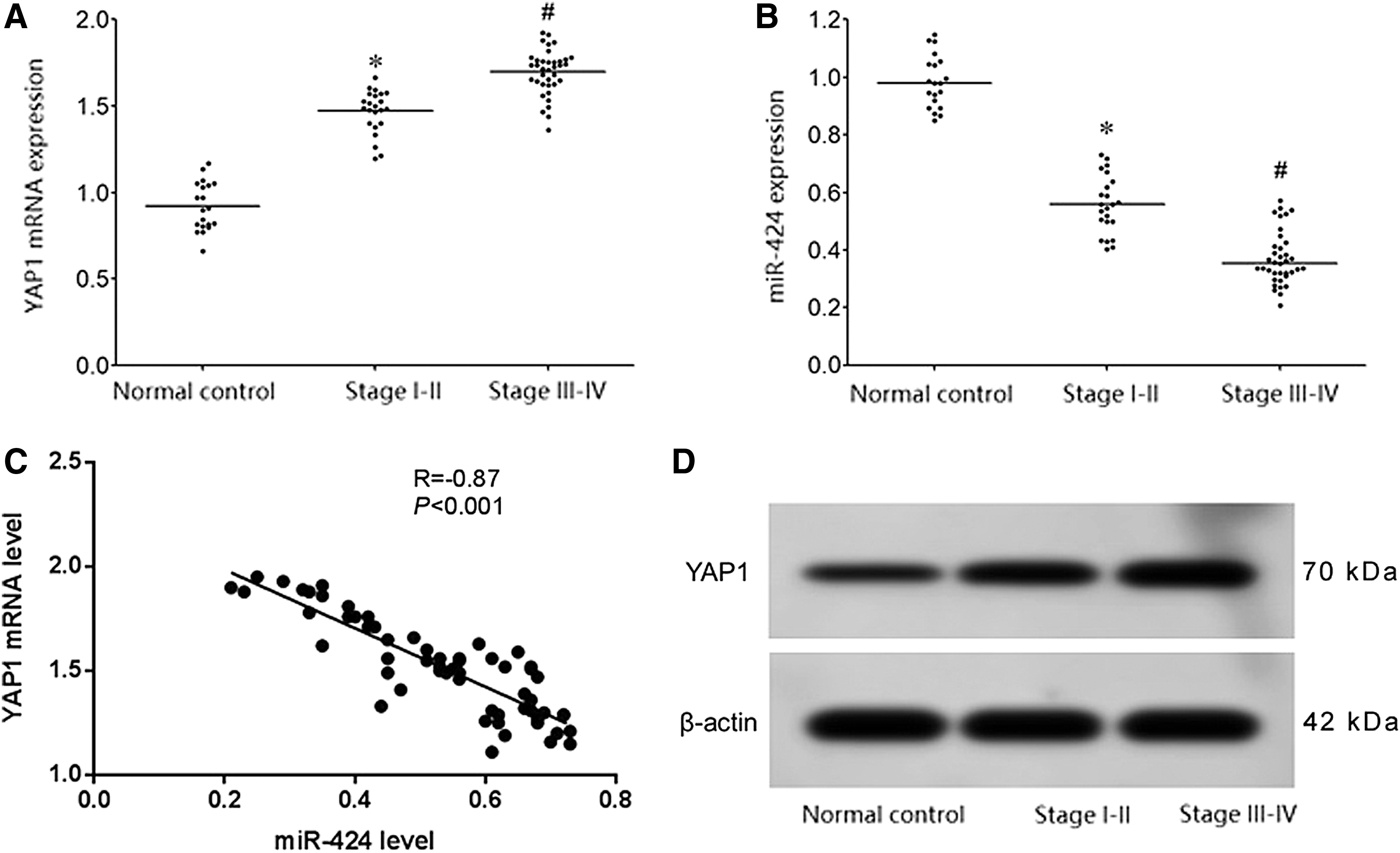
miR-424-5p expression was declined, while the YAP1 level was upregulated in HCC tissue.
miR-424-5p targeted regulated YAP1 expression

miR-424-5p targeted YAP1 expression.
miR-424-5p expression reduced, whereas YAP1 level was enhanced in HCC cells
qRT-PCR exhibited that compared with the human normal liver cell line HL-7702, YAP1 mRNA expression in HCCLM3 and MHCC97-L cells was significantly increased and its level in HCCLM3 cells with higher malignancy was obviously higher than MHCC97-L cells with lower malignancy (Fig. 3A). Compared with the human normal liver cell line HL-7702, miR-424-5p expression in HCCLM3 and MHCC97-L cells was markedly decreased and its content in HCCLM3 cells with higher malignancy was apparently lower than MHCC97-L cells with lower malignancy (Fig. 3B). The expression level of Foxo3a mRNA was significantly reduced (Fig. 3B). Western blot demonstrated that compared with HL-7702 cells, YAP1 protein expression in HCCLM3 and MHCC97-L cells was significantly enhanced and its expression in HCCLM3 cells with higher malignancy was obviously higher than MHCC97-L cells with lower malignancy (Fig. 3C)
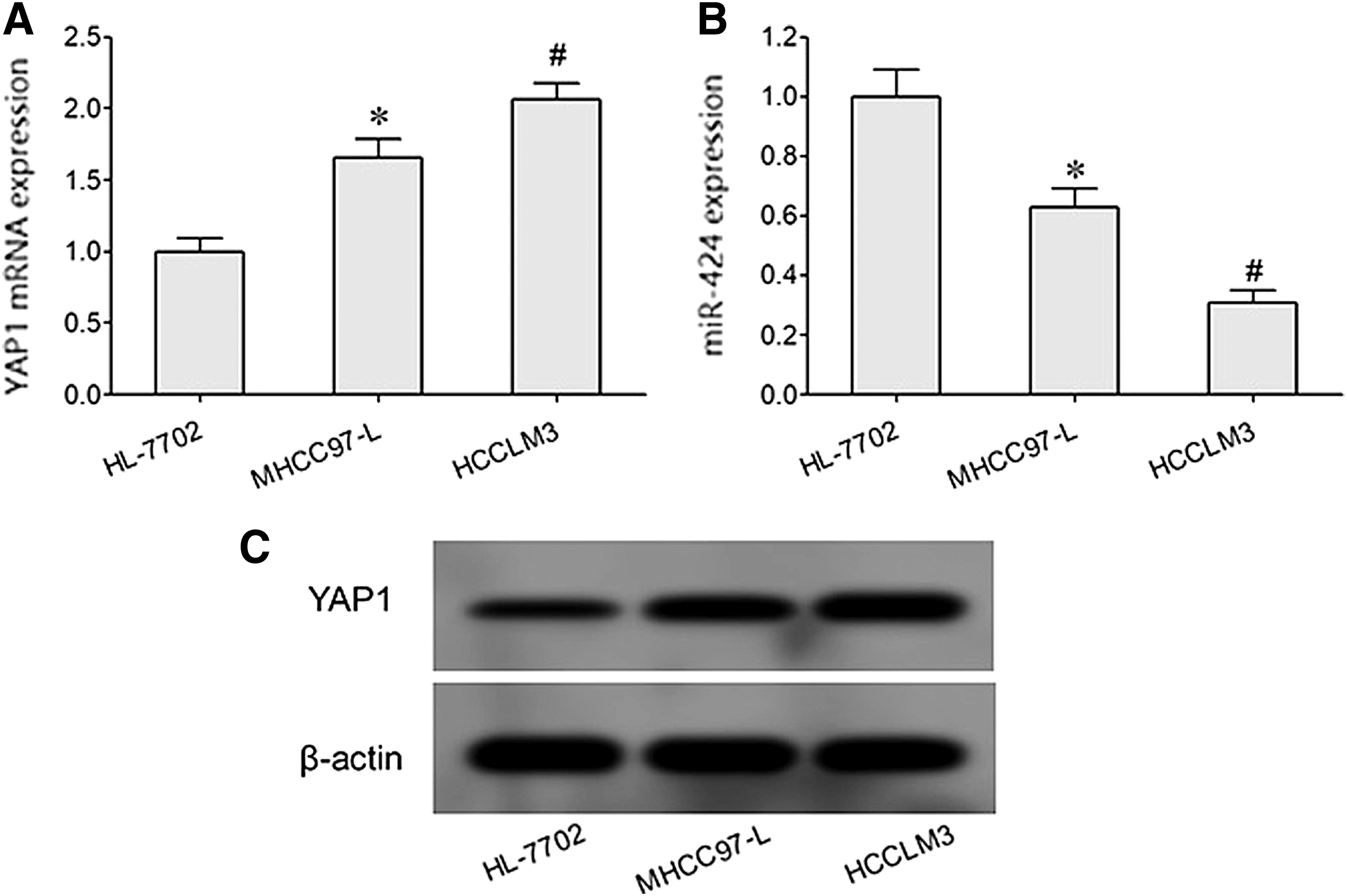
miR-424-5p expression was reduced, whereas the YAP1 level was enhanced, in HCC cells.
Overexpression of miR-424-5p inhibited YAP1 expression and cell proliferation, while it induced cell apoptosis
qRT-PCR revealed that the miR-424-5p level was significantly increased, while the YAP1 mRNA level was obviously declined in HCCLM3 and MHCC97-L cells transfected by miR-424-5p mimic compared with the miR-NC group (Fig. 4A, B). Western blot demonstrated that transfection of miR-424-5p mimic markedly downregulated YAP1 protein expression in HCCLM3 and MHCC97-L cells (Fig. 4C). Flow cytometry found that cell apoptosis was significantly enhanced (Fig. 4D), whereas cell proliferation was obviously inhibited (Fig. 4E) in the miR-424-5p mimic transfection group compared with the miR-NC group.
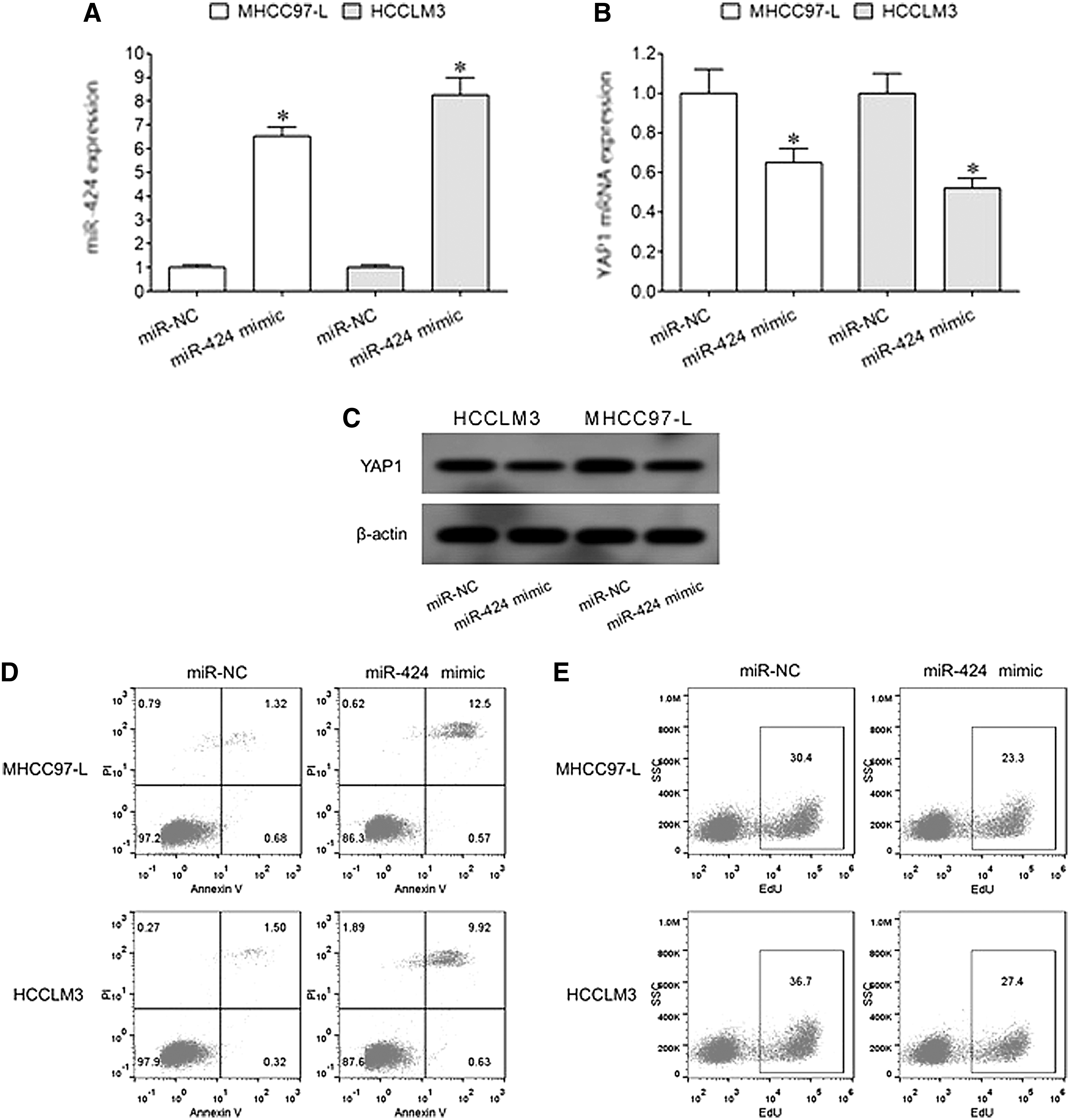
Overexpression of miR-424-5p inhibited YAP1 expression and cell proliferation, while it induced cell apoptosis.
Discussion
Pathogenesis of HCC is concealed with no obvious symptoms in the early stage. It is easy to be ignored and misdiagnosed. However, HCC progresses rapidly and most of the patients with symptoms were in middle and advanced stages, which show features of high malignancy, quick metastasis, difficulty in treatment, and poor survival. It is of great significance to explore the signal molecules or mechanisms of liver cancer to help early diagnosis, elevate efficacy, and improve prognosis.
The Hippo-YAP pathway plays an important regulatory role in tumor biology, thus it participates in occurrence, progression, and drug resistance of pancreatic, 15 ovarian, 16 colorectal, 17 prostate, 18 and gastric cancers. 19 The YAP1 gene is located at chromosome 11q13 position and encodes a protein of 65 kD size. 20 The functional activity of YAP1 is mainly regulated by a variety of tumor suppressor proteins at the upstream of the Hippo-YAP signaling pathway, such as mammalian Ste20-like protein kinase 1/2 (MST kinase 1/2), large tumor suppressor 1/2 (Lats kinase 1/2), and Mps one binder 1 (Mob1). 21
YAP1 mainly plays a role in regulating transcription and expression of various genes through binding with transcription factors. Numerous studies have shown that YAP1 is a cancer-promoting factor that plays a critical role in many types of tumors, such as esophageal, 6 gastric, 5 and nonsmall cell lung cancers. 22 It was observed that 8 –10 the expression and functional activity of YAP1 in HCC tumor tissues were significantly enhanced, suggesting that YAP1 is closely related to the occurrence and development of HCC. It was found that the expression of miR-424-5p significantly decreased in tumor tissues of HCC patients, revealing that miR-424-5p may play a role as a tumor suppressor gene in HCC. 13,14 Bioinformatic analysis demonstrated that there is a targeted complementary binding site between miR-424-5p and YAP1 mRNA, suggesting a potential regulatory effect. This study investigated the role of miR-424-5p in regulating YAP1 expression and affecting hepatoma cell proliferation and apoptosis.
The results of clinical specimens showed that compared with normal liver tissue, the expression of miR-424-5p significantly decreased in HCC patients with clinical stage dependence. Compared with normal liver tissue, expression levels of YAP1 mRNA and protein in tumor tissues of HCC patients were significantly upregulated with clinical stage dependence, suggesting that there may be a targeted inhibitory relationship between miR-424-5p and YAP1. Decreased expression of miR-424-5p may play a role in upregulating YAP1 expression and promoting the pathogenesis of HCC. The dual-luciferase assay revealed that miR-424-5p mimic or miR-424-5p inhibitor significantly declined or elevated the relative luciferase activity in HEK293T cells transfected by pMIR-YAP1-WT, further confirming the direct regulatory relationship between miR-424-5p and YAP1.
In cells cultured in vitro, expression of miR-424-5p in liver cancer cells markedly declined, while expression of YAP1 apparently increased compared with that in normal liver cells with malignancy dependence. Lu et al. 23 showed that compared with the adjacent tissues, expression of miR-424-5p in tumor tissues of HCC patients was significantly decreased and was related to survival and prognosis. In addition, miR-424-5p detection also has a high diagnostic value for HCC, with an area under the curve of up to 0.9768. Wu et al. 13 showed that expression of miR-424-5p was abnormally decreased in HCC tumor tissues compared with adjacent tissues, and the survival rate and prognosis of patients with lower expression of miR-424-5p were significantly worse. Yang et al. 24 showed that expression of miR-424-5p in tumor tissues of HCC patients was significantly lower than that in adjacent normal liver tissues. The lower the expression of miR-424-5p, the worse the prognosis of patients was. miR-424-5p expression was related to clinical features such as clinical stage, tumor size, and venous metastasis. This study observed that expression of miR-424-5p was decreased, while expression of the target gene YAP1 was abnormally elevated in hepatocarcinoma tissues. miR-424-5p may act as a tumor suppressor gene in HCC, while reduced miR-424-5p expression was an unfavorable factor in the pathogenesis of HCC.
To further investigate the effect of miR-424-5p on biological effects of HCC cells, this study overexpressed miR-424-5p in HCC cells cultured in vitro and observed the changes of cell proliferation and apoptosis. The results showed that transfection of miR-424-5p mimic significantly increased the expression of miR-424-5p and downregulated the expression of YAP1 in HCCLM3 and MHCC97-L cells, which obviously attenuated cell proliferation and enhanced apoptosis, confirming that miR-424-5p alleviates the malignant biological characteristics of HCC cells by targeting YAP1.
Yang et al. 24 found that overexpression of miR-424-5p in HCCLM3 and SMMC7721 cells significantly inhibited cell proliferation and clonality, induced cell cycle arrest, and weakened cell growth and tumorigenicity in BALB/c mice by targeting regulating Akt3 and E2F3 genes. Yu et al. 14 revealed that miR-424-5p can inhibit proliferation, migration, and invasion of HCC cells by targeting c-Myb, while downregulating the expression of miR-424-5p enhanced the malignant biological characteristics of HCC cells. Wu et al. 13 reported that overexpression of miR-424-5p significantly reduced migration and invasion of SK-HEP-1 and Huh-7 cells and inhibited cell proliferation, while downregulating miR-424-5p promoted HCC cell proliferation, migration, and invasion, thus confirming that miR-424-5p is a tumor suppressor gene in HCC. Zhang et al. 25 showed that overexpression of miR-424-5p can inhibit epithelial–mesenchymal transition, attenuate migration and invasion, induce apoptosis, and restrain tumorigenesis of HCC BEL7402, SMMC7721, and HepG2 cells through targeting the ICAT gene. In this study, miR-424-5p exhibited a tumor suppressing effect that attenuates the malignant features of HCC cells, which was similar to other studies.
At present, there are many studies focused on the relationship between miR-424-5p and HCC, but mainly targeting other genes. This study combined the targeted regulatory relationship between miR-424-5p and YAP1, revealing that the anticancer effect of miR-424-5p might be through targeted inhibition of YAP1 expression, suppressing proliferation and promoting apoptosis. However, future studies are required to confirm that the effects of miR-424-5p on cancer cell proliferation and apoptosis are indeed through regulation of YAP1 expression.
Conclusions
Decreased miR-424-5p expression and increased YAP1 expression levels are observed in patients with liver cancer. Increased miR-424-5p can inhibit YAP1 expression, attenuate hepatoma cell proliferation, and induce cell apoptosis.
Footnotes
Acknowledgment
This work was supported by the National Natural Science Foundation of China—Youth Science Foundation (No. 81301979).
Disclosure Statement
No competing financial interests exist.
Supplementary Material
Supplementary Figure S1
References
Supplementary Material
Please find the following supplemental material available below.
For Open Access articles published under a Creative Commons License, all supplemental material carries the same license as the article it is associated with.
For non-Open Access articles published, all supplemental material carries a non-exclusive license, and permission requests for re-use of supplemental material or any part of supplemental material shall be sent directly to the copyright owner as specified in the copyright notice associated with the article.