
Abstract
Cancer Biotherapy and Radiopharmaceuticals
officially retracts the paper entitled, “The Effect of PTCH1 on Ovarian Cancer Cell Proliferation and Apoptosis,” by Fang Zheng, Xinyi Xiao, and Chunmei Wang (Cancer Biother Radiopharm 2019;34:2;103–109; doi: 10.1089/cbr.2018.2626) due to the discovery that the paper was submitted from a paper mill which is a violation of the journal's proper protocols, and is considered an infraction against the rigorous standards of scientific publishing.
The Editor and Publisher of Cancer Biotherapy and Radiopharmaceuticals are committed to preserving the scientific literature and the community it serves and does not tolerate any violations of scientific misconduct.
Introduction
Ovarian cancer (OC) is one of the common malignant tumors in female reproductive organ. The incidence rate is followed by cervical cancer and endometrial cancer, ranking third among female reproductive system malignancies. 1 –3 The OC incidence ranks fifth in female malignant tumors. Epithelial OC is the main type accounting for more than 90%. 4 It was estimated that there are 225,000 new OC patients diagnosed each year worldwide, and the number of patients dying from OC is reached up to 145,000 per year. 5 Compared with developed countries, the incidence of OC in China is relatively low, but has kept increasing in recent years. 6 In the early stage of OC, there are no obvious clinical symptoms. However, it progresses rapidly, leading to most patients in the middle and late stages of the disease at the time of diagnosis. OC has the characteristics of high malignancy, easy invasion and metastasis, poor therapeutic effect, as well as high recurrence rate and mortality. Therefore, exploring the pathogenesis of OC has great significance in terms of diagnosis, elevating the therapeutic effect, and improving the prognosis of patients.
Hedgehog pathway is composed and activated by secreted Hh protein, which plays an important role in regulating embryonic development, cell proliferation, differentiation, and apoptosis. 7 –9 When the Hedgehog signaling pathway is inactive, Patched (PTCH) binds to Smoothened (SMO) and inhibits its activity, leading to GLI phosphorylation and subsequent ubiquitination degradation, thereby failing to enter the nucleus in full-length form and regulating transcription of downstream target genes. 10,11 When the Hedgehog signaling pathway is activated, HH ligand binds to the transmembrane protein receptor PTCH, which abolishes the inhibitory effect of PTCH on SMO and allows GLI enter the nucleus in full-length form, thus promoting the transcription and expression of various target genes, such as Myc and cyclin. It promotes cell proliferation, migration, and malignant transformation, leading to tumorigenesis. 12 Abnormal Hedgehog pathway is closely related to the occurrence, progression, metastasis, and recurrence of various tumors, such as breast cancer, 13 pancreatic cancer, 14 cervical cancer, 15 and gallbladder cancer. 16 The transmembrane protein receptor PTCH is an important component of the Hedgehog signaling pathway and negatively regulates Hedgehog signaling by inhibiting the transmembrane protein receptor SMO. 17,18 Considering the role of Hedgehog signaling in the pathogenesis of several cancers, whether PTCH1 involves in the development of OC remains poorly understood. In this study, the authors detected the expression of PTCH1 in OC tissues to analyze the relationship between expression of PTCH1 and prognosis, and to explore its role in regulating OC cell proliferation and apoptosis.
Materials and Methods
Main reagents and materials
Human normal ovarian epithelial cell line IOSE80, OC cell lines SKOV3, A2780, and Caov3 were purchased from Hunan Fenghui Biotechnology Co., Ltd. RPMI 1640 medium, FBS, and penicillin–streptomycin were purchased from Gibco. Liposome transfection reagent Lipofectamine 2000 was purchased from Invitrogen. ReverTra Ace qPCR RT Kit and SYBR dye were purchased from Toyobo (Japan). Rabbit antihuman PTCH1, Gli1, and β-actin antibodies were purchased from Abcam. HRP-conjugated secondary antibody was purchased from Sangon (Shanghai, China). Annexin V/PI Apoptosis detection reagent was purchased from Beyotime (Jiangsu, China). pIRES2 plasmid was purchased from BioVector (Beijing, China). EdU cell proliferation assay kit was purchased from RiboBio (Guangzhou, China).
Clinical information
Eighty patients with epithelial OC, diagnosed and treated in the hospital from January 2014 to October 2014, were enrolled with an average age of 49.7 (35–68) years old. Another 40 cases of normal ovarian tissue resected from benign ovarian cysts were used as controls with an average age of 51.1 (33–65) years old. All tissue specimens were confirmed by pathological examination. The samples were obtained with informed consent and reviewed by the hospital Ethics Committee (Approval number: 20130615E). Tissue specimens were stored in liquid nitrogen immediately after collection. Among them, 31 patients suffered from serous cystadenocarcinoma, 22 patients suffered from mucinous cystadenocarcinoma, and 27 patients suffered from mixed disease. OC were pathologically staged according to the International Federation of Obstetrics and Gynecology (FIGO, 1988) criteria, including 19 cases in stage I, 25 cases in stage II, 20 cases in stage III, and 16 cases in stage IV.
Cell culture
SKOV3, A2780, Caov3, and IOSE80 cells were maintained in RPMI 1640 medium containing 10% FBS and 1% penicillin–streptomycin and cultured in a 37°C and 5% CO2 incubator. The cells in logarithmic phase were used for experiments.
Overexpression plasmid construction and cell transfection
The pIRES2 was used as the eukaryotic expression plasmid, and XhoI and BamHI restriction sites were added to the ends of primers used to amplify the PTCH1 gene from cDNA using the primers as described previously. 19 The gel-recovery kit was adopted to collect the target fragment after gel electrophoresis separation. After ligated into the carrier, it was transformed into competent cell JM109. Next, the positive strain was selected by the ampicillin-resistant solid culture plate. Then the recombinant plasmid containing the target fragment was amplified, cultured, extracted, and sequenced.
pIRES2-Scramble and pIRES2-PTCH1 were transfected into SKOV3 and A2780 cells using Lipofectamine 2000 reagent according to manufacturer's instructions. The cells were divided into pIRES2-Scramble group and pIRES2-PTCH1 group. After 72 h, cells were collected for apoptosis detection or incubation with EdU for 48 h to detect cell proliferation.
Quantitative reverse transcription-polymerase chain reaction
The 10 μL reverse transcription system contained 1 μg RNA, 2 μL RT Buffer(5 × ), 0.5 μL oligo dT+Random Primer Mix, 0.5 μL RT Enzyme Mix, 0.5 μL RNase inhibitor, and ddH2O. The reverse transcription was performed at 37°C for 15 min and 98°C for 5 min. The primers used for polymerase chain reaction (PCR) were designed as follows: PTCH1PF: 5′-GAAGAAGGTGCTAATGTCCTGAC-3′, PTCH1PR: 5′-GTCCCAGACTGTAATTTCGCC-3′; β-actinPF: 5′-GAACCCTAAGGCCAAC-3′, β-actinPR: 5′-TGTCACGCACGATTTCC-3′. The PCR system contained 5.0 μL 2 × SYBR Green Mixture, 0.5 μL Forward Primer (2.5 μm/L), 0.5 μL Reverse Primer (2.5 μm/L), 1.0 μL cDNA, and 3.0 μL ddH2O. PCR conditions were predenatured at 95°C for 5 min, followed by 40 cycles of 95°C for 15 s and 60°C for 60 s on the Bio-Rad CFX96 Real-Time PCR Detection System.
Western blot
Cells and tissues were lysed by RIPA lysis buffer. Forty microgram proteins were separated by 10% SDS-PAGE gel and 4% concentrated gel followed by being transferred to PVDF membrane. Next, the membrane was blocked with 5% skim milk at room temperature for 60 min and incubated with primary antibody (PTCH1 [catalog number: ab53715; Abcam], GLI1 [catalog number: ab134906; Abcam], and β-actin [catalog number: ab8226; Abcam] at 1:2000, 1:1000, 1:5000, respectively) at 4°C overnight. After that, the membrane was incubated with HRP-conjugated Goat anti-Rabbit IgG (H+L) secondary antibody (1:10,000) at room temperature for 60 min followed by detection of protein band after addition of ECL chemiluminescence.
Flow cytometry detection of cell apoptosis
The cells were washed twice in PBS and digested by trypsin. After centrifugation, 100 μL binding solution was added into cells followed by addition of 5 μL of Annexin V-FITC and 5 μL of PI solution, and incubation for 10 min under dark. After supplementation with 400 μL binding solution, cell apoptosis was measured by flow cytometry (Beckman FC 500MCL).
Flow cytometry detection of cell proliferation
The cells were resuspended in DMEM complete medium with 10% FBS and incubated with 10 μM EdU for 2 h followed by culture for 48 h. After digestion by trypsin, cells were fixed in 100 μL fixative at room temperature for 15 min and then treated with 0.1% Triton X-100 solution at room temperature for 15 min. Next, 500 μL of Apollo solution was added into the cells and incubated at room temperature for 30 min under dark followed by analysis of cell proliferation on a Beckman FC 500MCL flow cytometer.
Statistical analyses
All data analyses were performed using SPSS 18.0 software. Measurement data are expressed as mean ± standard deviation and compared by Student's t-test or one-way ANOVA. PTCH1 mRNA expression was compared by Mann–Whitney U test. Survival curve was made using Kaplan–Meier method. p < 0.05 was considered as statistically significance.
Results
Decreased PTCH1 expression in OC tissue
Quantitative reverse transcription-polymerase chain reaction (qRT-PCR) showed that PTCH1 mRNA expression in OC tissue was significantly lower than that in normal ovarian tissue with clinical stage dependence (Fig. 1A). Consistently, western blot demonstrated that PTCH1 protein expression in OC tissue was significantly reduced compared with normal ovarian tissue with clinical stage dependence (Fig. 1B).
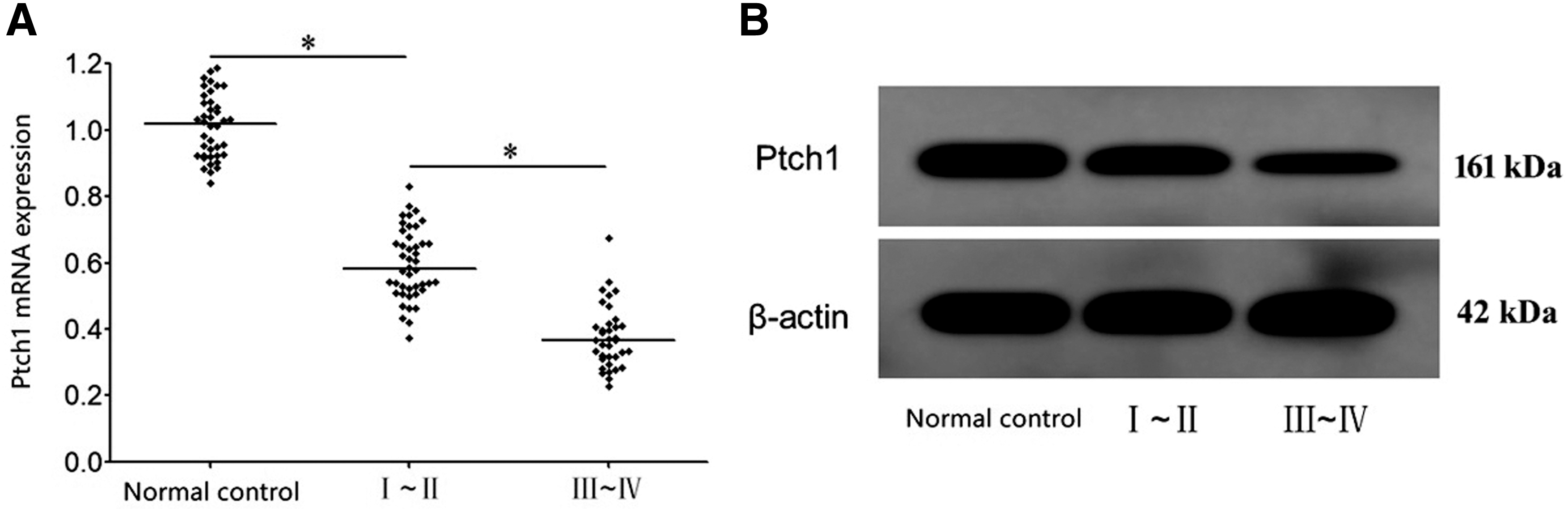
PTCH1 expression decreased in OC tissue. Total RNA and protein were isolated from OC tissues and control tissues followed by
The relationship between PTCH1 expression and clinical features in OC tissue
OC patients were divided into high PTCH1 expression and low expression based on the median PTCH1 protein expression to assess its relationship with clinical features. It was revealed that the expression level of PTCH1 was correlated with tumor tissue size, TNM stage, and pathological grade (p < 0.05), but not with lymph node metastasis, age, and pathological type (p > 0.05) (Table 1).
The Relationship Between Ptch1 Expression and Clinical Features in Ovarian Cancer Tissue
Bold values indicate p < 0.05 was considered statistically significance.
Low PTCH1 expression was related to poor prognosis of OC
The authors next evaluated the relationship of PTCH1 expression with prognosis. Survival curve analysis demonstrated that the prognosis of patients with low PTCH1 expression was significantly worse than that of patients with high PTCH1 expression (Log-rank test χ 2 = 4.213, p = 0.042) (Fig. 2).
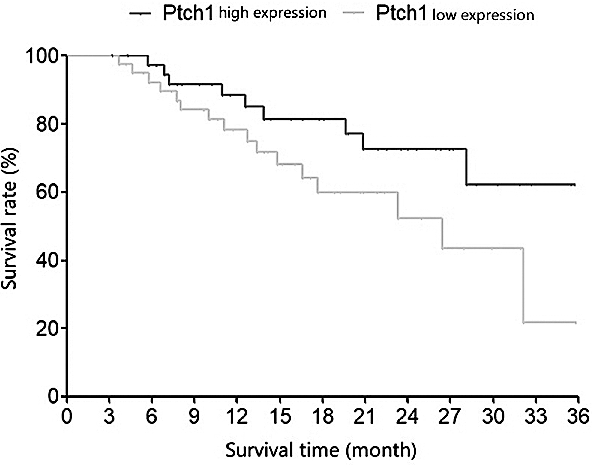
Patient survival rate comparison. OC patients were divided into high PTCH1 expression and low expression based on the median PTCH1 protein expression to assess its relationship with clinical features. OC, ovarian cancer; PTCH1, patched.
Reduced PTCH1 expression in OC cells
qRT-PCR demonstrated that PTCH1 mRNA level in OC SKOV3, A2780, and Caov3 cells was significantly lower than that in normal ovarian epithelial IOSE80 cells (Fig. 3A). In accordance with this, western blot showed that PTCH1 protein level in OC SKOV3, A2780, and Caov3 cells was significantly lower than that in normal ovarian epithelial IOSE80 cells (Fig. 3B).
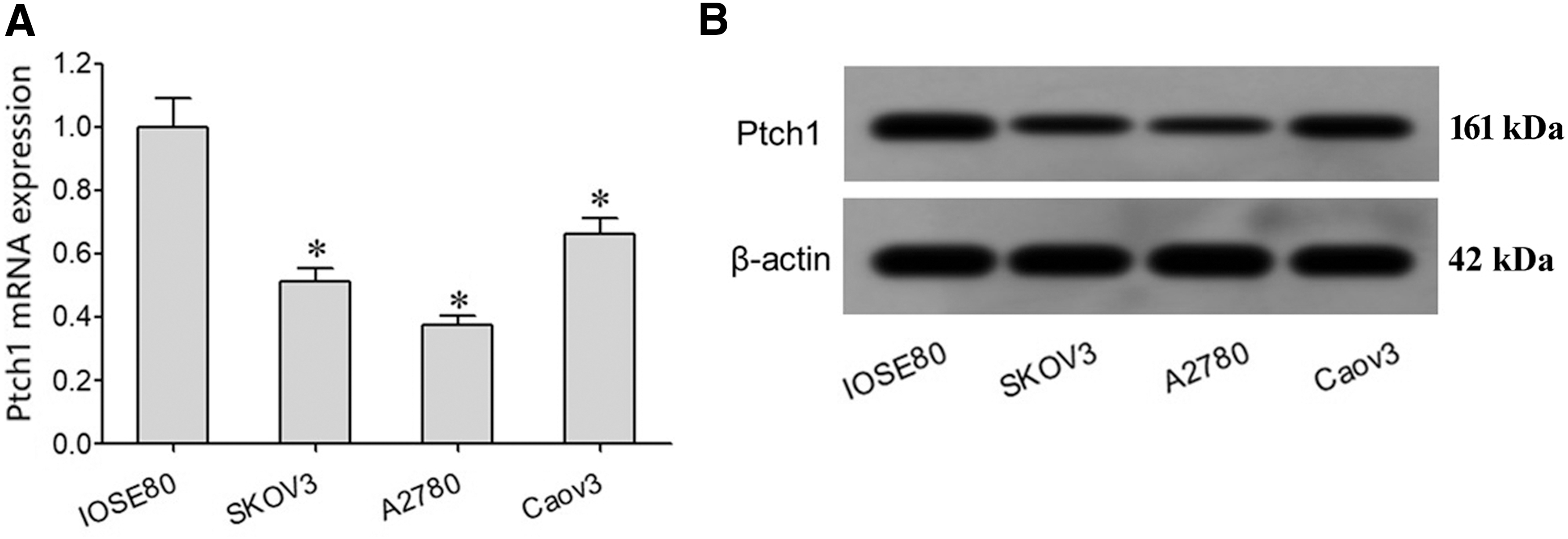
PTCH1 expression decreased in OC cells. Total RNA and protein were isolated from ovarian cell lines or control cell line followed by
PTCH1 overexpression inhibited GLI1 expression and OC cell proliferation, while induced cell apoptosis
The transfection efficiency was 73% for pIRES2-PTCH1 and 67% for Pires-scramble. qRT-PCR revealed that PTCH1 mRNA expression in OC SKOV3 and A2780 cells transfected by pIRES2-PTCH1 was significantly higher than that in pIRES2-Scramble group (Fig. 4A). Western blot demonstrated that, compared with pIRES2-Scramble group, PTCH1 protein level was significantly upregulated, while GLI1 protein expression was markedly declined in OC SKOV3 and A2780 cells transfected with pIRES2-PTCH1 (Fig. 4B). Flow cytometry exhibited that transfection of pIRES2-PTCH1 significantly attenuated cell proliferation (Fig. 4C) and increased cell apoptosis (Fig. 4D) in SKOV3 and A2780 cells.
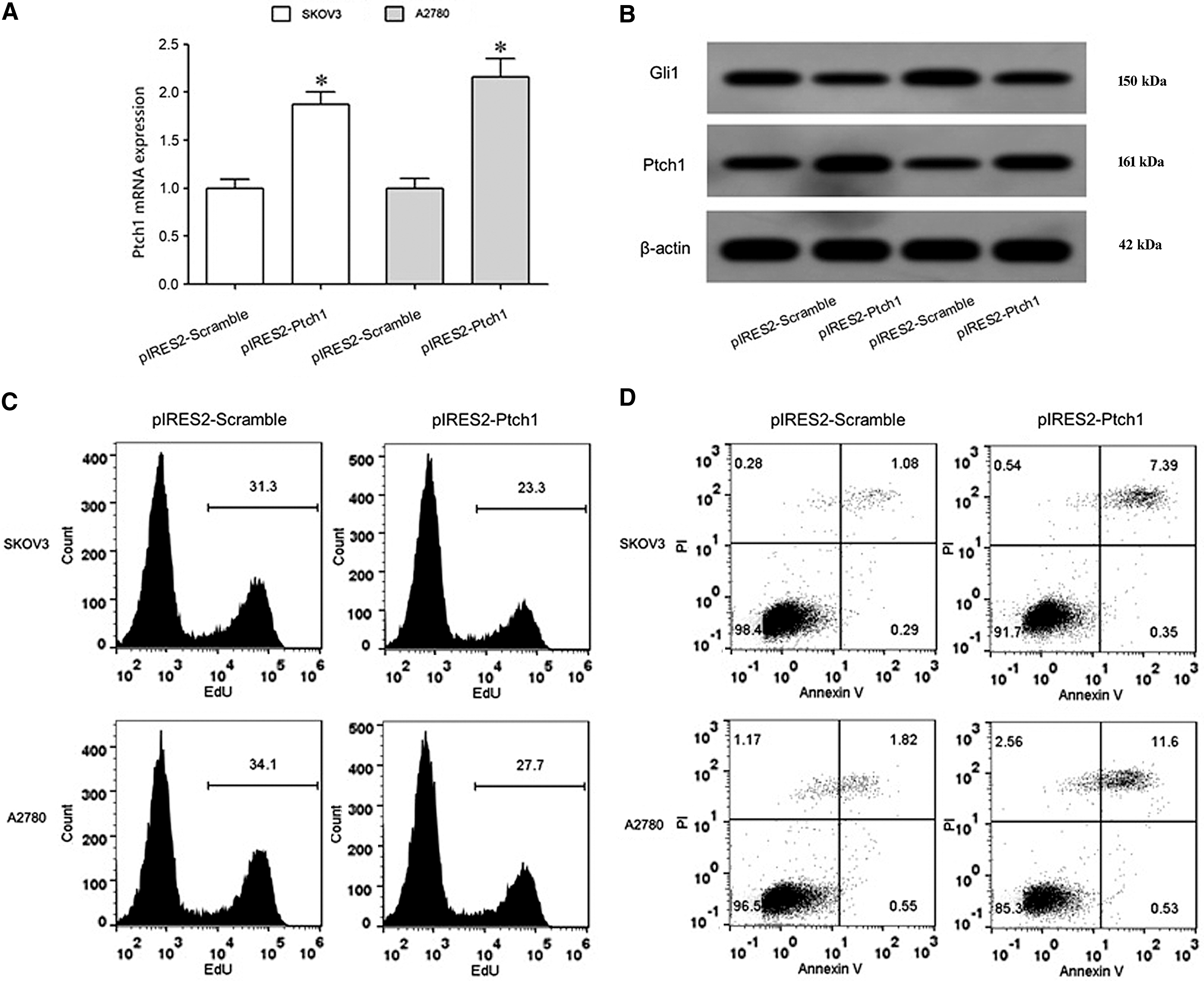
PTCH1 overexpression inhibited GLI1 expression and OC cell proliferation, while induced cell apoptosis.
Discussion
The Hedgehog signaling pathway includes extracellular signal ligand HH, transmembrane protein receptor PTCH, transmembrane protein SMO, transcription factor GLI, and downstream target genes. 20 –22 Ptch and Smo are two types of membrane protein receptor complexes. PTCH is a 12-transmembrane protein containing 1500 amino acids. There are two PTCH family members in mammals, namely PTCH1 and Ptch2. PTCH1 is the most important one of these two receptors of human Hedgehog protein. When Hedgehog binds to PTCH1, its inhibitory effect is eliminated. Essentially, PTCH1 is a tumor suppressor gene. It was confirmed that the expression of PTCH1 is associated with the occurrence, progression, and drug resistance of various tumors, such as intestinal cancer, 18 lung cancer, 23 and glioma. 17 This study was to detect the expression of PTCH1 in OC tissues to analyze the relationship between expression of PTCH1 and prognosis and to explore its role in regulating OC cell proliferation and apoptosis.
The authors' results showed that compared with normal ovarian tissue, PTCH1 mRNA and protein expressions in OC tissue were significantly lower than that in normal ovarian tissue with clinical stage dependence, consistent with a previous study showing reduced expression of PTCH1 in tumor tissues of OC patients. 24 In addition, the authors also found decreased PTCH1 expression and increased GLI1 expression in OC cell lines SKOV3 and A2780 cells, which was in accordance with a previous study demonstrating that compared with normal ovarian epithelial cells, the expression of PTCH1 was markedly decreased, while the expression of downstream molecule GLI1 was apparently enhanced in OC OVCAR-5, OV-202, and OV-167 cells. 25 These data suggest that decreased PTCH1 expression might play a role in the pathogenesis of OC through regulating Hedgehog signaling pathway via downregulation of GLI1. However, conflicts were found on hedgehog signaling activation in OC as demonstrated by a previous study 26 showing overexpression of PTCH and GLI1 protein in OCs correlated with poor survival of the patients and GLI1 expression is an independent prognostic marker. The contradictory results might be possibly due to the following reasons. First, immunohistochemistry was used to assess the expression of HH targets PTCH1 and GLI1 in the tumor specimens in the previous study, 26 whereas in the present study, the expression of HH target genes was measured by real-time PCR and western blot. Second, the involvement of HH signaling in human cancers may be context dependent, occurring in some tissues or cell lines, but not in others.
To analyze the relationship of PTCH1 expression with clinical features, OC patients were divided into high PTCH1 expression and low expression based on the median PTCH1 protein expression. It was revealed that the expression level of PTCH1 was correlated with tumor tissue size, TNM stage, and pathological grade, but not with lymph node metastasis, age, and pathological type. Survival curve analysis demonstrated that the prognosis of patients with low PTCH1 expression was significantly worse than that of patients with high PTCH1 expression.
To further investigate the effects of PTCH1 on the biological effects of OC cells, PTCH1 was overexpressed in OC cells to investigate its role in cell proliferation and apoptosis. It was shown that overexpression of PTCH1 significantly reduced the expression of Gli1, inhibited cell proliferation, and promoted apoptosis in OC cancer SKOV3 and A2780 cells, consistent with a previous study demonstrating that expression of PTCH1 in 293T cells inhibits cell proliferation. 27 Choi et al. 28 revealed that itraconazole can play an antitumor effect through inhibition of Hedgehog pathway. Steg et al. 29 observed that paclitaxel and tubastatin treatments can significantly upregulate the expression of PTCH1 gene, downregulate the expression of GLI1 and GLI2, inhibit cell proliferation, and decrease cell viability in OC cancer SKOV3TRip2 cells. Choi et al. 28 and Steg et al. 29 explore the effect of drug on the expression of PTCH1 and the proliferation activity of OC cancer cells. In contrast, this study directly observed that PTCH1 decreased Gli expression and attenuated the malignant biological characteristics of OC cells.
Conclusions
Overexpression of PTCH1 inhibited GLI1 expression, attenuated OC cell proliferation, and induced apoptosis, suggesting that manipulation of PTCH1 expression might be a novel approach for the treatment of OC.
Footnotes
Disclosure Statement
There are no existing financial conflicts.