




Abstract
Background:
Clear cell renal cell carcinoma (ccRCC), as the commonest type among renal cell cancers, is featured with easy relapse and metastasis. Despite mounting achievements on its treatment and diagnosis, the identification of new biomarkers remains urgent.
Purposes:
Present study aimed to explore the role of microRNA-4429 (miR-4429) in ccRCC.
Methods:
The expression of miR-4429 and cyclin-dependent kinase 6 (CDK6) was evaluated by real-time polymerase chain reaction and Western blot. Cell proliferation, migration and invasion was evaluated by MTT and transwell assays. The interaction between miR-4429 and CDK6 was assessed by luciferase reporter assay. Prognostic significance of miR-4429 was evaluated by Kaplan-Meier analysis. Correlation between miR-4429 and CDK6 was determined by Spearman's correlation analysis.
Results:
Firstly, the downregulation of miR-4429 and upregulation of CDK6 in ccRCC tissues and cells were uncovered by quantitative real-time polymerase chain reaction. The prognostic significance of miR-4429 in ccRCC patients was proved by Kaplan–Meier analysis. Gain- and loss-of-function assays validated the suppressive effect of miR-4429 on cell proliferation, migration, invasion, as well as epithelial-mesenchymal transition (EMT) progression. The interaction between miR-4429 and CDK6 was predicted by bioinformatics tool and confirmed by luciferase reporter assay. And the negative expression correlation between miR-4429 and CDK6 was verified by Spearman's correlation analysis. Rescue assays confirmed the role of miR-4429/CDK6 in proliferation, metastasis and EMT progression in ccRCC.
Conclusions:
Present study revealed that miR-4429 suppressed ccRCC tumor progression and EMT by targeting CDK6.
Introduction
Renal cell carcinoma (RCC) ranks as the most aggressive and commonest type of malignancy among all urological cancers, and clear cell renal cell carcinoma (ccRCC) presents the greatest prevalence among RCC. 1,2 ccRCC takes up around 80%–90% of RCCs, featured with higher relapse and metastasis rate than other RCC subtypes. 3 The morbidity of ccRCC keeps rising during the last three decades, especially in high income countries. 4 Although multiple molecular biomarkers have been explored by mounting studies in ccRCC, few of them are able to serve as effective prognostic and therapeutic targets. 5 –7 Therefore, identification of novel targets through molecular mechanism research in ccRCC is urgent.
microRNAs (miRNAs), recognized as an endogenous and conserved cluster of small noncoding RNAs, have been proved to modulate gene expression negatively via targeting the 3′-untranslated region (3′-UTR) of messenger RNAs (mRNAs). 8,9 Surging evidence has revealed the crucial roles of miRNAs in cellular processes, including carcinogenesis, cell proliferation, cell cycle, as well as metastasis. 10,11 Speaking of cancer metastasis, epithelial-mesenchymal transition (EMT) is a process closely related to metastasis through which terminal differentiated epithelial cells transform to migratory mesenchymal cells during embryo development. 12 During past years, the inhibition of EMT progression by miRNAs has been identified in numerous cancers, such as in hepatocellular cancer, 13 colorectal cancer, 14 and ccRCC. 15 Furthermore, numerous miRNAs have been identified in ccRCC to exhibit oncogenic or tumor-suppressive functions. 16 –21 microRNA-4429 (miR-4429) has been identified as a biomarker in papillary thyroid cancer. 22 However, its biological roles in many other cancers including ccRCC remain to be uncovered.
Cyclin-dependent kinase 6 (CDK6) is a member of CDKs family. Although regulation on cell cycle is considered to be the primary function of CDK6, it also exerts significant functions in proliferation, differentiation and metastasis. 23 –25 Upregulation of CDK6 has been shown to be related to the development of several types of human cancers, such as breast, colon, pancreatic, bladder, oral cancers. 26 –29 It has also been discovered to be suppressed by miR-1 to influence cell cycle and metastasis in ccRCC. 30 However, no study has revealed the correlation of CDK6 with miR-4429 to date. Present study attempted to explore the role of miR-4429/CDK6 in the tumor progression and EMT ccRCC.
Materials and Methods
Tissue samples
A total of 98 ccRCC tissues and paired non-tumor tissues, among which 59 of metastatic tissues and 39 non-metastasis tissues were dissected from ccRCC patients in the Fourth Affiliated Hospital, Zhejiang University School of Medicine. Before dissection, no patients received other treatment such as radiotherapy or chemotherapy. And each patient had handed in a written inform consent.
Cell lines and cell culture
Human ccRCC cell lines Caki-1, Caki-2, and 786-O, were cultured in Roswell Park Memorial Institute 1640 medium (RPMI-1640; Thermo Fisher Scientific, Waltham, MA). A-498 cell lines and normal renal epithelial cell line (HK-2) were incubated in Dulbecco's modified Eagle's medium (DMEM; Gibco; Thermo Fisher Scientific). And each media was treated with 10% fetal bovine serum (FBS; Hyclone Technologies, Logan, UT). All cell lines were purchased from ATCC (Manassas, VA), and were incubated at 37°C under the humid air with 5% carbon dioxide (CO2).
RNA isolation and quantitative real-time polymerase chain reaction
The isolation of whole RNA from tissues and cells was performed via TRlzol reagent (Takara, Dalian, China) in reference to the manufacturer's directions. The examination of mRNA levels was conducted by quantitative real-time polymerase chain reaction (qRT-PCR) utilizing SYBR Green PCR Master Mix (Takara) on the ABI 7500 Fast Real-Time PCR system. GAPDH served as endogenous control for analysis of CDK6 level and U6 for miR-4429 level. 2−ΔΔCT method was used to examine comparative quantification. Primers used were listed as follows:
miR-4429 Forward: 5′-GGCCAGGCAGTCTGAGTTG-3′ Reverse: 5′-GGGAGAAAAGCTGGGCTGAG-3′
CDK6 Forward: 5′-CGAATGCGTGGCGGAGATC-3′ Reverse: 5′-CCACTGAGGTTAGAGCCATC-3′ U6 Forward: 5′-GCTTCGGCAGCACATATACTAAAAT-3′ Reverse: 5′-CGCTTCACGAATTTGCGTGTCAT-3′ GAPDH Forward: 5′-GAAGGTGAAGGTCGGAGTC-3′ Reverse: 5′-GAAGATGGTGATGGGATTTC-3′
Cell transfection
miR-4429-mimic and miR-4429 inhibitor were utilized to overexpress and knockdown miR-4429, NC-mimic and NC-inhibitor as negative control. PcDNA3.1 subcoloned with CDK6 was utilized to overexpress CDK6, pcDNA3.1 as negative control. Specific small interfering RNAs targeting CDK6 (siCDK6) were used to silence CDK6 expression. The plasmid vectors were generated in Shanghai Integrated Biotech Solutions Co., Ltd. (Shanghai, China). Lipofectamine 2000 (Invitrogen, Carlsbad, CA) was employed for the transfection of the vectors into cells in the light of manufacturer's protocols. qRT-PCR analysis was carried out for the examination of transfection efficiency.
MTT assay
Cell proliferation assay was conducted under the manufacturer's protocols by using the MTT kit (Promega Corporation, Madison, WI). To be brief, cells were incubated in 96-well culture dishes at a density of 5 × 10 3 cells/well and cultured for 24, 48, 72 and 96 h. Twenty microliters of 5 mg/mL MTT was supplemented into each well and further cultured for 4 h in a humid incubator after 24 h transfection. After the removal of supernatant, 200 μL of dimethyl sulfoxide was utilized to dissolve the formazan. The optical density was determined at 490 nm.
Transwell migration and invasion assay
All cells were plated into the serum-free upper transwell insert in a 24-well format of 8 mm diameters (Corning, NY). Medium with 10% FBS was supplemented into the bottom compartment which served as a chemoattractant, and the inserts were placed into the 5% CO2 incubator for 48 h at 37°C. The cells located on the upper surface were wiped out by cotton-tipped swabs, and cells migrated or invaded to the lower chamber were stained for 2 h by 0.05% purple crystal. Then, the migrated and invaded cells were washed by phosphate buffered saline buffer, calculated in five random fields using a microscope. Procedures for migration and invasion assays were the same except that the upper compartment was precoated with matrigel for invasion assay.
Luciferase reporter assay
Luciferase reporter plasmids with wild type or mutant 3′-UTR of CDK6 were co-transfected with miR-4429-mimic or NC-mimic into ccRCC cells with the application of Lipofectamine 2000 (Invitrogen). Forty-eight hours after transfection, a dual-luciferase reporter assay system (Promega Corporation) was employed to determine the luciferase activity in accordance with manufacturer's protocols, Renilla fluorescence as the internal control.
Western blotting
The proteins were isolated in RIPA lysis buffer (Beyotime Biotechnology, China) with protease inhibitors (Roche, China). For the quantification of proteins, BCA™ Protein Assay Kit (Pierce, Appleton, WI) was applied under the manufacture's recommendations. Ten percent sodium dodecyl sulfate-polyacrylamide gel electrophoresis was utilized for loading and electrophoresing the proteins. After that, proteins were moved to polyvinylidene difluoride membranes which was later blocked for 1 h by skim milk and cultivated overnight at 4°C with primary antibodies. Then, the membranes were further incubated for 2 h under room temperature with the secondary antibodies (Santa Cruz Biotech, Santa Cruz, CA) and covered by Immobilon Western chemiluminescent HRP substrate (200 μL; Millipore). The protein density was calculated with the employment of Lab™ software (Bio-Rad, Hercules, CA). Primary antibodies used were: anti-CDK6 and anti-GAPDH (Abcam, Cambridge, MA); anti-E-cadherin, anti-N-cadherin, and anti-Vimentin (Cell Signaling Technology, Danvers, MA).
Statistical analysis
The data presentation from all assays was conducted by the mean ± standard deviation of the mean. And each assay was carried out in triplicate. The calculation of significant differences was conducted utilizing one-way multivariate analysis of variance for multiple group comparisons or the Student's t-test for two group comparisons. The correlation between miR-4429 and CDK6 was analyzed by Spearman's correlation analysis. And Kaplan–Meier analysis with log-rank test was performed to examine overall survival of ccRCC patients. Statistical analysis was conducted with the employment of GraphPad Prism version 5.0 (GraphPad Software, La Jolla, CA). Differences were counted as statistically significant when p < 0.05.
Results
miR-4429 was downregulated in ccRCC and was a prognostic mark in ccRCC patients
First, we investigated the association of miR-4429 with ccRCC through detecting its expression level in tissues and cell lines. qRT-PCR results revealed that miR-4429 level was lower in tumor tissues than in the corresponding non-tumor tissues, in metastatic tissues than in non-metastasis tissues (Fig. 1A, B). Also, miR-4429 was downregulated in ccRCC cell lines (Caki-1, Caki-2, 786-O, and A-498) compared with normal cell line (HK-2), and Caki-2 and 786-O presented the lowest miR-4429 level (Fig. 1C). Kaplan–Meier analysis revealed that low expression of miR-4429 indicated poor prognosis in ccRCC patients (Fig. 1D). Moreover, it was discovered that miR-4429 expression was closely related to clinical characteristics of ccRCC patients such as tumor, node, metastases (TNM) stage, lymph node metastasis and distant metastasis (Table 1). And miR-4429 expression was identified as an independent prognostic marker for ccRCC patients (Table 2). These results suggested that miR-4429 might function as a tumor suppressor in ccRCC and its downregulation was a target for poor prognosis of ccRCC.
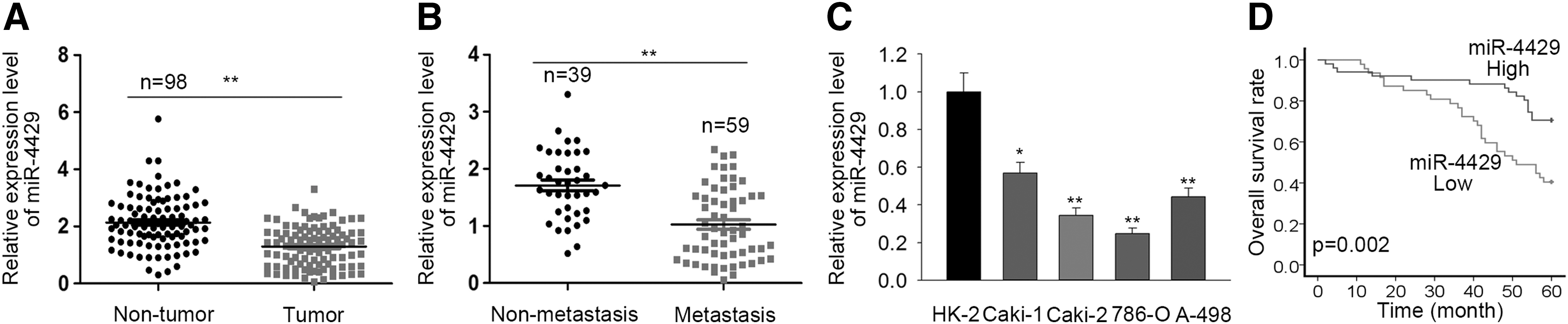
Downregulation of miR-4429 in ccRCC tissues and cell lines.
Correlation Between miR-4429 Expression and Clinical Characteristics (n = 98)
Low/high by sample mean. Pearson χ2 test. p < 0.01 was viewed as statistically significant.
TNM, tumor, node, metastases.
Multivariate Analysis of Prognostic Parameters in Patients with Clear Cell Renal Cell Carcinoma by Cox Regression Analysis
Proportional hazards method analysis revealed a positive, independent prognostic significance of miR-4429 expression (p = 0.03). p < 0.05, p < 0.01 was considered to have statistical significance.
miR-4429 inhibited cell proliferation, migration, invasion, as well as EMT progression
To examine whether miR-4429 exerted suppressive effect in ccRCC, we conducted gain- and loss-of-function assays were carried out. miR-4429 was overexpressed in two ccRCC cell lines with the lowest miR-4429 level (Caki-2 and 786-O) (Fig. 2A), and was knocked down in Caki-2 and A-498 cells which presented the highest miR-4429 level among all detected ccRCC cell lines (Supplementary Fig. S1A). MTT assay revealed that upregulation of miR-4429 reduced cell viability in both cell lines (Fig. 2B), whereas inhibiting miR-4429 exhibited opposite effect (Supplementary Fig. S1B). Transwell migration and invasion assay showed that ectopic expression of miR-4429 led to a reduction of the migrated and invaded cell number (Fig. 2C), while silencing of miR-4429 increased the number of migrated and invaded cells (Supplementary Fig. S1C). Additionally, to investigate the effect of miR-4429 on EMT progression, we detected the protein level of EMT markers under miR-4429 up- and down-regulation. Western blotting results revealed that miR-4429 overexpression caused the increase of E-cadherin and decrease of N-cadherin and Vimentin (Fig. 2D), whereas opposite results were observed under miR-4429 silencing (Supplementary Fig. S1D). Results above indicated that miR-4429 inhibited cell proliferation, migration, invasion, as well as EMT progression in ccRCC.
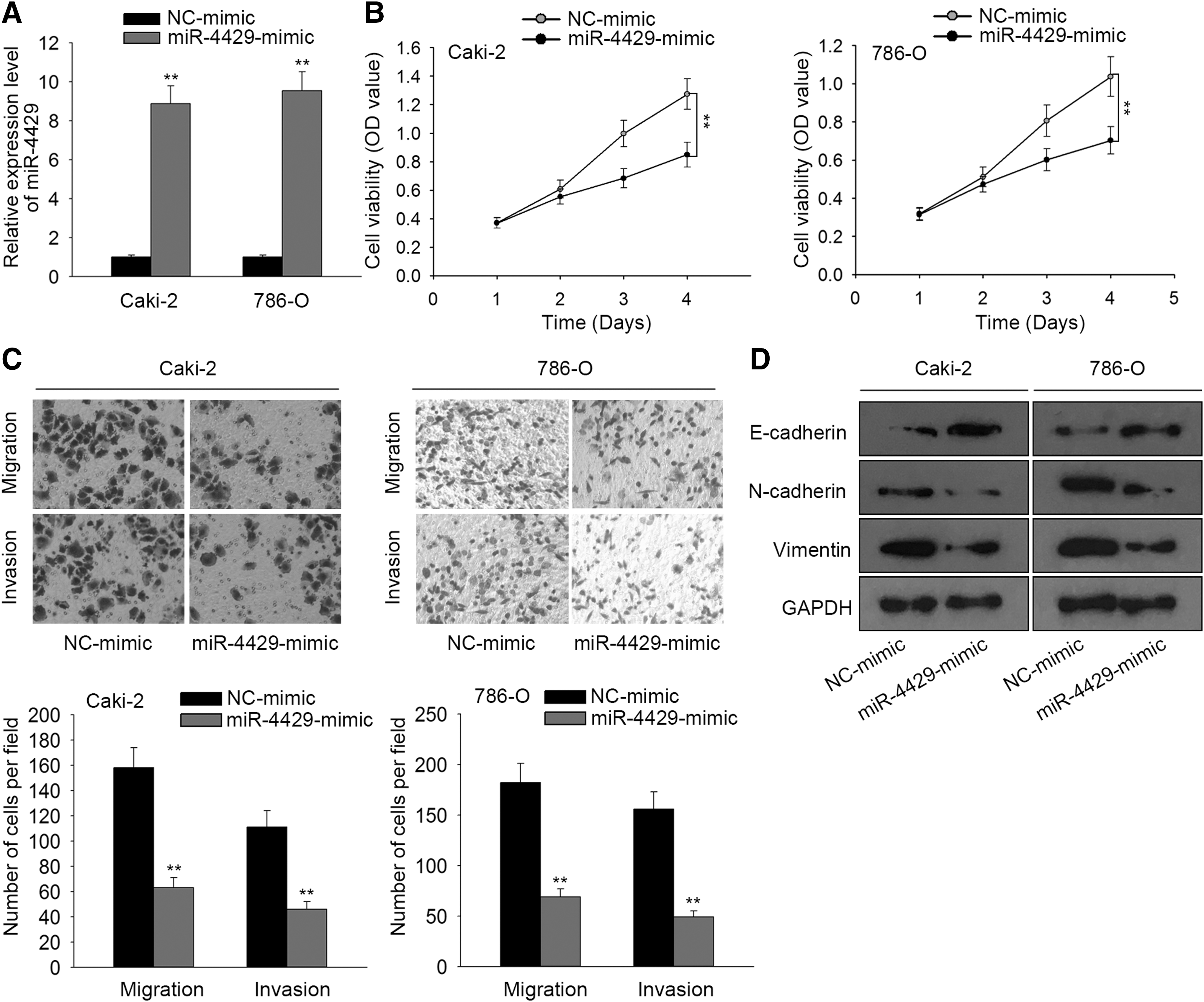
Overexpression of miR-4429 inhibited cell proliferation, migration, invasion, as well as EMT progression.
miR-4429 directly targeted CDK6 in ccRCC
It has been acknowledged that miRNAs can regulate cancer progression by binding to the 3′-UTR of target genes. 8,9 Thus, to further explore the mechanism by which miR-4429 regulated tumor progression and EMT in ccRCC, we searched for potential target of miR-4429 through TargetScan (v. 7.2). Among the candidate potential targets, we selected CDK6 because it was reported to be a promoter of cell proliferation and metastasis in various cancers, including ccRCC. 23 –25,30 Therefore, we hypothesized that miR-4429 might suppress progression and metastasis of ccRCC by targeting CDK6. To validate this hypothesis, we first examined the binding interaction between miR-4429 and CDK6 through luciferase reporter assay. The binding site and mutant site of CDK6 with miR-4429 were shown in Figure 3A. It was validated that overexpression of miR-4429 weakened the luciferase activity on wide type rather than mutant CDK6 compared with control (Fig. 3B), confirming CDK6 as a target of miR-4429.

miR-4429 directly targeted CDK6.
Then we investigated the effect of miR-4429 on CDK6 level. qRT-PCR and western blotting showed that upregulation of miR-4429 led to a diminishment on CDK6 mRNA and protein level (Fig. 3D, E). Furthermore, CDK6 was discovered to be upregulated in ccRCC tumor tissues and cell lines compared with normal tissues and cell line (Fig. 3F, G). Spearman's correlation analysis verified the negative correlation between CDK6 and miR-4429 expression (Fig. 3H). Results above implicated that miR-4429 targeted CDK6 in ccRCC.
miR-4429 inhibited cell proliferation, migration, invasion, and EMT progression by targeting CDK6 in ccRCC
To explore whether miR-4429 regulated ccRCC tumor progression and metastasis through targeting CDK6, we performed rescue assays. CDK6 was overexpressed in Caki-2 and 786O cells (Fig. 4A), and knocked down in Caki-1 and A498 cells (Supplementary Fig. S2A). MTT assays showed that upregulation of CDK6 restored the inhibitive effect of miR-4429 overexpression on cell viability (Fig. 4B). And silencing CDK6 reversed the inductive effect of miR-4429 inhibitor on cell viability (Supplementary Fig. S2B). Transwell migration and invasion assays demonstrated that forced expression of CDK6 attenuated the suppressive effect of miR-4429 overexpression on cell migration and invasion (Fig. 4C). Inversely, the miR-4429 inhibitor-facilitated migratory and invasive capacity of ccRCC cells was renovated by knocking down CDK6 (Supplementary Fig. S2C). Moreover, western blotting analysis revealed that CDK6 overexpression retarded the promotion of E-cadherin level and the suppression of N-cadherin and Vimentin level caused by miR-4429 upregulation (Fig. 4D). And CDK6 depletion countervailed the effect of miR-4429 inhibitor on the EMT markers above (Supplementary Fig. S2D). To sum up, results above suggested that miR-4429 inhibited cell proliferation, migration, invasion, and EMT by targeting CDK6 in ccRCC.
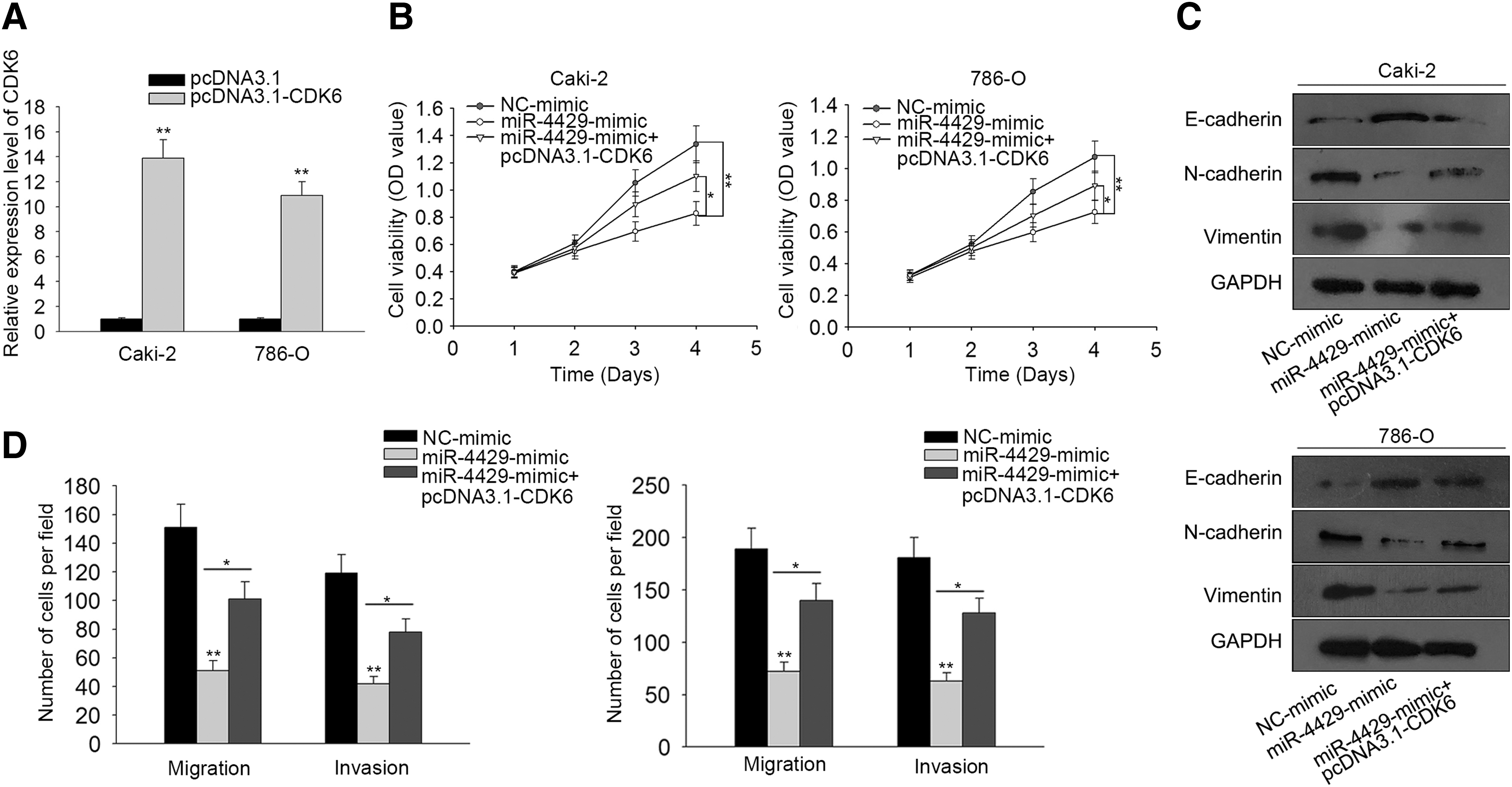
miR-4429 inhibited cell proliferation, metastasis and EMT progression by targeting CDK6 in ccRCC.
Discussion
ccRCC is the most prevalent subtype of renal carcinomas. 1 –3 Patients with this aggressive type of cancer are prone to poor overall survival in view of its easy metastasis and high reoccurrence rate. 3,4 Despite mounting effort in improving diagnosis and treatments for ccRCC through identifying molecular biomarkers, those eligible to serve as successful prognosis and therapeutic targets remains rare. 5 –7 Hence, it is of critical importance to recognize new targets via molecular mechanism research in ccRCC.
The role of miRNAs has caught increasing attention of the researchers. As a conserved class of small non-coding RNAs, they have been reported to participate in the regulation of multiple cancers, including ccRCC. 10,11,16 –21 miR-4429 is one of the miRNAs identified as tumor suppressor in papillary thyroid cancer, 22 but never has its role been touched in ccRCC. Thus, present study was the first to unveil the suppressive function of miR-4429 in ccRCC, which was in concordance with its tumor-suppressive role as revealed by previous study. We identified the upregulated level of miR-4429 in ccRCC tissues and cell lines. And we further uncovered through Kaplan–Meier analysis that miR-4429 could serve as a target for prognosis in ccRCC patients. Gain- and loss-of-function assays indicated that miR-4429 inhibited cell proliferation, migration, invasion and EMT.
For mechanism exploration, previous studies have presented that miRNAs could regulate gene expression though binding to their 3′-UTR in cancers. 8,9 Present study identified CDK6 as a target for miR-4429 via bioinformatics tools. CDK6 has been reported to influence not only cell cycle primarily, but also cell proliferation and, notably, cell metastasis in cancers. 23 –25 In our study, CDK6 was firstly revealed to be targeted by miR-4429. We validated the interaction between miR-4429 and CDK6 by luciferase reporter assay. Furthermore, the negative regulation of miR-4429 on CDK6 level was confirmed by qRT-PCR and western blotting. And upregulation of CKD6 in ccRCC tissues and cell lines, as well as its negative correlation with miR-4429 were verified respectively by qRT-PCR and Spearman's correlation analysis. Finally, we carried out rescue assays and revealed that CDK6 overexpression restored the inhibitive effect of miR-4429 on cell proliferation, metastasis as well as EMT progression. To sum up, present study revealed that miR-4429 suppressed ccRCC tumor progression and EMT via directly targeting CDK6, providing new molecular prognostic target for ccRCC, but more effort should be paid for deeper mechanism research and in vivo research in the future.
Footnotes
Acknowledgment
We express our gratitude to all participants.
Funding
This work was supported by Development and application of mobile phone software for maintenance hemodialysis patients' family diet management.
Disclosure Statement
It is declared by the author that no conflicts for interest exist in this article.
Supplementary Material
Supplementary Figure S1
Supplementary Figure S2
References
Supplementary Material
Please find the following supplemental material available below.
For Open Access articles published under a Creative Commons License, all supplemental material carries the same license as the article it is associated with.
For non-Open Access articles published, all supplemental material carries a non-exclusive license, and permission requests for re-use of supplemental material or any part of supplemental material shall be sent directly to the copyright owner as specified in the copyright notice associated with the article.
