
Abstract
Objective:
The aim was to investigate the lncRNA KB-1471A8.2 function in ovarian cancer progression and paclitaxel resistance.
Methods:
The expression and distribution of KB-1471A8.2 was detected by qRT-PCR. The cell proliferation, apoptosis, invasion, migration and chemoresistance were analyzed by CCK8 assay, flow cytometry and transwell assay. The expression of DEPTOR, whose sequence is reverse overlapped with KB-1471A8.2 was analyzed by qRT-PCR, western blot and immunofluorescence. The cell cycle and the cell cycle related gene expression were analyzed by flow cytometry and qRT-PCR, respectively.
Results:
KB-1471A8.2 was significantly downregulated in both ovarian cancer tissues and chemoresistant ovarian cancer cells. Overexpression of KB-1471A8.2 significantly inhibited the proliferation, invasion, migration, and paclitaxel resistance, and increased the apoptosis of ovarian cancer cells. KB-1471A8.2 was mainly distributed in the nucleus of ovarian cancer cells. KB-1471A8.2 overexpression significantly decreased the S phase cell ratio, increased the G0/G1 phase cell ratio, but not affected the expression and distribution of DEPTOR. However, cyclin-dependent kinase 4 (CDK4), which is an important regulator of G1/S transition, was significantly decreased in KB-1471A8.2-overexpressed ovarian cancer cells.
Conclusion:
KB-1471A8.2 could significantly inhibit the development and paclitaxel resistance of ovarian cancer cells, at least partly, by suppressing CDK4 expression.
Introduction
Ovarian cancer is the leading cause of deaths from gynecologic cancer. The median progression-free survival period is 16–22 months after primary debulking surgery and platinum/taxane-based chemotherapy. The 5-year survival rate of advanced ovarian cancer is only 27%. 1 Most patients with ovarian cancer develop chemoresistance and relapsed 1–2 year after treatment. Therefore, further exploration of the molecular mechanisms of ovarian cancer progression and chemoresistance is crucial for improving the survival of patients with ovarian cancer.
Several groups have reported on the long noncoding RNA (lncRNA) function in cancer development and chemoresistance. For instance, lncRNA HOXD-AS1 not only exerts its oncogenic function in cervical cancer but also promotes chemoresistance in cisplatin-resistant cervical cancer cells. 2 Downregulation of lncRNA BC200 promotes cell proliferation and increases the carboplatin resistance of ovarian cancer cells. 3 Several lncRNAs, such as PVT1, NEAT1, MALAT1, and PTAR, function by promoting or inhibiting ovarian cancer progression. 4 –7
In the current study, the expression and function of the novel lncRNA KB-1471A8.2 was first examined. Overexpression of KB-1471A8.2 significantly decreased the proliferation, invasion, and migration of ovarian cancer cells and increased the sensitivity of ovarian cancer cells to paclitaxel. Thus, KB-1471A8.2 was considered as one of the targets to oppose progression and paclitaxel resistance in ovarian cancer cells.
Materials and Methods
Clinical samples
A total of 10 normal ovarian tissue samples and 9 serous ovarian cancer tissue samples were obtained from 19 patients who underwent surgery at the Women's Hospital Affiliated to Nanjing Medical University from May 2015 to August 2017. All 10 normal ovarian tissues were obtained from the patients who underwent surgery for benign ovarian cancer or endometrial carcinoma. The normal ovarian tissues and serous ovarian cancer tissues were age-matched (49 ± 12.9 vs. 53 ± 17.5). All samples were quickly frozen in liquid nitrogen and stored at −80°C before use. The study was approved by the Ethics Committee of the Women's Hospital of Nanjing Medical University, and informed consent was obtained from each patient.
KB-1471A8.2 overexpression vector construction and plasmid transfection
The full length of KB-1471A8.2 (ENST00000523563.1) was ligated to the pcDNA3.1 vector located behind the cytomegalovirus promoter. For plasmid transfection, 2 μg/well KB-1471A8.2 overexpression vector and 2 μg/well control vector were transfected to the SKOV3 or A2780 cells in a six-well plate in accordance with the instruction provided by the manufacturer of Lipofectamine 2000 Transfection Reagent (Thermo Fisher Scientific, MA).
Cell culture
The human ovarian cancer cell lines A2780 and SKOV3 were purchased from the Jiangsu KeyGen Biotech Corp., Ltd. The A2780 cells were maintained in Dulbecco's modified Eagle's medium (DMEM; Thermo Fisher Scientific, MA), with 10% fetal bovine serum (FBS; Thermo Fisher Scientific), 100 μg/mL streptomycin, and 100 U/mL penicillin. SKOV3 cells were maintained in RPMI-1640 (Thermo Fisher Scientific) with 10% FBS, 100 μg/mL streptomycin, and 100 U/mL penicillin. All cells were cultured in a humidified atmosphere containing 5% CO2 at 37°C.
RNA extraction and quantitative real-time PCR
Total RNA from cultured cells, normal ovarian tissues, and epithelial ovarian cancer tissues were extracted using TRIzol (Invitrogen, Thermo Fisher Scientific) in accordance with the instructions provided by the manufacturer. cDNA was synthesized from 2 to 3 μg of total RNA by using the Reverse Transcription Kit (Applied Biosystems, Thermo Fisher Scientific) in accordance with the instructions provided by the manufacturer. The relative expression of KB-1471A8.2, DEPTOR, and the cell cycle-related genes were normalized to β-ACTIN. The primer sequences are listed in Table 1. PCR was conducted under the following conditions: 95°C for 5 min; 40 cycles of 95°C for 10 s; and 60°C for 30 s.
Primer Sequences
Nuclear and cytoplasmic RNA isolation
Nuclear and cytoplasmic RNA was isolated using the PARIS™ Kit (Invitrogen, Thermo Fisher Scientific) as instructed by the manufactures. The adherent cells (A2780 and SKOV3) were trypsinized and then collected after the trypsin was inactivated by FBS and centrifugation. After removing the supernatant, 300 μL of ice-cold cell fractionation buffer was added to the cells and gently resuspended by pipetting. After 10 min incubation on ice, the cell suspension was centrifuged for 5 min at 4°C and 500 g. The supernatant containing the cytoplasmic fraction and the nuclear fraction in the bottom was then collected, and the RNA of both the cytoplasmic fraction and nuclear fraction was isolated using the kit. The cDNA was synthesized in accordance with the aforementioned protocols. Quantitative real-time PCR (qRT-PCR) was used to quantify the lncRNA in both the nucleus and cytoplasm. Moreover, 45S rRNA and 12S rRNA, which were mainly distributed in the nucleus and mitochondria, were used as the nucleus and the cytoplasmic internal control as described in a previous study. 8 The primer sequences for 45S or 12S rRNA are listed in Table 1.
Cell proliferation and chemoresistance assay
The A2780 and SKOV3 cells transfected with the control vector (pcDNA3.1) and KB-1471A8.2 overexpression vector were seeded at a density of 5 × 10 3 /well in 96-well plates 24 h after transfection.
For chemoresistance assay, the cells were treated with paclitaxel (Sermoneta, Latina, Italy) at a gradient of 0, 2, 4, 8, 16, and 32 μM after 12 h. About 48 h after treatment, 10 μL Cell Counting Kit-8 (CCK8; Dojindo Molecular Technologies, Inc., Shanghai, China) was added to each well and then incubated for 1 h. The absorbance was measured using the Synergy H4 Hybrid Reader (BioTek Instruments, Inc., Shanghai, China) at 450 nm. The half-inhibitory concentration (IC50) was determined according the ODs detected in each well.
For cell proliferation analysis, CCK8 was added immediately after cell adhesion (as 0 h) and 24 h, 48 h, and 72 h after cell adhesion. Also, the absorbance was analyzed 1 h after CCK8 was added. The cell proliferation curve was generated according to the results above.
Cell migration and invasion assay
Cell invasion and migration were analyzed using Transwell chambers (Millipore) with or without Matrigel (BD Biosciences, CA) in accordance with the instructions provided by the manufacturer. Following this step, 3 × 10 5 cells in 200 μL serum-free medium were seeded on the upper chambers (for cell invasion assay, the chamber was coated with Matrigel, which was 1:5 diluted in the DMEM for A2780 cells and RPMI-1640 for SKOV3 cells). The lower chambers were added with 0.5 mL DMEM for A2780 cells or 0.5 mL RPMI1640 supplemented with 10% FBS and 100 μg/mL streptomycin and 100 U/mL penicillin for SKOV3 cells. After incubation for 48 h at 37°C with 5% CO2, the cells invading the lower surface were first fixed in 4% paraformaldehyde for 30 min, stained with crystal violet for 15 min, and photographed under microscope after removing the cells on the upper surface of the chamber. For the SKOV3 cells, both invaded and migrated cells were counted; each chamber had four fields. For A2780 cells, the cells in the upper chamber were removed, whereas those in the lower chamber were lysed in RIPA. Protein concentration was analyzed by measuring the absorbance using Synergy H4 Hybrid Reader (BioTek Instruments, Inc.) at 560 nm.
Cell cycle analysis
The A2780 and SKOV3 cells transfected with the control vector and KB-1471A8.2 overexpression vector were harvested 48 h after transfection, washed with ice-cold phosphate-buffered saline (PBS) three times, and fixed in 70% ethanol overnight. The cells were then pretreated with 5 mg/mL ribonuclease for 30 min at 37°C and stained with 100 μg/mL propidium iodide (PI) for 30 min under dark conditions.
Cell apoptosis assay
The A2780 and SKOV3 cells were plated at a density of 2 × 10 5 /well into the six-well plate. Transfection was performed as described in manufacturer protocol. The A2780 and SKOV3 cells transfected with the control vector and KB-1471A8.2 overexpression vector were cultured for 36 h, induced by the mixture of apoptosis inducer A and B in the Apoptosis Inducers Kit according to the Kit's instruction (Beyotime, Shanghai, China) for 12 h, harvested after digestion by 0.25% trypsin-EDTA (Thermo Fisher Scientific, MA), washed with PBS and stained with Annexin V and PI by using the Annexin V/PI Apoptosis Detection Kits (Dojindo Molecular Technologies, Inc.), and detected by flow cytometry (BD Biosciences).
Cell immunofluorescence
First, 5 × 10⁴ cells were seeded in the 12-well plate. After 24 h, cells were fixed with 4% paraformaldehyde for 15 min at room temperature. Followed by washing in PBS for three times, permeabilization in PBST (0.5% Triton X-100 added in the PBS) for 20 min, washing in PBS for three times, overnight incubation with DEPTOR antibody (Proteintech, Wuhan, China) at 4°C, washing in PBS for three times, incubation with Goat anti-rabbit Alexa Fluor 594 (diluted in PBS; Thermo Scientific, Carlsdad, CA) for 2 h, washing in PBS washing for three times, counterstaining with DAPI (Beyotime) for 5 min, and then fixed in 50% glycerol. It is noteworthy that from incubating with the secondary antibody, all the steps were conducted in dark.
Western blot
First, cells were lysed in protein lysis buffer containing 100 μL RIPA, 1 mM PMSF (Beyotime) and 1 × cocktail (Merck, MA), centrifugating at 4°C, 15 min and the concentration were determined by using BCA Protein Assay Kit (Thermo Scientific). Subsequently, protein samples were separated by SDS-PAGE, transferred to PVDF membrane (Millipore, Boston), blocked with blocking buffer (Beyotime) for 2 h, incubated with the primary antibodies at 4°C overnight, washed in PBST for three times, incubated with secondary antibodies for 2 h, and then scanned using an image acquisition system (FluroChem M, ProteinSimple, San Jose, CA). The primary antibodies used in this study were DEPTOR (Proteintech, Hubei, China), LC3B (Cell Signaling Technology, MA), and GAPDH (Santa Cruz, CA). The secondary antibodies were goat anti-rabbit IgG (Millipore, Frederick, MD).
Statistical analysis
All data were statistically analyzed using SPSS ver. 17.0 (Chicago, IL). All data are presented as the mean ± standard deviation from three independent repeats. Unless otherwise noted, the differences between the two groups were analyzed using Student's t-test.
Results
KB-1471A8.2 was significantly downregulated in the chemoresistant ovarian cancer cells as well as the epithelial ovarian cancer tissues
KB-1471A8.2 expression was first compared between normal ovarian tissues and epithelial ovarian cancer tissues by qRT-PCR. The results indicated that KB-1471A8.2 was significantly downregulated in the epithelial ovarian cancer tissues relative to the normal ovarian tissues (Fig. 1A).

KB-1471A8.2 is significantly downregulated in ovarian cancer tissues and in the chemoresistant ovarian cancer cells.
Chemoresistance predicts the poor prognosis of patients with cancer. Thus, the paclitaxel-resistant A2780 cells (A2780/PTX) and paclitaxel-resistant SKOV3 cells (SKOV3/PTX) were first established (SKOV3/PTX) in accordance with the protocols described previously. 9 The IC50 of A2780/PTX cells to paclitaxel was 9.651 ± 4.023 μg/mL, compared with 0.34 ± 0.227 μg/mL in A2780 cells, as reported in an earlier study. 9 The IC50 of SKOV3/PTX cells to paclitaxel was 4.327 μg/mL compared with 0.034 μg/mL in SKOV3 cells. Then KB-1471A8.2 expression was detected in the chemoresistant ovarian cancer cell lines (A2780/PTX and SKOV3/PTX) as well as their parental chemosensitive cell lines (A2780 and SKOV3) was detected by qRT-PCR. The results showed that KB-1471A8.2 decreased not only in the ovarian cancer tissues and ovarian cancer cell lines but also significantly decreased in the chemoresistant ovarian cancer cell lines compared relative to the chemosensitive ovarian cancer cell lines (Fig. 1B). Thus, the reduced KB-1471A8.2 expression might predict the poor prognosis of ovarian cancer.
KB-1471A8.2 overexpression increased the sensitivity of ovarian cancer cells to paclitaxel
Whether KB-1471A8.2 is involved in the progression and chemoresistance of ovarian cancer is a question. To determine the KB-1471A8.2 function on ovarian cancer cells, the KB-1471A8.2 overexpression vector was constructed and transfected into the A2780 and SKOV3 cells. As presented in Figure 2A, KB-1471A8.2 expression was significantly increased after transfection of the KB-1471A8.2 overexpression vector in both A2780 and SKOV3 ovarian cancer cells. Then the A2780 cells and KB-1471A8.2 overexpressed A2780 cells were treated with a gradient of paclitaxel (0, 2, 4, 8, 16 and cell 32μM), after 48 h treatment, the cell viability was detected by CCK8 assay. The results indicated that KB-1471A8.2 overexpression significantly increased the sensitivity of the ovarian cancer cells to paclitaxel (Fig. 2B).
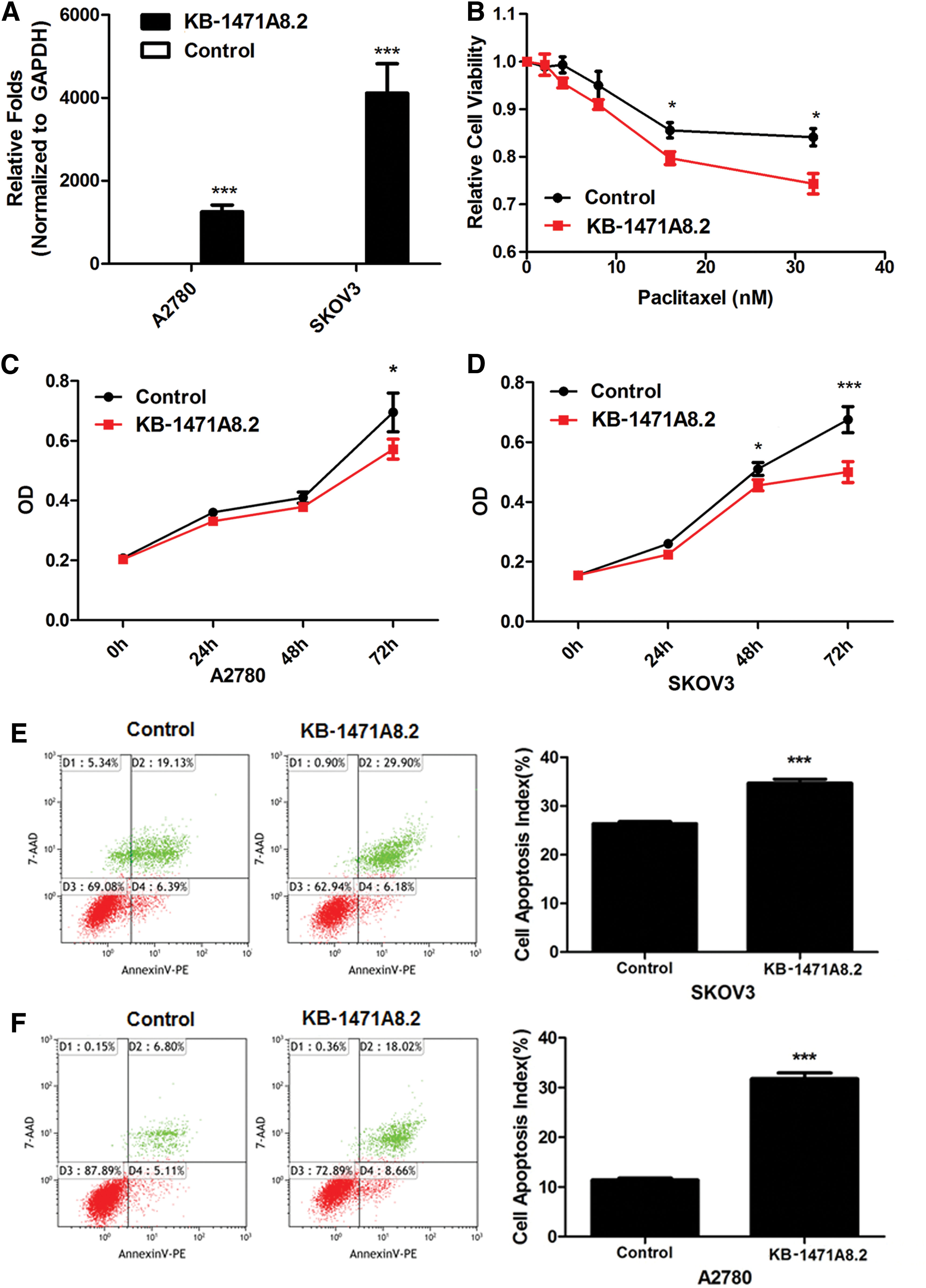
KB-1471A8.2 overexpression significantly inhibits cell proliferation and increases cell apoptosis and paclitaxel sensitivity of ovarian cancer cells.
KB-1471A8.2 overexpression decreased the proliferation, invasion, and migration and increased apoptosis of ovarian cancer cells
Subsequently, cell proliferation capacity was analyzed using the CCK8 assay kit following KB-1471A8.2 overexpression in A2780 and SKOV3 cells. And cell apoptosis was analyzed by flow cytometry. The results indicated that KB-1471A8.2 overexpression significantly decreased cell proliferation 72 h after transfection in A2780 cells, 48 and 72 h after transfection in SKOV3 cells relative to the cells transfected with the empty vector (Fig. 2C, D). Also, 12 h after treatment with the apoptotic inducer, the apoptotic cells were significantly increased in KB-1471A8.2-overexpressed A2780 and SKOV3 cells (Fig. 2E, F). In addition, invasion and migration was also significantly decreased in the KB-1471A8.2-overexpressed A2780 and SKOV3 cells (Fig. 3).
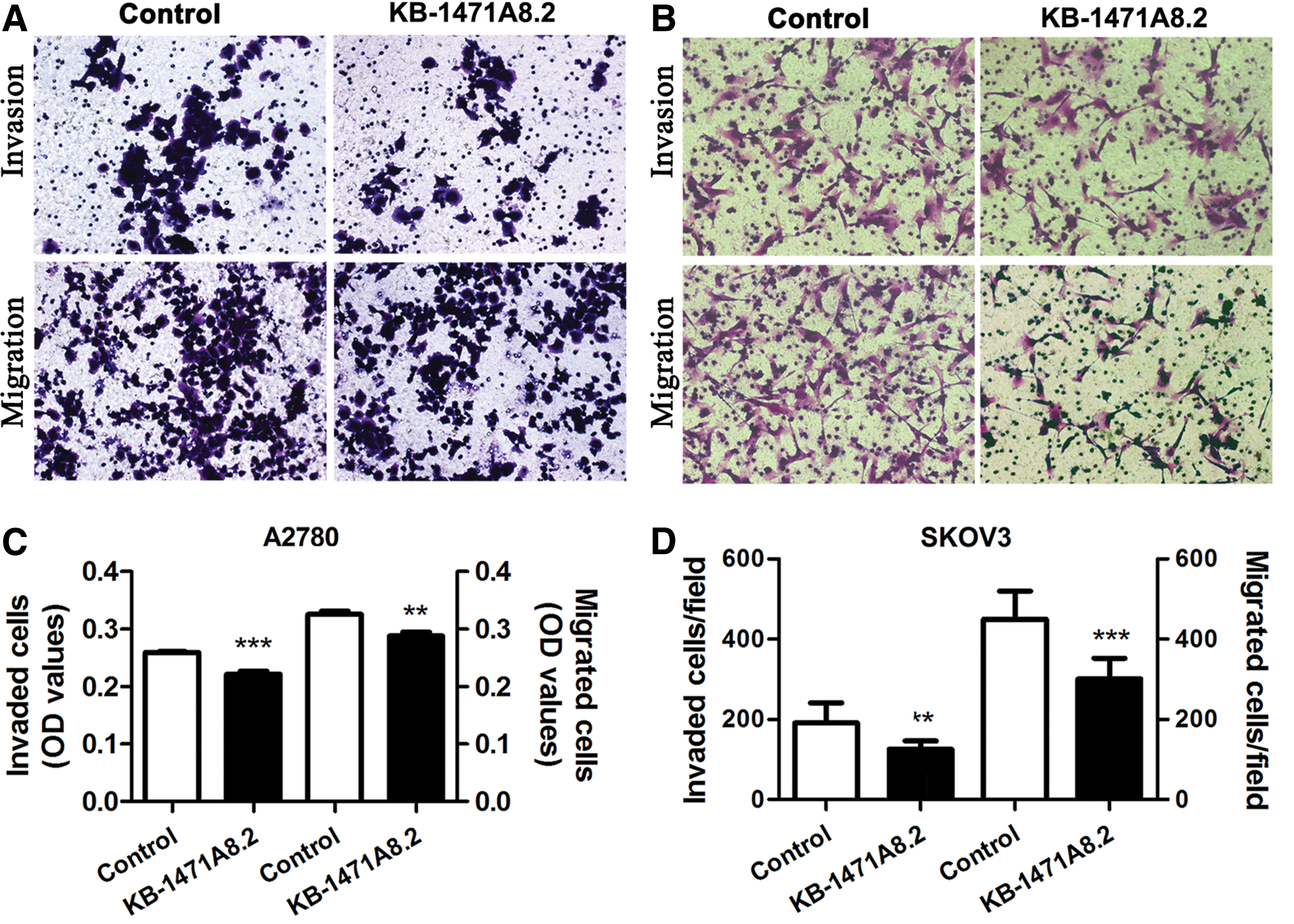
KB-1471A8.2 significantly inhibits the invasion and migration of ovarian cancer cells.
KB-1471A8.2 mainly distributed in the nucleus of the ovarian cancer cells
The distribution of genes or lncRNAs often relates their potential functions. For example, the genes or lncRNAs distributes in the nucleus often function as transcription regulators 10,11 or recruit chromatin-modifying molecules, such as PRC2, PRC1, and HDAC, 12,13 or function as enhancers 14 ; in addition, the lncRNAs distributed in the cytoplasm often function as miRNA sponges, 15,16 alter mRNA stability, 16 change protein trafficking, or stability. 17 –21 Thus, KB-1471A8.2 expression in the cytoplasmic and nuclear RNA fraction was detected by qRT-PCR. The results showed that KB-1471A8.2 was mainly distributed in the nuclear (Fig. 4).
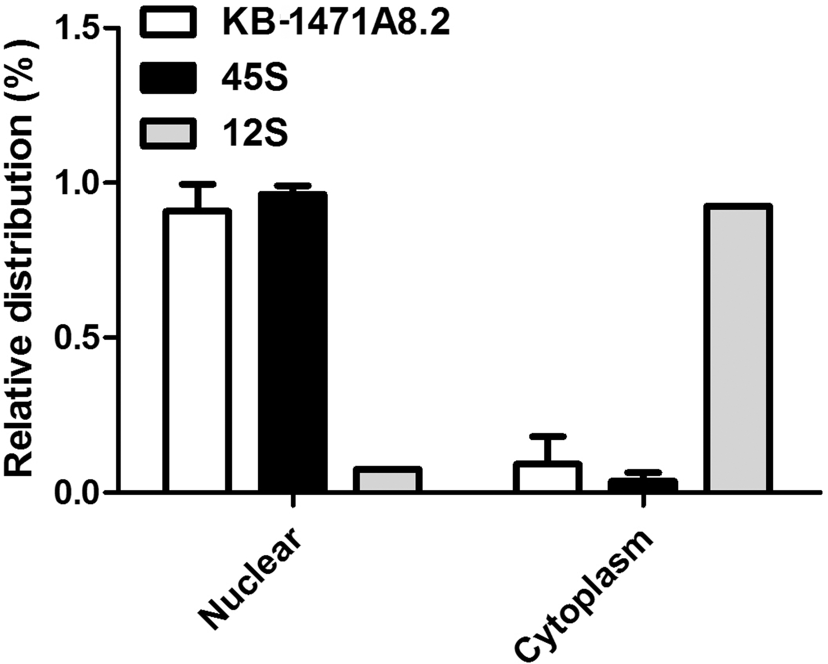
KB-1471A8.2 is mainly distributed in the nucleus of ovarian cancer cells. Using 45S as the nuclear localized control and 12S as the cytoplasmic localized control, the results indicate that KB-1471A8.2 is mainly distributed in the nucleus of ovarian cancer cells.
KB-1471A8.2 overexpression exerted no effect on the expression and distribution of DEPTOR
KB-1471A8.2 (also a transcript of KB-1471A8.1) is a newly identified lncRNA, which is reverse complemented with DEP domain-containing mTOR-interacting protein (DEPTOR), an endogenous inhibitor of mTORC1 and mTORC2 with key roles in numerous biologic and pathologic processes, including autophagy. 22 A large number of studies have demonstrated the function of DEPTOR on metabolism and chemoresistance of cancer cells. 22,23 Thus, DEPTOR expression and distribution was further analyzed in both the control and KB-1471A8.2-overexpressed ovarian cancer cells. Immunofluorescence analysis indicated that DEPTOR expression and distribution in the ovarian cancer cells remained unchanged in KB-1471A8.2-overexpressed cells (Fig. 5A, B). In addition, qRT-PCR and western blot analysis showed that both the mRNA expression and protein expression were not significantly changed in the KB-1471A8.2-overexpressed SKOV3 ovarian cancer cells (Fig. 5C, D).
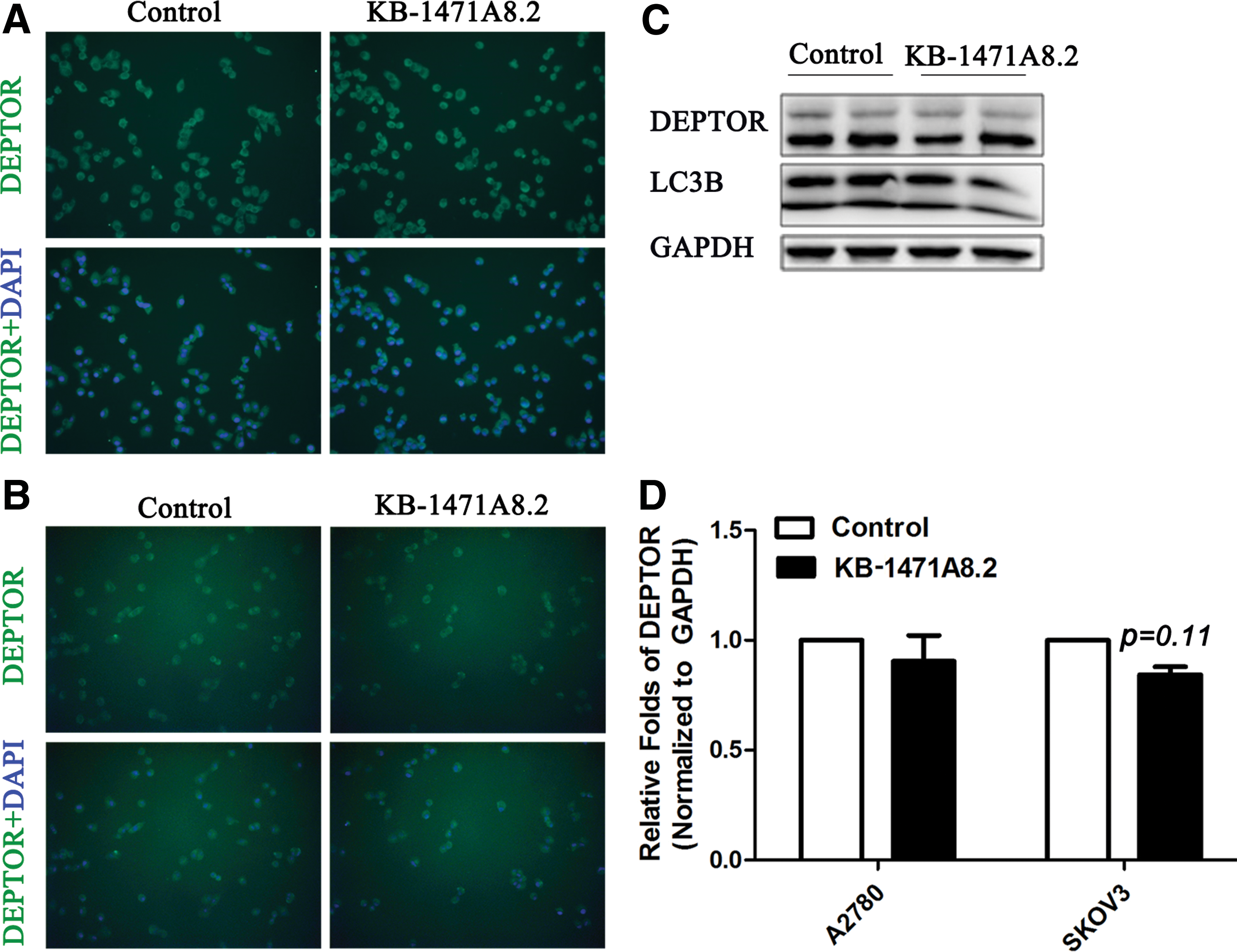
KB-1471A8.2 overexpression exerts no effect on DEPTOR expression and distribution.
KB-1471A8.2 overexpression significantly inhibited S-phase entry by downregulating CDK4 expression
Paclitaxel mainly exerts its antitumor activity by blocking cell mitosis and promoting cancer cell apoptosis. To determine whether KB-1471A8.2 overexpression affects the cycle of ovarian cancer cells, flow cytometry was performed. The results showed that KB-1471A8.2 overexpression significantly decreased the S-phase cell ratio and increased the G0/G1 phase cell ratio (Fig. 6A). Thus, the expression of cell division-related genes (CDC25C, PLK1, PTTG1, and ESPL1), cyclins (CCNA1, CCNB1, CCND1, and CCNE1), cyclin-dependent kinases (CDK1, CDK2, and CDK4), and cyclin-dependent kinase inhibitors (CDKN1A and CDKN1B) were analyzed by qRT-PCR. Only cyclin-dependent kinase 4 (CDK4) was significantly downregulated in KB-1471A8.2-overexpressed SKOV3 cells (Fig. 6B).

KB-1471A8.2 overexpression significantly inhibits G1/S transition by suppressing the expression of cyclin-dependent kinase 4 (CDK4) in ovarian cancer cells.
Discussion
With the development of high-throughput sequencing, an increasing number of lncRNAs are increasingly being discovered, from which thousands of differentially expressed lncRNAs are identified in chemoresistant cancers compared with chemosensitive cancers. However, only about 30 lncRNAs' functions in chemoresistance have thus far been elucidated. Their functions cover the resistance to gemcitabine, 5-FU, cisplatin, docetaxel, adriamycin, and so on. 24
Several studies indicated that several lncRNAs functions by regulating the genes that are reverse-complemented with the lncRNAs. For instance, the lncRNA GMAN, which is inversely complemented with EFNA1, regulates the translation of EFNA1 by binding competitively to the antisense GMAN RNA. 25 The lncRNA ZEB1-AS1 can significantly induce ZEB1 transcription in prostate cancer cells and cervical cancer cells. 26,27 Notably, KB-1471A8.2 is reverse-complemented with DEPTOR, an important regulator of autophagy. So the DEPTOR expression was analyzed in the control and KB-1471A8.2 overexpressed ovarian cancer cells. However, DEPTOR expression and distribution were not significantly changed in the KB-1471A8.2 overexpression cells. Therefore, although KB-1471A8.2 and DEPTOR are close to each other on the chromosome and reverse-complemented with each other, no direct regulatory relationship exists between them.
Previous studies have reported that inhibition of CDK4 by palbociclib enhanced the paclitaxel activity of ovarian cancer cells by inducing apoptosis. 28 Another study also indicated that patients with high CDK4/6-specific activity exhibited shorter progression-free survival (p = 0.024). 29 Serum starvation can induce G1 arrest via CDK2 and CDK4 inhibition in ovarian cancer cells. 30 All results obtained in the current study are consistent with these studies. Therefore, KB-1471A8.2 can inhibit cell proliferation, invasion, and migration, and increase the apoptosis and paclitaxel sensitivity of ovarian cancer cells, at least in part, by suppressing CDK4 expression.
Footnotes
Acknowledgments
This study was financially supported by the National Natural Science Foundation of China (No. 81572556 & No. 81602285), Nanjing Medical Science and technique Development Foundation (No. ZKX15046 & No. ZDX16015 & No. QRX17159 & No. JQX17009), Research Innovation Program for Graduates of Jiangsu Province (No. JX22013446), Jiangsu Provincial Medical Talent (Xuemei Jia).
Disclosure Statement
No competing financial interests exist.