
Abstract
Background:
Ruyiping is an effective traditional Chinese herbal medicine formula for preventing postoperative recurrence and metastasis of breast cancer. However, the exact function and underlying mechanism of Ruyiping in breast cancer remain unclear.
Materials and Methods:
After breast cancer cell lines MDA-MB-231 and MDA-MB-468 were treated with Ruyiping, the CCK8, colony formation, wound-healing, and transwell invasion assays were used to examine cell proliferation, migration, and invasion, respectively. Flow cytometry was performed to examine the effect of Ruyiping on cell cycle distribution. Western blot was performed to examine the expression of related proteins, and the activity of MMP9 was detected using Gelatin zymography assay.
Results:
Ruyiping treatment significantly inhibited cell proliferation and viability of MDA-MB-231 and MDA-MB-468 cells. Ruyiping was also revealed to trigger cell cycle arrest at the G2 phase in MDA-MB-231 and MDA-MB-468 cells. Moreover, Ruyiping suppressed the migration and invasion abilities of MDA-MB-231 and MDA-MB-468 cells in vitro. Furthermore, Ruyiping blocked the activity of MMP9 in MDA-MB-231 and MDA-MB-468 cells. Additionally, western blotting showed that Ruyiping attenuated epithelial-to-mesenchymal transition (EMT) of breast cancer through downregulation of N-cadherin, Vimentin, Snail1, and Snail2 and upregulation of E-cadherin. The authors observed that the components of Ruyiping Pseudobulbus Cremastra seu pleiones polysaccharide and curcumol showed significant suppression in the growth and invasion of breast cancer cell.
Conclusions:
The observations of this study suggest the antitumor properties of Ruyiping in cell growth and invasion of breast cancer, which are modulated by induction of cell cycle arrest and reduction of MMP9 and EMT.
Introduction
Breast cancer is the second leading cause of cancer death among women worldwide, with ∼40,290 deaths per year in the United States. 1,2 In particular, the highly malignant triple-negative breast cancer is characterized by rapid progression, frequent recurrence, and metastasis, resulting in poor prognosis of patients. Although the advances in early diagnosis, radical surgery, and adjuvant treatment in breast cancer have improved the survival rate and prognosis of patients in recent years, however, the mortality rate of breast cancer patients is still high, and the prognosis of patients with advanced stage remains poor owing to the aggressive behavior of the tumor. 3 –5 With the development of medicine and the continuous updating of knowledge, cancer treatment methods have become more diversified, developing a comprehensive cancer treatment strategy.
Traditional Chinese medicine (TCM), a therapy with a history of thousands years, has received increasing attention as an important component of complementary and alternative medicine, especially in cancer treatment. 6,7 Increasing studies have confirmed that numerous traditional Chinese herbal medicines can be used as anticancer drug in cancer treatment. 8 –10 It has been demonstrated that traditional Chinese herbal medicines play important roles in the recovery period, radiotherapy, or chemotherapy period of tumor surgery as adjuvant therapy and also inhibit tumor cell proliferation and survival in vitro. 11,12
Ruyiping is an effective traditional Chinese herbal medicine formula from clinical experience, with the function of detoxifying and dissipating the knot. Ruyiping is composed of Pseudobulbus Cremastra seu pleiones (Shancigu), Nidus Vespae (Lufengfang), Curcuma zedoaria (ezhu), raw seeds of Coix lacryma-jobi L. var. mayuen (Roman.) Stapf (Shengyiyiren), and Akebiae Fructus (Bayuezha). Indeed, in clinic, Ruyiping decoction has been used for the prevention of postoperative recurrence and metastasis of breast cancer. 13 It has been reported that Ruyiping inhibits pulmonary metastasis of breast cancer in animal model. 14 However, to the best of the knowledge of the authors, there are no reports on the effects of Ruyiping on cellular behaviors of breast cancer, and the underlying mechanism of Ruyiping in preventing breast cancer recurrence and metastasis remains unclear.
In the current study, the authors aimed to investigate the effective mechanism of Ruyiping on breast cancer. For the first time, the data showed that Ruyiping inhibited cell growth by inducing cell cycle arrest at the G2 phase. Moreover, it was observed that Ruyiping suppressed the migration and invasion abilities of breast cancer cells, which might be regulated by diminishing MMP9 and epithelial-to-mesenchymal transition (EMT) phenotype. This study might provide a novel perspective for the therapeutic role of Ruyiping in breast cancer.
Materials and Methods
Ruyiping decoction
The Ruyiping decoction was prepared by the pharmacy of Affiliated Hospital of Shandong University of Traditional Chinese Medicine, with fixed origin and quality control standards. Specifically, it consists of Pseudobulbus Cremastra seu pleiones (Shancigu), 12 g; Nidus Vespae (Lufengfang), 12 g; Curcuma zedoaria (ezhu), 12 g; raw seeds of Coix lacryma-jobi L. var. mayuen (Roman.) Stapf (Shengyiyiren), 12 g; and Akebiae Fructus (Bayuezha), 9 g. According to the traditional decoction method, the decoction of Ruyiping was concentrated at 3.2 g/mL and dried at 80°C to form coarse powder. The coarse powder was immersed in 90% ethanol (50–100 mL) at room temperature. After 1 week, the extract was collected, and the sediment was extracted once with 90% ethanol and mixed with the extract. Then, the ethanol extract was centrifuged at 3000 rpm for 10 min to collect the supernatant. The supernatant was placed in a boiling water bath to volatilize the ethanol and concentrated to a thick paste. The extract was dissolved in 1 mL of dimethyl sulfoxide and stored at 4°C. The Ruyiping decoction was diluted to the set concentration with phosphate buffer and filtered with a 0.2-μm filter before use.
Cell culture and treatment
The human breast cancer cell lines MDA-MB-231 and MDA-MB-468 were obtained from the Cell Bank of Chinese Academy of Sciences (Shanghai, China). Cells were cultured in Dulbecco's Modified Eagle Medium (DMEM) (Hyclone, Logan, UT), which contained 10% fetal bovine serum (FBS; Gibco, New York) and antibiotics (100 U/mL penicillin, 0.1 mg/mL streptomycin; Sigma–Aldrich, St. Louis, MO; Germany) at 37°C with 5% CO2. Cells were seeded to 24-well plates and incubated in DMEM medium for 24 h followed by incubation with Ruyiping medium or different reagents. Phosphate-buffered saline (PBS) was used as negative control (NC). The Pseudobulbus Cremastra seu pleiones polysaccharide (PCSPP) was purchased from Science Biotech (Xian, China), and curcumol was obtained from Aladdin (Shanghai, People's Republic of China).
CCK8 assay
Screen for appropriate drug concentration: cells were seeded into a 96-well plates for 24 h at a density of 1 × 10 3 cells per well. Ruyiping decoction was diluted in different concentrations (0%, 1%, 2.5%, 5%, 10%, 20%, 30%, 40%, 45%, and 50%) and then added into the plates. After 24 h of treatment, Cell Counting Kit-8 (CCK8, 10 μL/well; Beijing Solarbio Science & Technology, Beijing, China) reagent was added into each well, followed by incubation at 37°C for 90 min. The optical density was measured at 450 nm to identify the appropriate concentration of Ruyiping for breast cancer cells.
For assessment of cell viability, cells were seeded into 96-well plates at a density of 1 × 10 3 cells per well and cultured at 37°C for 24 h. The cell viability was assessed every 24 h by adding Ruyiping to the plate, and 10 μL of CCK8 reagent was added per well before the test.
Colony formation assay
Cells were seeded into 6-cm culture dishes at a density of 500 cells per dish followed by the treatment with Ruyiping. After 1 week of incubation at 37°C, visible colonies were fixed in 5 mL of 4% paraformaldehyde for 30 min and then stained with 0.1% crystal violet for 30 min. The colonies were counted and photographed. PBS was used as NC.
Cell cycle analysis
Flow cytometry was performed to examine the effect of Ruyiping on cell cycle distribution. After being treated with Ruyiping or PBS for 24 h at 37°C, cells were collected and fixed in precooled 70% ethanol at −20°C overnight. Then, cells were centrifuged and resuspended in the buffer. Subsequently, cells were stained with propidium iodide for 30 min at 37°C; afterward, cell cycle distribution was analyzed by a flow cytometer (BD FACSCanto II; BD Biosciences) and calculated using BD FACSDiva software (BD Bioscience).
Wound healing assay
The MDA-MB-231 and MDA-MB-468 cells were grown in six-well plates at a density of 5 × 10 5 cells per well for 12 h. Subsequently, the wound was generated with a pipette tip, and cells were cultured in a serum-free medium supplemented with Ruyiping for 24 h. After that, the wound was photographed and the wound closure was measured using ImageJ software (National Institutes of Health).
Transwell invasion assay
Transwell chambers (Millipore, MA) coated with Matrigel (BD Bioscience, CA) were used for the assessment of cell invasion according to the manufacturer's instructions. MDA-MB-231 and MDA-MB-468 cells treated with Ruyiping for 24 h were trypsinized and resuspended in serum-free medium at a concentration of 1 × 10 6 cells/mL. Next, the upper compartment of Transwell chamber was seeded with 100 μL of cell suspension and the lower chamber was filled with complete medium with 10% FBS as the chemoattractant. After 24 h of incubation, cells that invaded through the Matrigel were fixed with 4% paraformaldehyde for 30 min and stained with 0.1% crystal violet for 20 min. The stained cells were imaged (magnification, × 40) and counted under the microscope.
Western blotting assay
After 48 h of treatment with Ruyiping, cells were collected and lysed using RIPA Lysis Buffer (CWBIO, Beijing, People's Republic of China) on ice for protein extraction. The protein concentration was determined using a BCA kit (CWBIO). Equal amount of protein from each sample was electrophoresed by 10% sodium dodecyl sulfate-polyacrylamide gel electrophoresis (SDS-PAGE) gel and then electrotransferred onto a polyvinylidene fluoride membrane (Millipore, Billerica, MA). The membrane was blocked with 5% nonfat milk for 1 h at room temperature before incubation with primary antibodies (1:1000; Proteintech Group, IL) at 4°C overnight. After washing with TBS buffer containing 0.01% Tween-20 for three times, membranes were incubated with horseradish peroxidase-conjugated secondary antibodies (1:3000; Proteintech Group) in blocking buffer for 1 h at room temperature. Finally, the protein bands were visualized using the enhanced chemiluminescence kit (CWBIO). GAPDH was used as an internal control.
Gelatin zymography
After being treated with Ruyiping for 24 h, cells were washed with serum-free medium and cultured in serum-free medium for 24 h at 37°C. Culture supernatants were collected and centrifuged to remove cells. The supernatants diluted by the loading buffer without mercaptoethanol were added into electrophoresis bath followed by electrophoresing by SDS-PAGE containing 0.5 mg/mL gelatin (Sigma–Aldrich, St. Louis, MO). After that, the gel was eluted, incubated, and stained with Coomassie Blue R250. After being decolorized, the gel was scanned and analyzed.
Statistical analysis
All data were expressed as mean ± standard deviation from triplicate independent experiments. GraphPad Prism 7.0 (GraphPad Soft-ware, Inc., La Jolla, CA) was employed for statistical analysis. The difference between two groups was estimated with Student's t test. p-Value of <0.05 was considered statistically significant.
Results
Ruyiping inhibits the proliferation and viability of breast cancer cells
To assess the effect of Ruyiping on tumor growth, Ruyiping was diluted to different concentrations (0%, 1%, 2.5%, 5%, 10%, 20%, 30%, 40%, 45%, and 50%) to treat breast cancer cell lines MDA-MB-231 and MDA-MB-468 for 24 h. As evidenced in Figure 1A, Ruyiping significantly inhibited the viability of MDA-MB-231 cells when the concentration of Ruyiping was more than 40% (p < 0.05). Similarly, Ruyiping also had a significant inhibitory effect on the viability of MDA-MB-468 cells (p < 0.05, Fig. 1B). The IC50 values of Ruyiping on MDA-MB-231 and MDA-MB-468 cells were 47.41% and 43.38%, respectively. Hence, 40% extract of Ruyiping was used in subsequent experiments due to its appropriate effect.
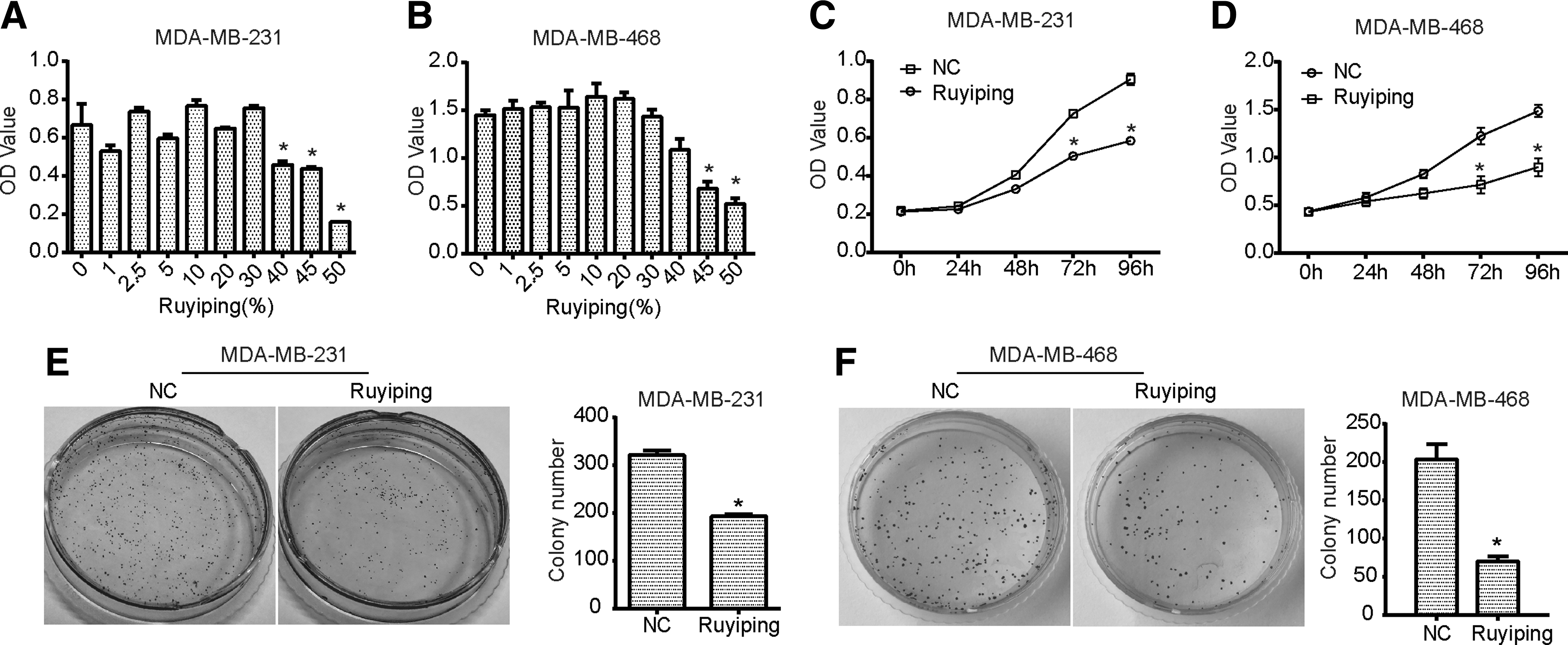
Ruyiping inhibits the viability and colony formation of breast cancer cells. The CCK8 assay for MDA-MB-231
As shown by the CCK8 results, in comparison with NC cells, a significant inhibition in proliferation of MDA-MB-231 and MDA-MB-468 cells was observed in a time-dependent manner followed by the treatment with 40% of Ruyiping (Fig. 1C, D). Moreover, the colony formation assay was performed to further confirm the antiproliferation activity of Ruyiping in breast cancer. As expected, Ruyiping significantly inhibited the colony formation ability of MDA-MB-231 and MDA-MB-468 cells with obvious decreased number of cell colonies compared with the NC group (p < 0.05, Fig. 1E, F).
Ruyiping triggers cell cycle arrest at the G2 phase in breast cancer cells
To further investigate the effect of Ruyiping, flow cytometry was performed for assessing the cell cycle distribution after treatment with Ruyiping for 24 h. The authors observed that compared with the control group, the proportion of cells in the G2 phase was increased in MDA-MB-231 cells treated with Ruyiping, at the same time, the proportions of cells in the G1 and S phases were both decreased (p < 0.05; Fig. 2A). In MDA-MB-468 cells, Ruyiping also increased the proportion of cells in the G2 phase, whereas it reduced the proportion of cells in the G1 phase (p < 0.05; Fig. 2B).
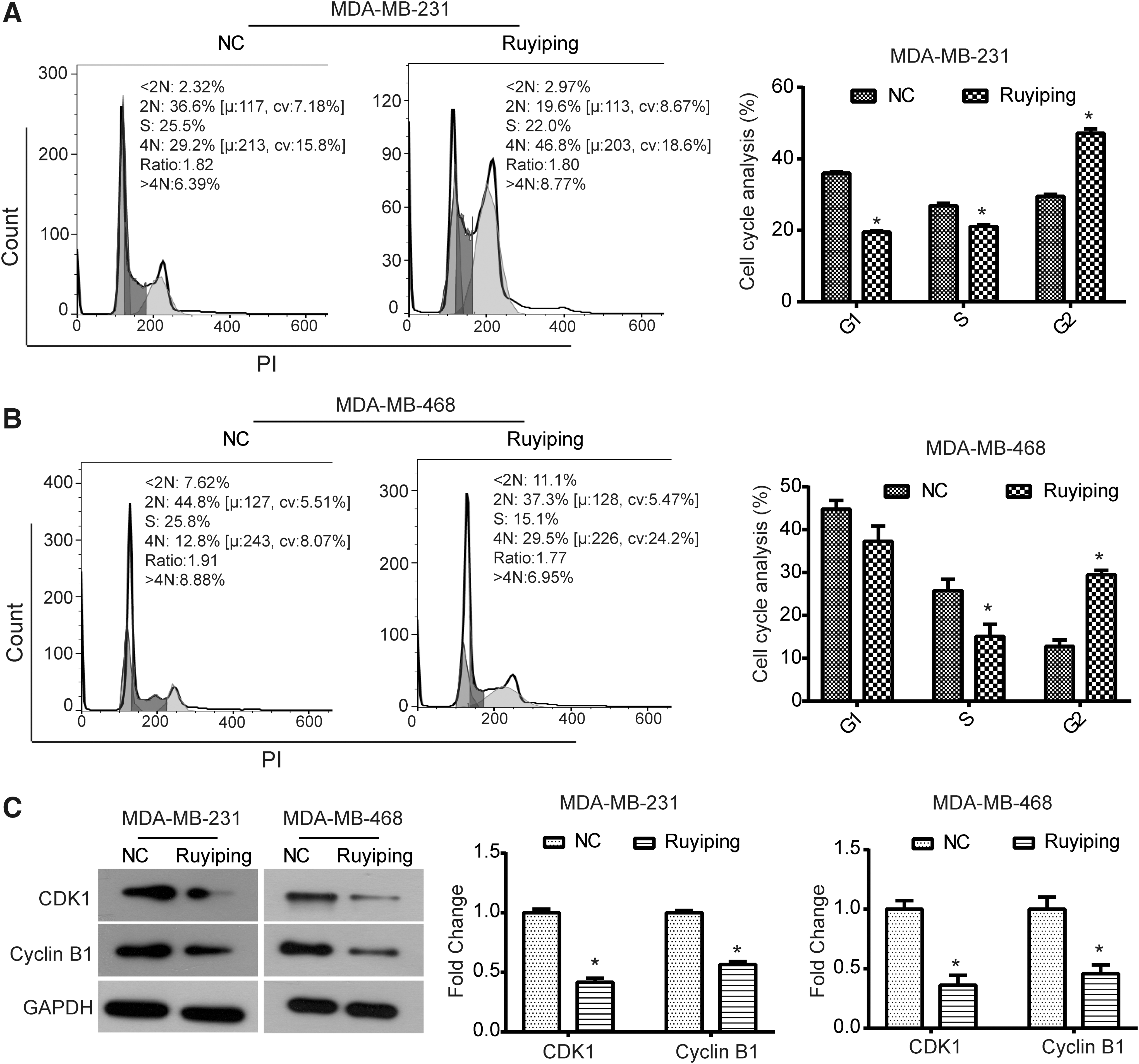
Ruyiping triggers cell cycle arrest at the G2 phase in breast cancer cells. MDA-MB-231
In addition, the expression of G2 phase-related proteins was detected by western blot to further investigate the mechanism of the cell cycle arrest caused by Ruyiping. As shown in Figure 2C, the expression of CDK1 and cyclin B1 was significantly decreased by Ruyiping in both MDA-MB-231 and MDA-MB-468 cells (p < 0.05). These results indicated that Ruyiping induced cell cycle arrest at the G2 phase in MDA-MB-231 and MDA-MB-468 cells by the regulation of CDK1/cyclin B1 axis.
Ruyiping suppresses the migration and invasion abilities of breast cancer cells
A wound healing assay was performed to examine whether Ruyiping impacts the cell migration ability of breast cancer cells. After being treated with Ruyiping for 24 h, MDA-MB-231 cells displayed a significant depression in the ability to migrate into the empty space compared with the NC group (p < 0.05; Fig. 3A, B), which was also observed in MDA-MB-468 cells (p < 0.05; Fig. 3A, B). Moreover, Transwell assay revealed that when exposed to Ruyiping for 24 h, the number of invasive cells was notably decreased in both MDA-MB-231 and MDA-MB-468 cells (p < 0.05; Fig. 3C, D). These results suggested that Ruyiping could suppress the migration and invasion abilities in breast cancer cells.

Ruyiping suppresses cell migration and invasion abilities of breast cancer cells.
Ruyiping diminishes the MMP9 and EMT in breast cancer cells
Further study was performed to explore the molecular mechanism of Ruyiping in suppressing the migration and invasion abilities of breast cancer cells. As known, the MMP9 and EMT have been proven to play crucial roles in tumor adhesion and invasion. 15,16 Herein, the gelatin zymography analysis demonstrated that Ruyiping significantly decreased the activity of MMP9 in MDA-MB-231 and MDA-MB-468 cells compared with NC cells (p < 0.05; Fig. 4A, B).

Ruyiping diminishes the MMP9 and EMT in breast cancer cells.
Since EMT is a pivotal process involved in tumor metastasis, the expression of EMT marker proteins was further examined by western blot to investigate whether EMT participates in the functional role of Ruyiping. As illustrated in Figure 4C, the expression of E-cadherin was significantly upregulated by Ruyiping in MDA-MB-231 and MDA-MB-468 cells, whereas the expression of N-cadherin, Vimentin, Snail1, and Snail2 was downregulated (p < 0.05). Taken together, these data shed light on the fact that the antitumor metastasis activity of Ruyiping may be regulated by the inhibition of MMP9 and EMT.
PCSPP and curcumol are the critical drug monomers regulating the growth and invasion of breast cancer
Since the Ruyiping formula components are complicated, MDA-MB-231 and MDA-MB-468 cells were treated with PCSPP (0.6 mg/mL) and curcumol (0.4 μM/mL), which are active components of Ruyiping, to further understand the functional exact component. As shown in Figure 5A and B, when exposed to PCSPP, the proliferation of MDA-MB-231 and MDA-MB-468 cells was significantly decreased compared with the NC group. Similarly, curcumol also caused a significant decrease in the proliferation of MDA-MB-231 and MDA-MB-468 cells (Fig. 5A, B). Moreover, the invasion ability of breast cancer cells was inhibited by PCSPP and curcumol (Fig. 5C, D). Furthermore, the authors found that the expression of E-cadherin was upregulated by PCSPP and curcumol compared with the NC group, whereas the expression of N-cadherin was significantly downregulated (Fig. 5E, F). In general, the above results demonstrate that PCSPP and curcumol are the main functional components of the anticancer activity of Ruyiping.
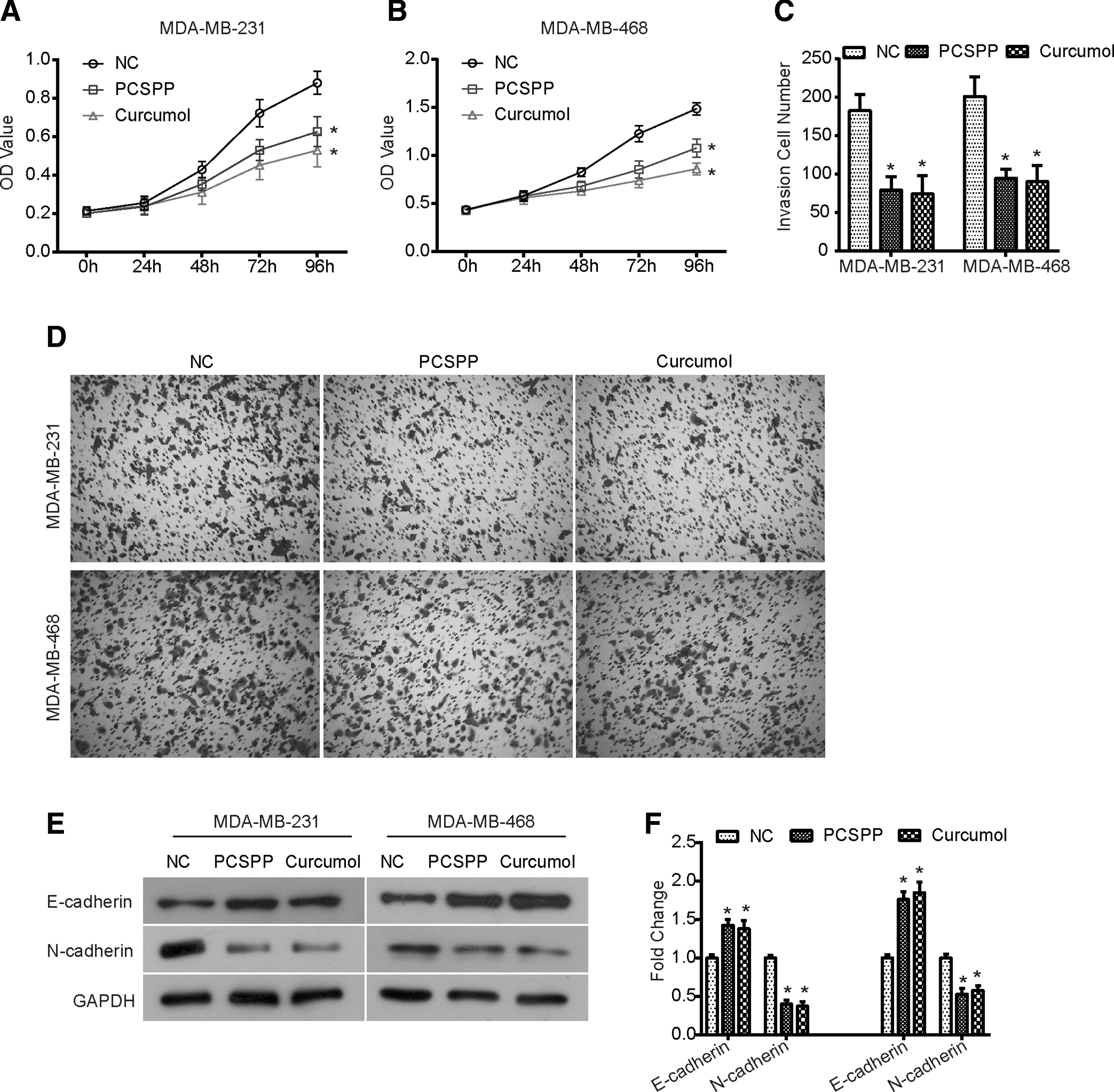
PCSPP and curcumol inhibit the growth and invasion of breast cancer cells.
Discussion
TCM has been developed for thousands of years and provides a wealth of potential chemopreventive and therapeutic drugs for the treatment of cancers. 8 For example, Xiong et al. reported that Ke formula can inhibit cell growth and induce cell cycle arrest and apoptosis through suppressing the PI3K/Akt pathway in non-small cell lung cancer. 8 The extract of Tubeimu has been reported to inhibit the growth and metastasis of triple-negative breast cancer both in vitro and in vivo. 17 Therefore, further understanding of the roles of traditional Chinese herbal medicines in tumor progression may provide a new perspective for the development of novel cancer treatment strategies.
Ruyiping formula is developed from several traditional Chinese herbal medicines and has been used clinically in China to inhibit postoperative recurrence and metastasis of breast cancer. The active components of the Ruyiping formulation are PCSPP, nidus vespae protein, curcumol, coixenolide, and Akebiae Fructus alcohol. In the present study, the authors evaluated the anticancer properties of Ruyiping in breast cancer in vitro and have also investigated the underlying mechanism.
For the first time, the authors demonstrated that Ruyiping inhibited the proliferation, viability, and clonogenic ability of breast cancer cells MDA-MB-231 and MDA-MB-468, suggesting an antigrowth activity of Ruyiping in breast cancer. Moreover, to gain insight into the relevant mechanism by which Ruyiping contributes to the inhibition on cell growth, cell cycle distribution was analyzed using flow cytometry. It is well known that uncontrolled proliferation is the most basic biological feature of malignant tumors, which is caused by the frequently occurring cell cycle dysregulation during tumorigenesis. 18,19 Therefore, blocking cell cycle progression may be an effective strategy for eliminating cancer cells, 18 which is also the main mechanism of several antitumor agents underlying the inhibition of tumor cell proliferation. 8,12,20
In this study, the authors illustrated that Ruyiping triggered cell cycle arrest at the G2 phase in breast cancer cells. The activation of CDK1/cyclin B1 complex is a pivotal regulator to promote cell cycle from G2 to M phase. 21 The authors observed that Ruyiping treatment significantly downregulated the expression of CDK1 and cyclin B1, providing a molecular-level explanation for Ruyiping-triggered cell cycle arrest at the G2 phase in breast cancer cells. As the active components of nidus vespae, nidus vespae protein named NVP (1) has been reported to inhibit cell proliferation and induce cell cycle arrest in HepG2 cells by downregulating the expression of cyclin B1. 22 Wang et al. found that curcumol, the active component of Curcuma zedoaria, decreases the proliferation of colorectal cancer LoVo cells through regulating the p38 MAPK signaling pathway. 23 Curcumol also induces cell cycle arrest at the G0/G1 phase via the downregulation of Akt/GSK3β/cyclin D1 pathways. 24 In this study, the data also demonstrated that PCSPP and curcumol displayed antiproliferation activity in breast cancer. Taken together, the regulation of cell cycle distribution might be involved in the antiproliferation activity of Ruyiping in breast cancer, which may be modulated by PCSPP and curcumol.
The MDA-MB-231 and MDA-MB-468 cells are triple-negative breast cancer cell lines with high metastatic potential. In respect to the research, the authors observed that Ruyiping resulted in significant suppression in migration and invasion abilities of MDA-MB-468 and MDA-MB-231 cells, suggesting the antimetastasis property of Ruyiping. Further study was performed to investigate the mechanism of Ruyiping underlying the suppression on the metastasis of breast cancer. MMP9, belongs to the matrix metalloproteinase (MMP) family, is revealed to be upregulated in tumors and involved in tumor metastasis. 25 –27 The data of this study suggested that Ruyiping significantly hindered the activity of MMP9 in MDA-MB-231 and MDA-MB-468 cells.
Additionally, EMT frequently occurs in the progression of epithelial cell carcinomas, which is the main process that promotes tumor metastasis and invasion. 28,29 Numerous studies have reported that some TCM can inhibit EMT in cancers. For example, Zhang and colleagues revealed that Jianpi Huayu decoction can suppress the metastasis of hepatocellular carcinoma SMMC7221 cells by blocking the EMT via regulating the Smad3/Smad7 cascade. 30 Xuan et al. showed that JianPi JieDu recipe can inhibit the migration and invasion of colorectal cancer LoVo cells by inhibiting the transforming growth factor-β-induced EMT. 31 At the molecular level, the process of EMT is characterized by downregulation of epithelial cell markers and upregulation of mesenchymal markers. 32,33 These marker proteins have been proven to play key roles in regulating EMT, such as E-cadherin and Snail, 34 which are considered as potential targets for TCM.
As shown in the data of this study, Ruyiping could upregulate the expression of E-cadherin in breast cancer cells and downregulate the expression of N-cadherin, Vimentin, Snail1, and Snail2, indicating that Ruyiping inhibited the process of EMT via regulating these EMT-associated proteins. Curcumol has been revealed to inhibit the migration and invasion abilities of nasopharyngeal carcinoma cell NPC5-8F through upregulating the expression of E-cadherin and downregulating the expression of N-cadherin. 35 The authors observed that PCSPP and curcumol displayed a significant depression in breast cancer cell invasion and EMT process, which was consistent with the antimetastatic function of Ruyiping.
Collectively, these data suggested that Ruyiping attenuates the migration and invasion abilities of breast cancer cells through suppressing the MMP9 and EMT; PCSPP and curcumol, as key active components, may be involved in the antimetastasis role of Ruyiping. However, the roles of other active components, coixenolide and Akebiae Fructus alcohol, in the antitumor activity of Ruyiping have not been well studied, which will be further investigated in the authors' future research.
Conclusions
In summary, for the first time, the observations of this study provide evidence that Ruyiping inhibits the growth and metastasis of breast cancer through triggering cell cycle arrest and diminishing the MMP9 and EMT. The present study suggests a reliable experimental basis for the application of Ruyiping in the clinical treatment of breast cancer.
Footnotes
Acknowledgments
This work was supported by a grant from the Natural Science Foundation of Shandong Province (no. ZR2017BH107) and the National Natural Science Fund (no. 81573989).
Authors' Contribution
Z.L. performed the experiments and wrote the article. X.S., X.L., and Z.S. also performed the experiments. J.L. collected and/or assembled data. All co-authors have reviewed and approved the article prior to submission.
Disclosure Statement
No competing financial interests exist.