
Abstract
Background:
Early detection of apoptosis is very important for therapy and follow-up treatment in various pathologic conditions. Annexin V interacts strongly and specifically with phosphatidylserine, specific biomarkers of apoptosis with some limitations. Small peptides are suitable alternatives to annexin V. A reliable and noninvasive in vivo technique for the detection of apoptosis is in great demand. Based on our previous studies, three new peptide analogs of LIKKPF (Leu-Ile-Lys-Lys-Pro-Phe) as apoptosis imaging agents were developed.
Materials and Methods:
Aoa-LIKKP-Cl-F, Aoe-LIKKP-Pyr-F, and Aoe-LIKKP-Nap-F were synthesized, functionalized with aminooxy, and radiolabeled with 18F-FDG. Their biologic properties were evaluated in vitro using apoptotic Jurkat cells. 18F-FDG-Aoe-LIKKP-Pyr-F peptide was injected into normal and apoptotic mice models for biodistribution and in vivo positron emission tomography/computed tomography imaging studies.
Results:
18F-FDG-Aoe-LIKKP-Pyr-F peptide showed higher affinity for apoptotic cells. The localization of peptide in apoptotic liver mice was confirmed in biodistribution and imaging studies.
Conclusion:
The results showed that Aoe-LIKKP-Pyr-F peptide is an auspicious agent for molecular imaging of apoptosis.
Introduction
Apoptosis is defined as programmed cell death, which keeps the balance between cell proliferation and cell death. Disturbance of this balance often leads to pathologic accumulation of cells such as cancer. In apoptosis, cells lose plasma membrane asymmetry, and phosphatidylserine (PS) is exposed on the outer leaflet of plasma membrane, while the integrity of the membrane kept is intact. PS on the outer cell surface of apoptotic cells acts as a tag for specific identification by macrophages. Regarding the role of apoptosis in various pathologic conditions such as cardiovascular diseases, autoimmune and neurodegenerative diseases (increase in apoptosis), or tumor growth (decrease in apoptosis), early detection of apoptosis is imperative for therapy and follow-up treatment. Ran and Thorpe, showed that PS is also a biomarker of tumor vasculature and a potential target for cancer therapy. 1 Considering the fact that PS is a specific biomarker of apoptosis, ligands with affinity for PS are good candidates for the detection of apoptosis. 2 –10
Annexin-V, an endogenous phospholipid protein, interacts strongly and specifically with PS in the presence of mM of Ca2+ (Kd = ∼10 nM). Since annexin V cannot pass through phospholipid bilayer of membranes, it does not bind to normal cells. Annexin V and its derivatives have been labeled with radionuclides (such as [99mTc] and [18F]) and fluorescence dyes and have been used in several studies. Yagle et al. 11 radiolabeled recombinant human annexin V with N-succinimidyl-4-18F-fluorobenzoic acid 18F-SFB and evaluated in an animal model of apoptosis. They could quantify cell death in vivo. However, annexin V has some limitations, such as inability to differentiate between apoptosis and necrosis, and the high cost of production. 12,13
To surmount these limitations, small peptides were developed as alternatives to annexin V. Peptides are less effective immunogens with favorable pharmacokinetic properties, including high signal to background ratio due to rapid clearance from the blood and low cost of production. 14
In recent years, some peptides have been introduced through phage display technologies for the detection of apoptosis. 15 –19 One of these peptides is LIKKPF (Leu-Ile-Lys-Lys-Pro-Phe) that has shown high affinity for PS in ELISA using phage display (Kd = 2.5 nM), but has low sensitivity as an imaging agent. 16 In the authors' previous studies, they synthesized and radiolabeled LIKKPF with 18F-FDG and [99mTc]. The biologic properties of radiolabeled peptides were assessed in vitro and in vivo. 20 –22 LIKKPF showed less affinity for PS compared to original phage display. To improve the affinity of LIKKPF for PS, the authors developed three new analogs based on the hydrophobic, steric, and electronic properties of peptides. The amino acid phenylalanine (F) was replaced with three synthetic amino acids, 9-fluoroenylmethoxycarbonyl (Fmoc)-D-Phe (4-Cl)-OH, Fmoc-1-naphthyl-D-Ala-OH, and Fmoc-(4-pyridyl)-D-Ala-OH to produce three new analogs: LIKKP-Cl-F, LIKKP-Pyr-F, and LIKKP-Nap-F, respectively. Radiolabeling of small peptides with 18F can be done using prosthetic groups such as 18F-SFB and 18F-FBAM. 23,24 Recently, the authors described the radiolabeling of LIKKPF with 18F-FDG as a prosthetic group. 21 The new analogs were functionalized with aminooxy (Aoe) and radiolabeled with 18F-FDG. 25 In this study, they reported development, synthesis, radiolabeling with 18F-FDG, and biologic evaluation of three new analogs of LIKKPF in vitro and in vivo.
Materials and Methods
Peptide synthesis
Peptides were manually synthesized using the Fmoc solid phase synthesis starting from Wang resin. The synthesis and cleavage processes of the reference peptide, Aoe-LIKKPF, were described in detail in the literature. 20 –22
Synthesis of LIKKP-Cl-F, which was functionalized with aminooxy, Aoa-LIKKP-Cl-F (Fig. 1A), was done as follows:

Chemical structures of synthesized peptides.
0.5 mg Wang resin was swelled, and 4–6 equivalent (eq) of N-α-Fmoc protected amino acids (Fmoc-D-Phe (4-Cl)-OH) were first attached to the resin with 2 eq hexafluorophosphate azabenzotriazole tetramethyl uranium (HATU) and 3 eq of diisopropylethylamine (DIPEA) as coupling reagents. Removal of the protecting group was achieved with 10% piperidine in dimethylformamide (DMF). Then, the next N-α-Fmoc protected amino acids were coupled to the attached amino acid with 2 eq N-hydroxybenzotriazole (HOBT) and diisopropylcarbodiimide (DIC) as coupling reagents. The cycle was repeated until the last amino acid was coupled with the same procedure. Coupling of Aoa to the last amino acid was done with 2 eq of HATU, 3 eq of DIPEA, and 2 eq Eei-Aoa-NHS (N-(1-ethoxyethyidene)-2-aminooxyacetic acid N-hydroxysuccinimidyl) in dry DMF. 21 The completeness of the coupling reaction was checked by Kaiser test. The cleavage of peptide from resin was carried out using cocktail of trifluoroacetic acid (TFA)/triisobutylsilane (TIS)/H2O (95:2.5:2.5) for 45 min. The solvents were evaporated, and the peptide was precipitated with diethyl ether.
Synthesis of LIKKP-Pyr-F and LIKKP-Nap-F functionalized with aminooxy, Aoe-LIKKP-Pyr-F (Fig. 1B), and Aoe-LIKKP-Nap-F (Fig. 1C) was done as described above employing Fmoc-(4-pyridyl)-D-Ala-OH and Fmoc-(1-naphthyl)-D-Ala-OH as N-α-Fmoc-protected amino acids, respectively. The identity of peptides was confirmed by liquid chromatography-mass spectrometry (LC-MS, Triple Quad 6410 Agilent Technologies using series 1200 HPLC system, Tokyo, Japan); column: C-18, 250 × 4.6 mm, 5 μm, mobile phase: A: H2O + 0.1% TFA, B: acetonitrile, flow rate: 1 mL/min, 20 μL, total run time: 40 min).
Peptides radiolabeling
Radiolabeling of aminooxy functionalized peptides with 18F-FDG was carried out with a modification to the procedure reported previously 21,26 (Fig. 2). Two hundred micrograms of peptide immersed in 40 μL of 96% ethanol was incubated with 18F-FDG (37 MBq/200 μL) momentarily at 100°C for 30 min, pH (2–2.5, 5–6). Radiolabeled peptides were purified using Sep-Pak C18 cartridge. Radiochemical purity (RCP) was determined using radio-thin layer chromatography (TLC) and radio-high-performance liquid chromatography (HPLC). TLC chromatography was performed on TLC silica gel 60 F254, acetonitrile:water (95:5) as a mobile phase, over a path of 8 cm. HPLC radiochromatogram was obtained by injection of 20 μL of purified mixture to LC-MS. The outlet of LC-MS column was disconnected from mass and connected to a fraction collector. Forty fractions (1 mL/min) were collected, and activity was measured.

Radiolabeling of peptides functionalized with aminooxy with 18F-FDG. 25
Partition coefficient of radiopaptides and stability of peptides and radiopeptides in human plasma were determined based on the authors' previous publications. 20 –22,27
Induction of apoptosis
The biologic activity of 18F-FDG-labeled peptide was evaluated on human leukemia cells (Jurkat J6 cells; Pastur Institute, Tehran, Iran). Detailed information on this is in their previous work. 22
Binding studies of 18F-FDG-labeled peptides
Apoptosis binding was tested using camptothecin-treated Jurkat cells. The cells were washed twice with phosphate-buffered saline (PBS) and resuspended in annexin V-binding buffer (HEPES 10 mM, NaCl 140 mM, bovine serum albumin 1 mg/mL, CaCl2 2.5 mM, and pH7.4). Increased concentrations of radiolabeled peptides (200–4000 nM) were added to the cell suspension and incubated at room temperature for 30 min. At the end of incubation, the cells were centrifuged (700 g, 5 min) and washed with PBS. The radioactivity of the pellet was measured in a γ counter as total binding (TB). Jurkat J6 cells (not treated with camptothecin) were used as control cells. For each radioligand concentration, nonspecific binding (NSB) was determined by incubation of cells with excess amount of cold peptide (100 × of maximum concentration of radiolabeled peptide). The NSB at a particular concentration of radioligand was subtracted from the TB at that concentration to compute the specific binding (SB [SB = TB − NSB]). The data were analyzed using GraphPad Prism software. To determine Bmax and Kd, the data were incorporated into the equation using nonlinear regression analysis, binding saturation, and one-site SB. All experiments were carried out in triplicate.
In vivo studies
All animal studies were conducted in accordance with the guidelines established by the Shahid Beheshti University of Medical Sciences (EC approval No.IR.SBMU.PHNM.1395.605, approval date: March 7, 2017).
Liver apoptosis was induced by intraperitoneal injection (IP) of lipopolysaccharide (LPS, Escherichia coli, Serotype 055:B5; Sigma) dissolved in normal saline at a dose of 0.5 mg/kg for 12 h before commencement of experiment. 28 The pretreated mice received 500 μg cold peptide 30 min before injection with radiolabeled peptide.
Small animal imaging with fluorescent annexin
One hundred microliters of FITC annexin V was injected via the tail vein of normal and apoptotic mice models. The animals were sacrificed 30 min after injection (n = 3 for each time point). Organs of interest, including liver, heart, lungs, and spleen, were collected and examined under small animal imaging (Kodak, FPro, USA) using 470 nm excitation and 535 nm emission wave lengths.
Biodistribution studies of 18F-FDG-Aoe-LIKKP-Pry-F
One hundred microcuries of 18F-FDG-Aoe-LIKKP-Pry-F in100 μL saline was injected via the tail vein of normal and apoptotic mice models, pretreated (with 500 μg cold peptide injection) and nontreated. The animals were sacrificed 15 min and 1 h after injection (n = 3 for each time point). Organs and tissues of interest were separated, weighed, and counted. The results were reported as percentage of injected dose per gram of organ (%ID/g).
Positron emission tomography/computed tomography imaging of normal and apoptotic mice
The positron emission tomography/computed tomography (PET/CT) imaging was performed using the Siemens Biograph 6 clinical PET/CT scanner. The mice were placed in supine position and CT scan were performed for anatomical reference and attenuation correction (spatial resolution 1.25 mm, 80 kV, 150 mAs) with a total CT scanning time of 20 s. PET acquisition was performed in List Mode Format for 45 min after injection with 18F-FDG-Aoe-LIKKP-Pry-F. PET images were reconstructed in three sets of emission images starting from 1, 16, and 31 min after radiotracer injection. The maximum activity concentrations (Bq/cc) of the liver, kidneys, and bladder were measured in both apoptotic and normal mice. Reconstruction was performed using the TrueX algorithm with attenuation correction. The reconstruction settings were 2 iterations and 21 subsets in a 336 × 336 matrix, with a postfiltration of 5 mm. Transmission data were reconstructed into a matrix of equal size by means of filtered back projection, yielding a coregistered image set. The reconstructed emission images were reformatted into maximum intensity projection image set and fused with CT images.
Results
All experiments were conducted in triplicate. The data are presented as mean ± SD. The peptides were synthesized using standard Fmoc strategy, functionalized with Aoe at the N-terminal and analyzed by LC-MS. Aoe-LIKKP-Cl-F calculated for C40H66ClN9O9: 851.47; found at m/z = 852 [M+H]+. Analytical RP-HPLC: Rt = 17.23 min, 20% A: 50% B (Fig. 3). Aoe-LIKKP-Nap-F calculated for C44H69N9O9: 867.5; found at m/z = 868 [M+H]+. Analytical RP-HPLC: Rt = 17.97 min, 20% A: 50% B (Fig. 3). Aoe-LIKKP-Pyr-F calculated for C39H66N10O9: 818.5; found at m/z = 819 [M+H]+. Analytical RP-HPLC: Rt = 6.9 min, 20% A: 80% B (Fig. 3). The peptides were stable in human plasma at 37°C for 24 h.
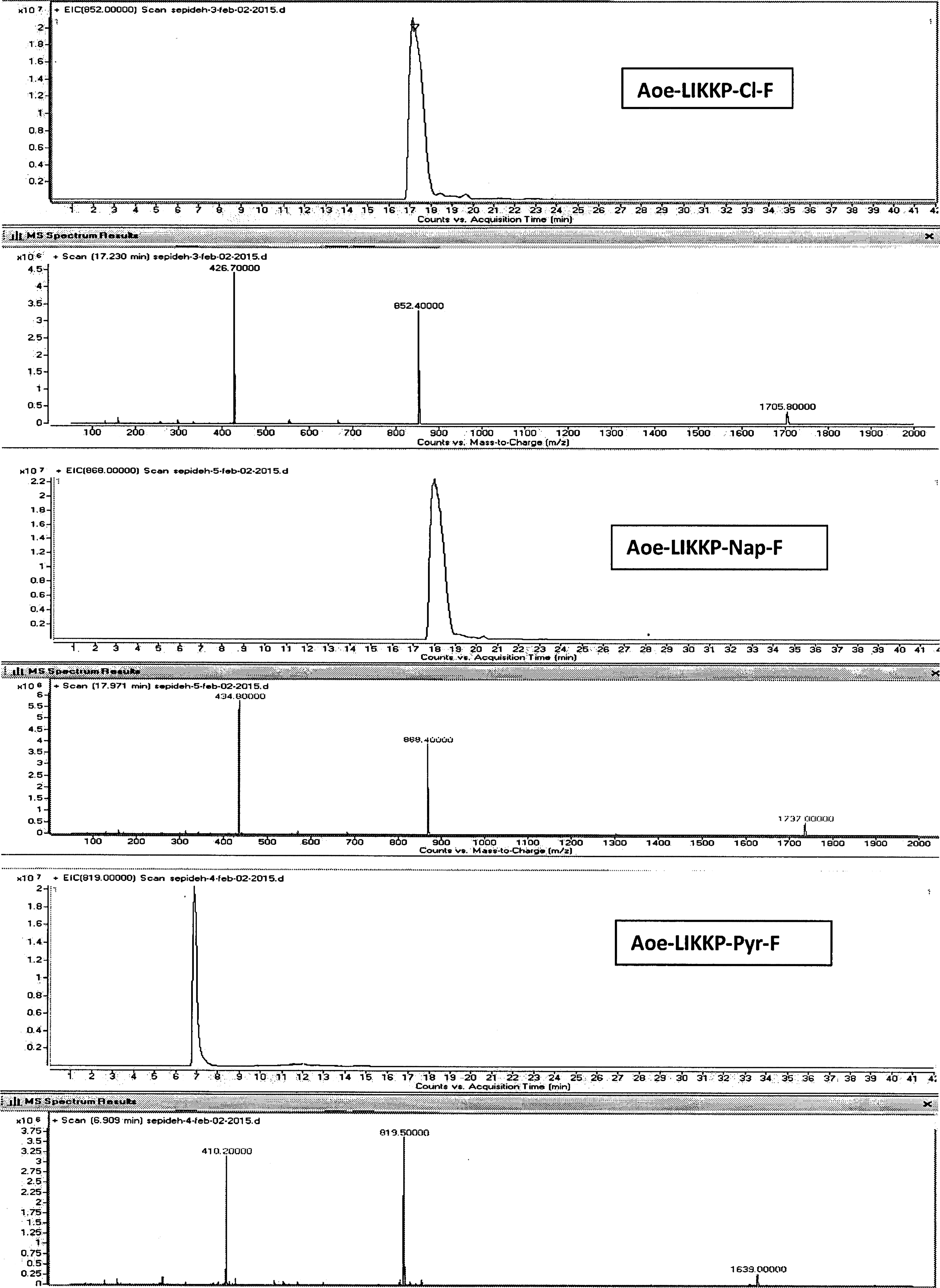
Liquid chromatography-mass spectrometry chromatogram of Aoe-LIKKP-Cl-F, Aoe-LIKKP-Nap-F, Aoe-LIKKP-Pyr-F.
The peptides were radiolabeled with 18F-FDG with specific activities of (1–1.2) Ci/mmole. The optimal labeling conditions with 18F-FDG was 200 μg peptide, 37 MBq 18F-FDG, pH = 5–5.5, 100°C, incubation time of 30 min, and RCP of more than 95% for all of them. TLC-SG, acetonitrile:water (95:5), radiopeptides remained at origin (Rf = 0), while 18F-FDG moved up with solvent (Rf = 0.5) (Supplementary Fig. S1).
The HPLC radiochromatogram of 18F-FDG-Aoe-LIKKP-Cl-F, 18F-FDG-Aoe-LIKKP-Nap-F, and 18F-FDG-Aoe-LIKKP-Pyr-F showed retention times (Rt) of 17, 18, and 6 min, respectively (Supplementary Figs. S2 and S3). Radiolabeled peptides remained more than 99% intact after 2 h in human fresh plasma.
The affinity of peptides for PS was determined in saturation binding studies using camptothecin-treated Jurkat J6 cells. Induction of apoptosis and the percentage of apoptotic cells were examined and measured by flow cytometry using ITC annexin V and PI. 21,22 The LogP, dissociation constants (Kd) and number of binding sites per cell (Bmax) are shown in Table 1 and Figure 4. The data presented were incorporated into a model of nonlinear regression analysis, binding saturation, and one-site SB by using GraphPad Prism software to determine the Bmax and Kd values.
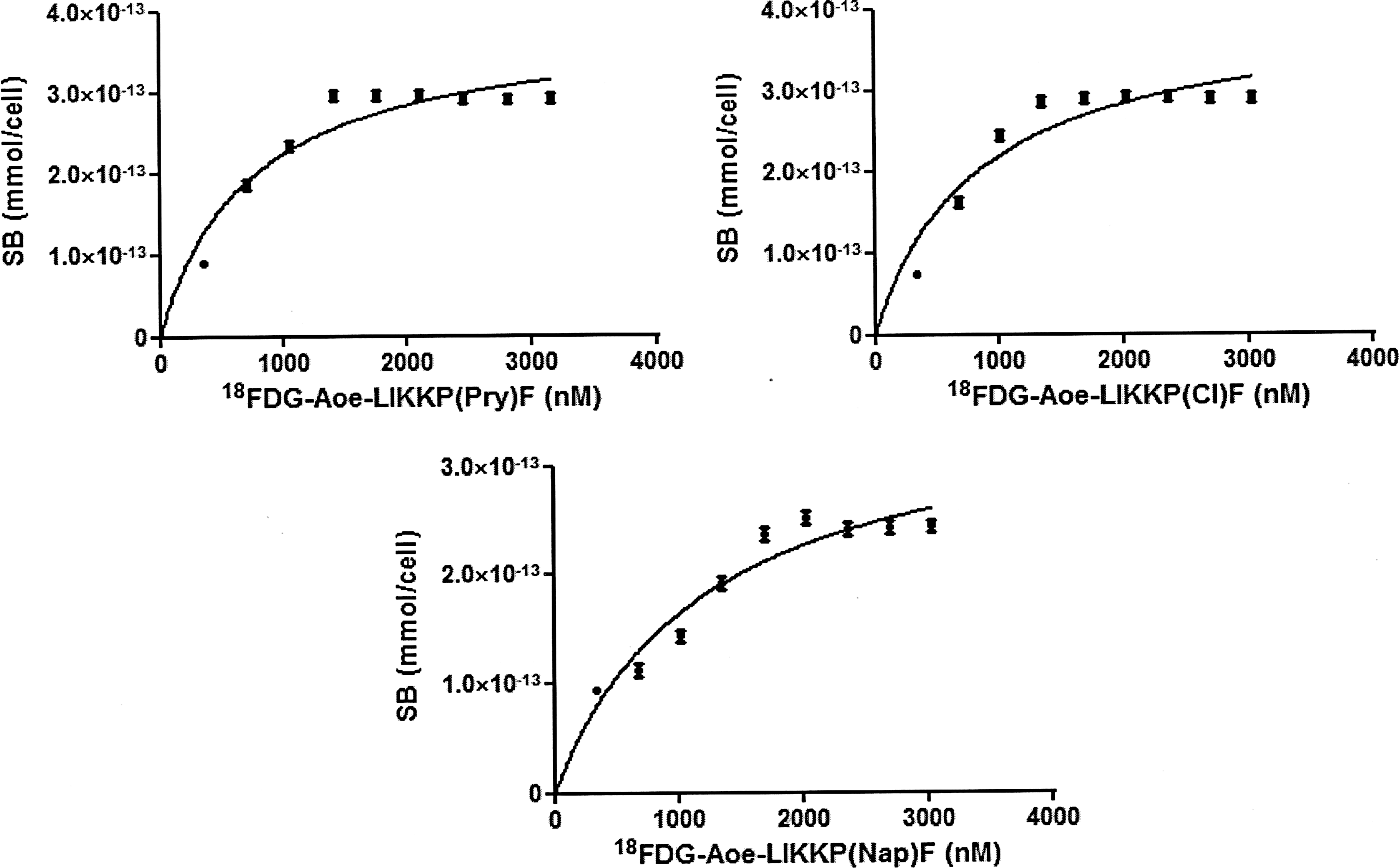
Saturation binding studies of 18F-FDG-Aoe-LIKKP-Cl-F, 18F-FDG-Aoe-LIKKP-Nap-F, and 18F-FDG-Aoe-LIKKP-Pyr-F in camptothecin-treated Jurkat cells. SB, specific binding.
Biologic Characteristics of Radiolabeled Peptides
n = 3, Mean ± SD.
Induction of apoptosis in mouse liver was investigated and imaged by small animal image system. Thirty minutes after injection with FITC annexin V, animals (normal and apoptotic) were sacrificed; liver and other organs such as heart, lungs, and spleen were removed and imaged. FITC annexin V was accumulated in apoptotic liver compared with normal liver and other organs (Supplementary Fig. S4).
The biodistribution of the radiolabeled peptide 18F-FDG-Aoe-LIKKP-Pry-F in normal and pretreated and nontreated apoptotic mice models was determined 15 min and 1 h after injection. The uptake in nontarget tissues was similar in the mice models. The most prominent uptake by organs in mice was observed in the kidneys (15 min: 2.68 ± 0.14, 60 min: 2.05 ± 0.13) and bladder (15 min: 6.38 ± 0.2, 60 min: 7.21 ± 0.26). It is apparent that radioactive elements were rapidly cleared from the blood and excreted in the urine, suggesting renal clearance. The biodistribution pattern of radiopeptide in the pretreated apoptotic mice was similar to normal mice (data not shown).
Uptake of 18F-FDG-Aoe-LIKKPF and 18F-FDG-Aoe-LIKKP-Pry-F in the liver of nontreated apoptotic mice models were significantly higher than normal mice at 15 and 60 min postinjection (p < 0.001). The uptake values of 18F-FDG-Aoe-LIKKPF were 2.58% ± 0.08% at 15 min (fivefold the normal value) and 0.86% ± 0.02% at 1 h (twofold the normal value) postinjection. The uptake values of 18F-FDG-Aoe-LIKKP-Pry-F were 2.84% ± 0.15% at 15 min (5.6-fold the normal value) and 1.31% ± 0.01% at 1 h (13-fold the normal value) postinjection.
Uptake of 18F-FDG-Aoe-LIKKP-Pry-F in the liver of nontreated apoptotic mice models was 1.5 times the uptake of 18F-FDG-Aoe-LIKKPF 1 h postinjection, which was statistically significant p < 0.0001 (Fig. 5).
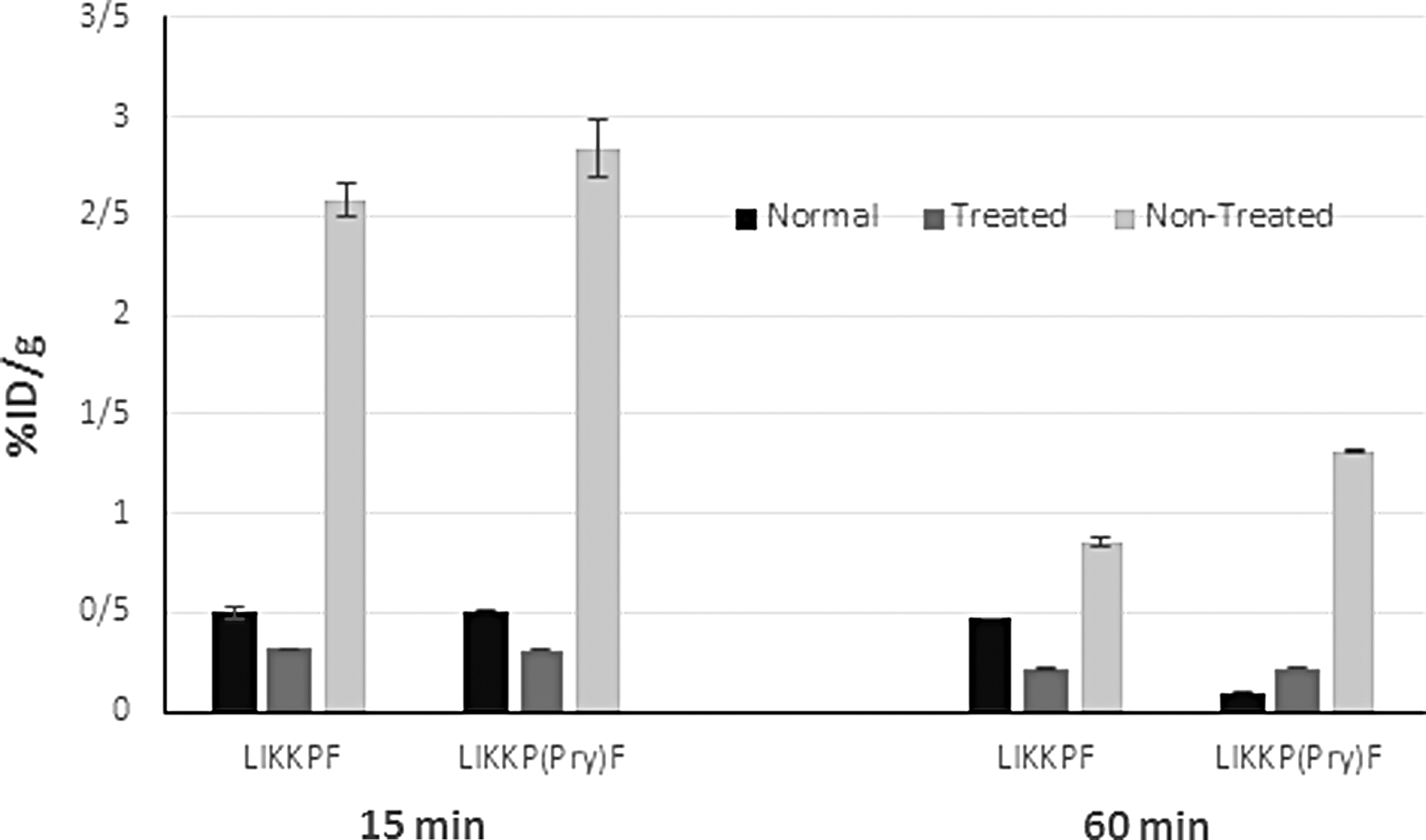
Biodistribution study of 18F-FDG-Aoe-LIKKP-Pyr-F in normal, pretreated, and nontreated mouse model of liver apoptosis at 15 and 60 min postinjection (n = 3). Radioactivity is shown in terms of %ID/g. 100 μCi of 18F-FDG-Aoe-LIKKP-Pyr-F (1 μCi/1 μL in saline) was injected via the tail vein. %ID/g, percentage of injected dose per gram of organ.
One hundred microcuries of 18F-FDG-Aoe-LIKKP-Pyr-F (1 μCi/1 μL in saline) was injected via the tail vein of normal and apoptotic mice models. As shown in Figure 6, the PET/CT fused images of injected mice clearly showed the activity concentration of 18F-FDG-Aoe-LIKKP-Pry-F in kidneys and bladder as expected. For quantitative evaluation of uptake behavior, maximum activity concentrations (Bq/cc) were measured for liver, kidney, and bladder in all imaging studies. In Figure 6A, the maximum activity concentrations in normal mouse were 3.5 × 10 4 , 2.0 × 10 5 , 2.1 × 10 5 , and 4.6 × 10 6 (Bq/cc) for liver, right kidney, left kidney, and bladder 30 min after 18F-FDG-Aoe-LIKKP(Pry)F injection, respectively. In Figure 6B, PET/CT image of the apoptosis rat 30 min after injection, showed maximum activity concentration values of 1.1 × 10 5 , 1.7 × 10 5 , 1.9 × 10 5 , and 9.0 × 10 6 for liver, right kidney, left kidney, and bladder, respectively.
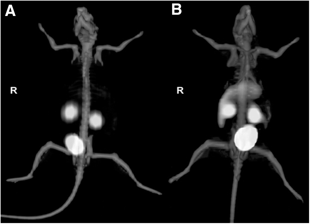
Positron emission tomography/computed tomography fused images 30 min after injection of 75 μCi 18F-FDG-Aoe-LIKKP-Pyr-F
Discussion
In this study, three new peptide analogs based on LIKKPF structure binding to PS in apoptotic cells and a mouse model of liver apoptosis were designed, synthesized, and radiolabeled with 18F-FDG.
In the authors' previous studies, LIKKPF peptide was synthesized and radiolabeled with 18F-FDG and [99mTc]. The biologic properties of radiolabeled peptides were evaluated in vitro and in vivo. 20 –22 Radiolabeled LIKKPF peptide showed less affinity for PS compared to original phage peptide. The interaction of phage peptide included more than one copy of peptide as well as superior and more suitable conformation than single synthesized peptide. The aim of this study was to make minor changes to the structure of LIKKPF as a lead compound to produce analogs and assess the impact of these structural changes on biologic activity of the peptide. Phenylalanine (F) amino acid of LIKKPF was selected for structural changes since it could easily be replaced with unnatural amino acids. Three new analogs of LIKKPF were designed based on the replacement of phenyl ring of phenylalanine with parachlorophenyl, 4-pyridyl, and naphthyl rings (Fig. 1). The analogs have different hydrophobic, electronic, and steric properties, which affected their interaction with PS as target. Phenyl and naphthyl are electron rich, while parachlorophenyl and pyridyl are electron deficient. Among these rings, pyridyl can form hydrogen bond; naphthyl is bulky and lipophilic.
The new peptide analogs LIKKP-Cl-F, LIKKP-Pyr-F, and LIKKP-Nap-F were successfully synthesized and functionalized with aminooxy at N-terminal. The structures were confirmed with LC-MS. The peptides were stabilized in human plasma at 37°C for 24 h. The peptides were radiolabeled with 18F-FDG (RCP >95%). Radiolabeled peptides remained more than 99% intact after 2 h in human fresh plasma. The LogP of radiolabeled peptides showed fast clearance of peptides from the blood and excretion through kidneys. Binding of radiolabeled peptides to PS was investigated in saturation binding studies using camptothecin-treated Jurkat cells (apoptotic cells). Apoptosis was confirmed in cells by flow cytometry using FITC annexin V and PI. 22
Table 1 provides the Kd values of LIKKPF and its analogs. The binding affinity of LIKKPF (Kd = 0.86 μM) and parachlorophenyl analog (Kd = 0.83 μM) were similar. The incorporation of chlorine (Cl) atom into phenyl ring resulted in electron deficiency of the ring. The replacement of phenyl with pyridine (an electron deficient ring) increased affinity, while naphthyl (an electron rich ring) reduced the affinity. The lone pair electrons of nitrogen atom in pyridine ring can form hydrogen bond with–OH group of PS. The authors assumed that the higher affinity of 18F-FDG-Aoe-LIKKP-Pyr-F compared to other analogs was due to hydrogen bond, which provided better interaction of peptide with PS and the low affinity of naphthyl analog was due to steric hindrance induced by the bulky naphthyl ring, which hindered ideal interaction with PS. The chlorine atom also induced steric hindrance, but not as much as naphthyl ring. Both pyridine and parachlorophenyl are electron deficient rings. Based on the results, the difference between the affinity of pyridine and parachlorophenyl analogs could possibly be due to the hydrogen bond and steric hindrance of chlorine atom. Naphthyl and phenyl are electron rich and the low affinity of naphthyl derivative in comparison with the phenyl analog could be due to steric hindrance of the bulky ring of naphthyl. It can be concluded that hydrogen bond and steric hindrance had higher impacts on the interaction of peptide with PS compared to the electronic properties of the rings.
Affinity was measured in μM and not nM. The 18F-FDG solution used for radiolabeling contained glucose which competed with 18F-FDG for radiolabeling of peptide and resulted in low specific activity of 18F-FDG-labeled peptide. The computation of affinity (Kd) is based on 18F-FDG-labeled peptide bound to PS (SB). Since some of the PS were occupied with unlabeled peptide, the Kd value was higher than expected. The low specific activity was responsible for the high Kd values (μM of Kd). They anticipated that radiolabeling of peptide with [99mTc] would induce better affinity because of the higher specific activity of [99mTc] radiolabeled peptide.
Among three new analogs of LIKKPF, 18F-FDG-Aoe-LIKKP-Pyr-F with the highest affinity (Kd = 0.52 μM) was selected for in vivo studies in mice. To confirm the affinity of peptide to PS, biodistribution and in vivo imaging studies were performed in normal and mouse model of liver apoptosis. Apoptosis was induced in mouse liver by injection of LPS intraperitoneally 12 h before experiment. Apoptotic cells in liver were detected and imaged by small animal image system (Supplementary Fig. S4).
Biodistribution of 18F-FDG-Aoe-LIKKPF and 18F-FDG-Aoe-LIKKP-Pry-F in normal and apoptotic mice models were compared. Results showed that uptake of radiopeptides in liver of nontreated apoptotic mice 15 min after injection was 5 to 6 times higher than normal mice, but 1 h after injection; the uptake of 18F-FDG-Aoe-LIKKP-Pry-F in liver was 13 times the normal, while the uptake of 18F-FDG-Aoe-LIKKPF was 2 times the normal. The uptake of 18F-FDG-Aoe-LIKKP-Pry-F in liver of nontreated apoptotic mice models was 1.5 times the uptake of 18F-FDG-Aoe-LIKKPF 1 h postinjection (p < 0.0001) (Fig. 5). The uptake of 18F-FDG-Aoe-LIKKPF and 18F-FDG-Aoe-LIKKP-Pry-F in normal and treated groups 15 and 60 min postinjection was statistically significant p < 0.05. The higher uptake of radiopeptides in liver of normal mice compared with treated mice can be explained by considering the fact that there are always some cells at apoptosis stage in normal tissues such as liver. The excess cold peptides (treated mice) block the PS in liver and decrease the uptake of radiopeptides in treated group (Fig. 5).
PET/CT fused images of 18F-FDG-Aoe-LIKKP-Pyr-F (Fig. 6) showed a significant uptake of radiopeptide in the liver of apoptotic mouse in comparison with normal mouse. The maximum activity concentration ratio of radiopeptide in the liver of apoptotic mouse to normal mouse 30 min after injection was 3. As the maximum activity concentration ratio of liver to kidneys attained 0.17 and 0.61 in normal and apoptotic mouse, respectively (about four times more uptake in the apoptotic mouse), the liver uptake was more pronounced in the apoptotic mouse than the normal one.
Although 18F-FDG-Aoe-LIKKP-Pyr-F peptide showed high Kd 0.52 μM (520 nM) in cell studies, there was increased liver localization in imaging studies (Fig. 6).
Conclusion
In this study, three new peptides based on LIKKPF structure were developed. The peptide 18F-FDG-Aoe-LIKKP-Pry-F had higher affinity for PS compared to other analogs. This new analog will have potential in the diagnosis and therapy monitoring of apoptosis-related pathologies
Footnotes
Acknowledgment
This research was supported by the School of Pharmacy, Shahid Beheshti University of Medical Sciences, and Tehran University of Medical Sciences, Tehran, Iran.
Authors' Contribution
All coauthors have reviewed and approved of the article before submission.
Disclosure Statement
There are no existing financial conflicts.
Supplementary Material
Supplementary Figure S1
Supplementary Figure S2
Supplementary Figure S3
Supplementary Figure S4
References
Supplementary Material
Please find the following supplemental material available below.
For Open Access articles published under a Creative Commons License, all supplemental material carries the same license as the article it is associated with.
For non-Open Access articles published, all supplemental material carries a non-exclusive license, and permission requests for re-use of supplemental material or any part of supplemental material shall be sent directly to the copyright owner as specified in the copyright notice associated with the article.