
Abstract
Background:
Kinesin Family Member 3B (KIF3B) is one of the most ubiquitously expressed KIFs, which is related to numerous physiological responses. KIF3B has also been implicated in carcinogenesis such as in hepatocellular carcinoma cells. However, the expression of KIF3B has not been studied in pancreatic cancer along with its clinical significance.
Methods:
Immunohistochemical assays were performed to detect the expression levels of KIF3B in the tumor tissues and adjacent non-tumor tissues. Patients were sequentially divided into different expression levels of KIF3B group based on the staining intensity of FKIF3B in tumor tissues. The link between KIF3B expression and clinical characteristics were investigated, and the role of KIF3B on pancreatic cancer cell proliferation was detected by colony formation and 3-(4,5-dimethyl-2-thiazolyl)-2,5-diphenyl-2-H-tetrazolium bromide, respectively. And the proliferation related proteins such as Ki67 and proliferating cell nuclear antigen (PCNA) were detected by Western blot. The possible effects of KIF3B on tumor growth were assessed in vivo.
Results:
KIF3B was highly expressed in human pancreatic cancer tissues. We also found KIF3B was significantly associated to the pTNM stage (*p = 0.018), lymph node metastasis (*p = 0.040) and vascular invasion (*p = 0.034). We reported that increased expression of KIF3B was significantly correlated with poor clinical outcome in our clinical cohort of pancreatic cancer. Furthermore, functional assays revealed that knockdown KIF3B in vitro and in vivo might inhibit cancer cells proliferation by affecting Ki67 and PCNA.
Conclusions:
Our data suggested that KIF3B was associated with pancreatic cancer malignant progression especially proliferation. Hence, KIF3B might serve as a potential therapy target of pancreatic cancer in clinical treatment.
Introduction
Pancreatic cancer is predicted to be the second cause of cancer-associated death in the next decade in the United States. 1,2 Estimated new cancer cases of pancreatic cancer in the United States are 55,440, of whom 44,330 cases will die in 2018. 1 Five-year survival rate of pancreatic cancer is 3%, and during the years 2011–2015, death rate rose in men by 0.3% per year. 3 In spirit of the improved clinical technology, the prognosis of pancreatic cancer is still poor. To improve the effect of therapy, it is very crucial to identify and investigate new genes and pathways involved in pancreatic cancer development.
Many activities within the cell are closely related to cytoskeleton, which composes of microtubule, microfilament, and intermediate filament in eukaryotic cell. Kinesin superfamily proteins (KIFs) are a group of microtubule-dependent molecular motor proteins, which are ubiquitous in all eukaryotes, which play an important role in intracellular transport, and they can transport cargos along microtubules by ATP hydrolysis. 4 KIFs also participate in mitosis and meiosis, and transport macromolecules. Kinesin Family Member 3B (KIF3B) is one of the most ubiquitously expressed KIFs, which is related to lots of physiological responses. 4,5 It is noted that KIF3B has been implicated in proliferating carcinogenesis such as human hepatocellular carcinoma cells. 4
However, the expression of KIF3B has not been researched in pancreatic cancer as well as the clinical significance. So the authors contrasted the expression of KIF3B in pancreatic ductal adenocarcinoma tissues with the adjacent noncancerous tissues by immunohistochemical staining. They also proved effects of KIF3B on pancreatic ductal adenocarcinoma cell proliferation in vivo and vitro. Owing to the data they presented, KIF3B might serve as a novel target in the clinical treatment.
Materials and Methods
Patients and tissue samples
Pancreatic cancer tissues were obtained from 82 patients. The clinicopathologic and follow-up data were completely selected and are mainly shown in Table 1. Paired cancer and adjacent noncancerous tissue samples, which were located more than 1 cm away from the tumor, were obtained through the open surgeries in Xingtai people's hospital. The paraffin-embedded tissue samples were stained with hematoxylin and eosin and confirmed by experienced again. Informed consents were obtained from all the patients.
Relationships of KIF3B and Clinicopathological Characteristics in 82 Patients with Pancreatic Ductal Adenocarcinoma
p < 0.05.
KIF3B, Kinesin Family Member 3B.
Immunohistochemistry
The method of immunohistochemistry and the antibody of KIF3B, as well as the evaluation of immunohistochemical staining were the same as those reported previously 4 : pancreatic cancer tissues were cut in 5 μm thickness and pretreated in Tris-buffered saline. After antigen retrieval, samples were incubated with primary antibody rabbit KIF3B polyclonal antibody (1:300 dilution, PA5-52386; Invitrogen, New York) at 4°C overnight. Next, sections were washed twice with Tris-buffered saline and incubated for 30 min at room temperature with the blocking buffer of 5% conjugated secondary antibody. Subsequently, diaminobenzidine was dripped on in 3–5 min.
The expression of KIF3B protein is located in the cytoplasm of tumor cells. Positive cells: the percentage of positive tumor cells <10% scores 0; the percentage of positive tumor cells occupying 10%–30% scores 1. The positive percentage of tumor cells that was 30%–70% scores 2. The positive percentage of tumor cells that was >70% scores 3. Meanwhile, the staining intensity of tumor cells with positive staining was evaluated: 0 for negative staining, 1 for weak positive staining, 2 for moderate positive staining, and 3 for strong positive staining. Results high expression (2–3) or low expression (0–1) were determined according to the score of positive cell percentage score and staining intensity score. The sections of each patient were observed within five visual fields, and two experienced pathologists analyzed the sections without getting the pathological grade and clinical data. The results were judged by a double-blind method.
Cell culture
Human pancreatic ductal adenocarcinoma cell line PANC-1 and BxPC-3 were obtained from the Committee of Type Culture Collection of the Chinese Academy of Sciences (Shanghai, China). The PANC-1 or BxPC-3 cells were cultured in high-glucose Dulbecco modified Eagle medium or Roswell Park Memorial Institute 1640 (RPMI 1640) supplemented with 10% fetal bovine serum in a humidified atmosphere of 5% CO2 at 37°C.
Stable cell line generation
The human KIF3B targeting small hairpin RNA was generated from Vigene (Cat# SH890974; Vigene Biosciences, Inc., Maryland) for human KIF3B (RefSeq number: NM_004798) downregulation in pAV-U6-GFP vector. The shRNA for KIF3B: CCCATGAATGGCAAGGAAAAGGC. Cells were transfected with virus according to the manufacturer's instructions. Real time quantitative polymerase chain reaction (RT-qPCR) and Western blotting were performed to confirm the effect of KIF3B silencing.
RNA analysis
Total RNA was extracted using TRIzol reagent (Invitrogen) and confirmed the RNA integrity. PrimeScript RT reagent kit (Invitrogen) was used for cDNA generation. RT-qPCR was performed with SuperReal PreMix (Tiangen Biotech) by the following program: 95°C for 3 min in 1 cycle; 95°C for 5 s, 58°C for 30 s, and 72°C for 30 s in 35 cycles. To ensure the DNA production, a melting curve analysis was performed according to ABI StepOne system. The relative gene expression was normalized to the internal standard β-actin using 2-ΔΔCt method. All experiments were performed independently thrice. Primers used for the analysis are as follows:
KIF3B F: 5′-ATCCTGGAGCAGAAACGACAGG-3′,
KIF3B R: 5′-GTTCCAAGGTCTCCTCATCTCG-3′;
β-actin F: 5′-CAGCTCACCATGGATGATGATATC-3′,
β-actin R: 5′-AAGCCGGCCTTGCACAT-3′.
Western blot assay
Cells were lysed in lysis buffer (60 mM Tris-HCl, pH6.8, 2% sodium dodecyl sulfonate (SDS), 20% glycerol, 0.25% bromophenol blue, 1.25% 2-mercaptoethanol, and protease inhibitor cocktail). Lysates used the BCA Protein Assay (Thermo Fisher Scientific) to measure concentration followed by manufacturer's instructions. Next, the equal amounts of protein (30–50 mg) were electrophoresed by 10% SDS-polyacrylamide gel electrophoresis gels and transferred onto polyvinylidene fluoride membranes (Merck Millipore). Then, using nonfat milk to block the membrane, the membranes were incubated with antibodies at 4°C overnight. The antibodies used are shown as follows: the rabbit KIF3B polyclonal antibody (0.4 μg/mL, PA5-52386; Invitrogen, New York), mouse anti-β-actin (1:1000 dilution, ab8226; Abcam, Cambridge, United Kingdom), rabbit anti-Ki67 (1:1000 dilution, ab16667; Abcam), and mouse anti-proliferating cell nuclear antigen (PCNA) (1:500 dilution, ab29; Abcam).
Cell proliferation assay by colony formation
Cells were incubated in six-well plates, 500 cells per well before being fixed in methyl hydrate for 10 min. Then, the colonies were stained and counted with an optical microscope.
3-(4,5-dimethyl-2-thiazolyl)-2,5-diphenyl-2-H-tetrazolium bromide assay
To observe and compare cell proliferation ability, a 3-(4,5-dimethyl-2-thiazolyl)-2,5-diphenyl-2-H-tetrazolium bromide (MTT) assay was used; 5 × 10³ cells were plated into a well of 96-well plates at 37°C in 5% CO2. After three days, the medium was removed and 20 μL MTT solution was added into each well at 37°C for 4 h. Then, dimethyl sulfoxide was added (150 μL/well) into wells to solubilize the formazan crystals. This plate was incubated at room temperature for 10 min on a shaking table. The results were measured in 570 nm by spectrophotometry.
Xenograft assays in vivo
All surgeries were performed under sodium pentobarbital anesthesia. Athymic Balb/c nude mice (aged 5 weeks, 18–20 g) were provided by Slac Laboratory Animal Co. Ltd. (Shanghai, China). Mice were housed in a pathogen-free animal facility and randomly divided into two groups according to the cell groups described above. There were eight mice per group. Around 0.1 mL serum-free RPMI 1640 with 2 × 10⁶ cells was injected into the right flank subcutaneously of each mouse. The tumors were measured by vernier caliper on day 14, 17, 21, 23, 26, and 29. Mice were killed at 29 d postinoculation. The final volume of tumor tissues was calculated with the following equation: Tumor volume (mm3) = tumor length (mm) × tumor width (mm) × tumor height (mm)/2.
Statistics
All statistical analyses were performed using SPSS 22.0 software. Kaplan-Meier method was used to perform survival analysis. The association between KIF3B expression and clinicopathological features was studied using the χ 2 test. Statistical significance was defined as p-values <0.05.
Results
Increased expression of KIF3B connected with poor clinical outcomes in pancreatic cancer patients
In their clinical cohort of 82 pancreatic adenocarcinoma patients, the authors divided them into two groups: the low group (n = 42) and high group (n = 40) based on the protein expression level of KIF3B (Table 1). Immunohistochemistry was used for detecting the KIF3B expression with low and high intensity in stained pancreatic cancer specimens (Fig. 1A), while the KIF3B expression was negative/low in the tissue adjacent to carcinoma (Fig. 1B). Particularly, patients with high KIF3B protein expression level were strongly connected to vicious pTNM stage (p = 0.018), higher probability of lymph node metastasis (p = 0.040), and vascular invasion (p = 0.034) when compared to the low expression group. No significant clinical connection was determined between KIF3B expression and other clinicopathological features, including age, gender, tumor grade, and tumor size (Table 1). To better investigate the KIF3B clinical feature, Kaplan-Meier survival analysis was performed to determine the cancer overall survival (OS) and disease-free survival (DFS) in their 30-month follow-up time after surgery. The data showed that high KIF3B protein expression level was preferred to gain a short cancer OS time and DFS time (Both p < 0.05, Fig. 1C).
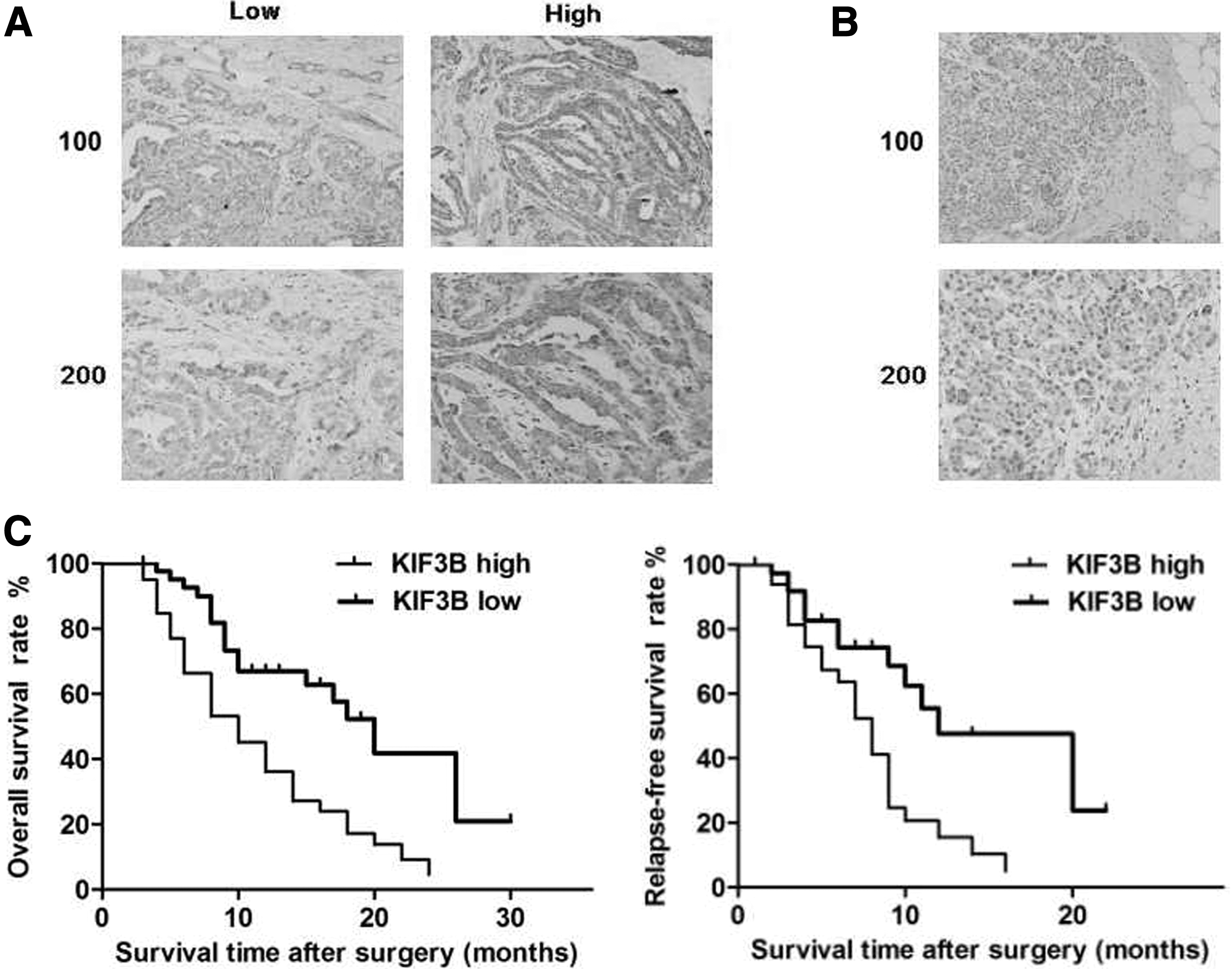
Increased expression of KIF3B connected with poor clinical outcomes in the clinical cohort.
Construction of lentivirus-based KIF3B stable knockdown cell lines of PANC-1 and BxPC-3
PANC-1 and BxPC-3 cells were transfected with recombinant lentivirus to silence KIF3B expression in vitro. Through RT-qPCR (Fig. 2A) and Western blotting (Fig. 2B) validation, the authors generated stable cell lines with both KIF3B mRNA and protein level decreased significantly when compared with control cells (Fig. 2). Using this model, they could efficiently study the KIF3B role in cancer cells.
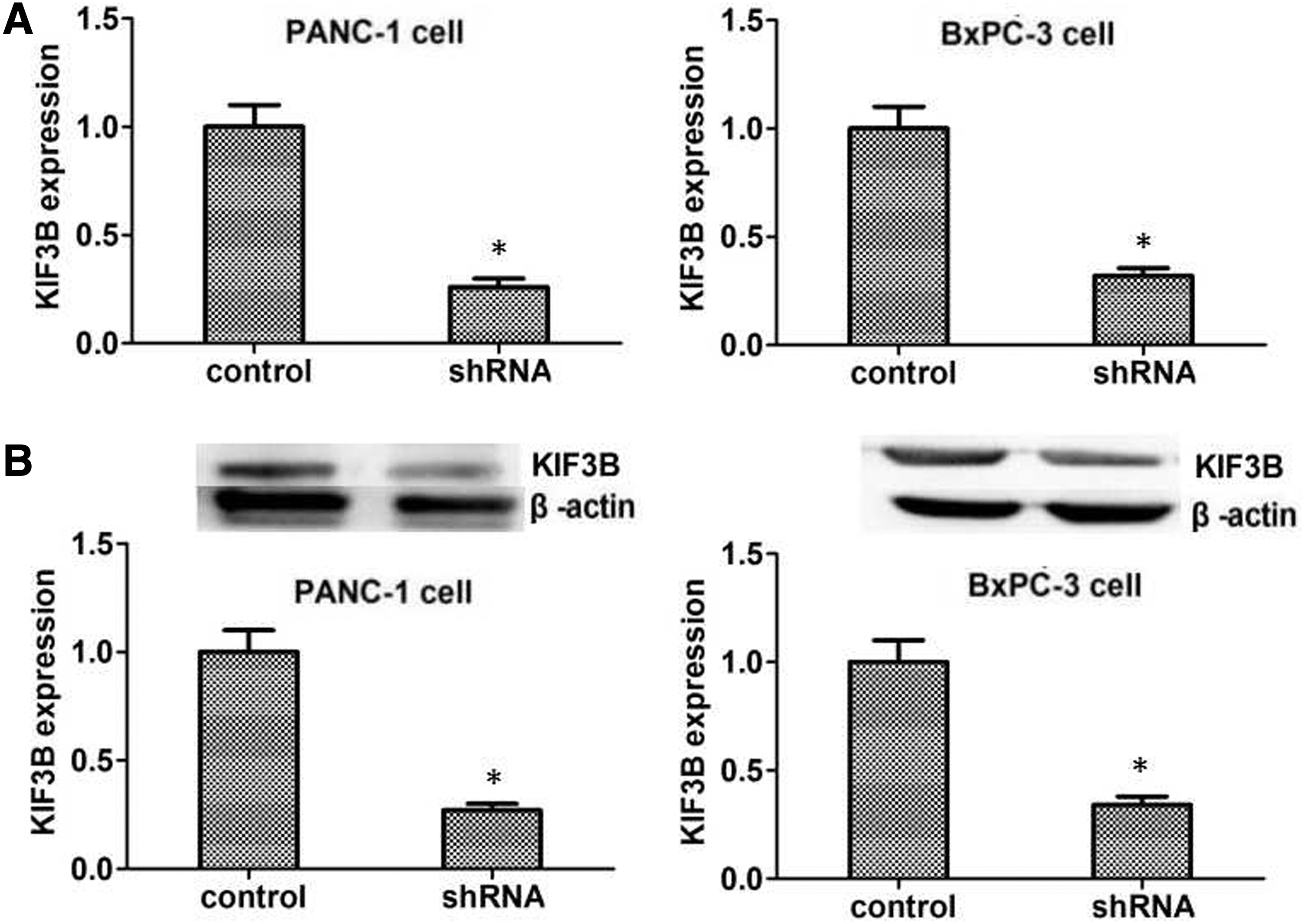
The mRNA and protein expression level of KIF3B in generated stable cell lines. Both mRNA and protein expression level of KIF3B were significantly decreased by RT-qPCR
Knockdown KIF3B reducing proliferation through regulating Ki67 and PCNA proteins in PANC-1 and BxPC-3 cell lines
To investigate the biological effect of KIF3B, the authors conducted colony formation assay and MTT assay to evaluate the possible role for proliferation in pancreatic cancer cell lines PANC-1 and BxPC-3. Compared to control cells, stable knockdown KIF3B in PANC-1 and BxPC-3 cells by recombinant lentivirus significantly decreased the ability of proliferation in vitro compared to control cells (p < 0.05, Fig. 3A, B). The above results may imply that knockdown KIF3B reduced proliferation ability in cancer cells. To further investigate the mechanism of partially eliminated proliferation, they checked proliferation-related proteins such as Ki67 and PCNA in vitro. Western blotting revealed that Ki67 and PCNA protein were significantly downregulated in the silencing KIF3B cell lines compared with the control, respectively (p < 0.05, Fig. 3C, D). Taken together, the above data suggested that knockdown KIF3B may downregulate Ki67 and PCNA protein to reduce the proliferation in pancreatic cancer cells.
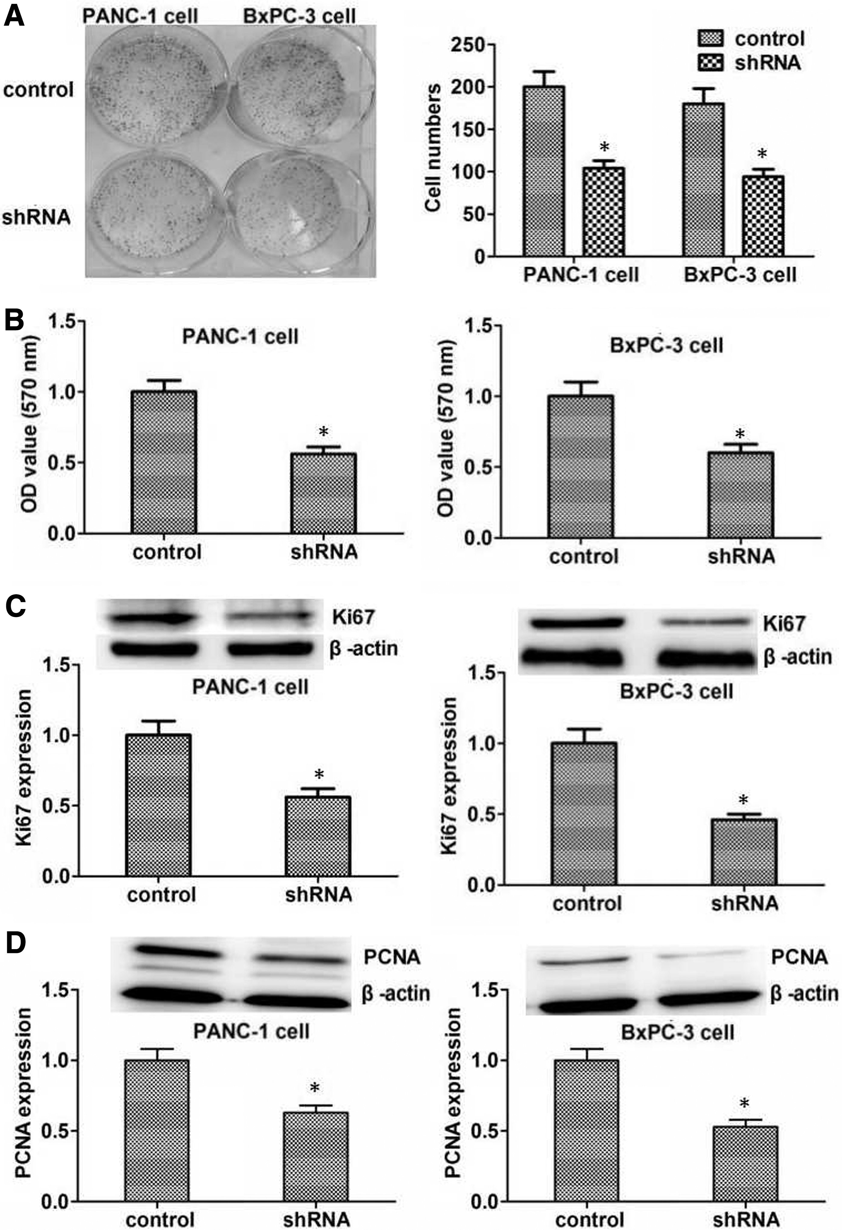
Role of KIF3B on the proliferation in PANC-1 and BxPC-3 cell lines.
In vivo inhibitory role of knockdown KIF3B
Based on the results above, downregulated KIF3B in pancreatic cancer cell lines significantly inhibited the ability of cell proliferation in vitro. To better investigate the above findings in vivo, xenograft assay was performed. The tumor volume of downregulated KIF3B group was observably smaller than control after a 4-week management (p < 0.05, Fig. 4A). In addition, to further certify the effect of KIF3B silencing, Western blotting and immunohistochemistry assays were performed to consolidate the KIF3B silencing effect in mice tumors. The following data showed that KIF3B protein was dramatically reduced by the recombinant lentivirus (p < 0.05, respectively, Fig. 4B, C). Taking together, the authors' results suggested that knockdown KIF3B might exert inhibitory effect in vivo.

Inhibitory role of KIF3B silencing in vivo.
Discussion
Rapid spreading in patients of pancreatic cancer and difficulty to detect in the early stages made pancreatic cancer become the leading cause of cancer-related death. 6 The cancer mortality is extremely high, although substantial improvement has been achieved in the fields of early diagnosis, surgical techniques, combination chemotherapies, and other anticancer treatments for patients with pancreatic cancer. 7,8 In common, there are no apparent symptoms or signs in patients unless this malignancy progresses into an advanced stage. 9 Once in advanced stages, the chance of surgery has been lost. Therefore, it is of vital importance that clinical physicians should identify the patients with high risk. The cancer-specific 5-year OS is less than 5%. 10 Moreover, patients proceeded curative surgical resection till relapse in the following 2 years. Hence, the mechanisms of cancer development and progression were needed to be unveiled and for cancer treatment, potential biomarkers and novel anticancer strategies are urged to be identified. 11
KIF3B forms a heterotrimeric motor complex with KIF3A and KAP3 (kinesin-associated protein), which is a member of the kinesin-2 subfamily of KIFs. 4,5 During mitosis, chromosomes are equally segregated into two daughter cells and errors in this process may result in cancer. 12 –18 KIFs play critical roles in the process of cell proliferation, 12,14 –18 including Kid being important for chromosome alignment and orientation. 18 –20 One of the most ubiquitously expressed KIFs, Kinesin-2, is a complex composed of a KIF3A/3B heterodimer and KAP3, which are linked together in various cargoproteins and play an important role in ATP metabolism for the progression of mitosis. 5,21 –23 It was reported that multiple mitotic spindle poles in KIF3B-knockdown cells resembling phenotypes observed following expression of a dominant negative mutant of KIF3B were associated with ARHGEF10-RhoA and another motor protein KIFC5A in the molecular mechanisms and signaling pathways. 4,5,12 –18
Recent studies have suggested that KIF3B was overexpressed in various types of tumor, which showed to be closely bound up with the carcinogenesis of cancer, and related to the clinical progression. 4,24 –29 Therefore, KIF3B might play a role in pancreatic cancer carcinogenesis. This study revealed that KIF3B was highly expressed in pancreatic cancer tissue compared with the normal tissue adjacent to carcinoma, since KIF3B was not or low expressed in the normal epithelia pancreatic tissue. Also, the result was consistent with a recent study in human hepatocellular carcinoma: KIF3B protein level was increased in HCC tissues compared with the adjacent nontumorous tissues. 4 Furthermore, their results also showed that KIF3B was significantly associated with histological differentiation, tumor size, and the level of alpha fetal protein. 4 The authors' clinical data showed that patients with high KIF3B expression by immunohistological analysis were more likely to harbor vicious pTNM stage, higher probability of lymph node metastasis, and vascular invasion. These results were different from the authors' owing to the different types of cancer. In addition, their results indicated that OS time and DFS time were decreased compared with the low expression group, which was similar to the results that overexpression of KIF3B was correlated with poor survival. 4
Considering the account sources of potential bias or imprecision, in this study, there are also some limitations. First of all, the results of immunohistochemistry are only single centered and the number should be expanded. Then, the authors only used two kinds of cell lines, and the further mechanism should be explored in the future study (the findings about the upstream and downstream genes or proteins). At last, but not the least, it is better to use the pancreatic cancer in situ model or the fresh human pancreatic cancer tissues.
The recent study pointed that KIF3B might not only promote cancer cell growth but also induce apoptosis of cells through the release of HepG2 cells from serum starvation by upregulating the expression of KIF3B. 4 To confirm this clinical finding the authors observed, they generated KIF3B targeting shRNA with recombinant lentivirus to silence its expression in pancreatic cancer cells, and then validated by Western blot assay. They investigated some proliferation-associated protein expression, such as Ki67 and PCNA, while KIF3B silencing. Consistent with the above report, the mechanism of proliferation suggested that KIF3B regulated Ki67 and PCNA to influence the cancer proliferation abilities. 4 Furthermore, they proved this in vivo. In summary, this study demonstrated that silencing KIF3B in pancreatic cancer cell lines might inhibit the cell proliferation by regulating the concerning proteins. However, they could not find similar results with the above study about apoptosis in pancreatic cancer (data not shown). Moreover, although the authors'results showed that high KIF3B expression had significantly higher percentage of lymph node metastasis, in accordance with the study concerning the function of KIF3B in metastasis process, 29 they could not find the function of KIF3B on invasion or migration in vitro in pancreatic cancer (data not shown). Therefore, the key signaling pathway and molecular mechanism of KIF3B still needed to be elucidated.
Conclusions
Taken together, the authors first demonstrated that elevated KIF3B expression was significantly correlated with poor clinical outcome in pancreatic cancer. Then they studied that functional role of KIF3B by knockdown in vitro and in vivo significantly inhibits cancer cell proliferation. Hence, KIF3B might be a potential target of pancreatic cancer in the clinical treatment.
Ethics Approval and Consent to Participate
All applicable international, national, and/or institutional guidelines for the care and use of human specimens and animals were followed. The animal study was carried out in accordance with the guidelines approved by the animal experimentation ethics committee of Xingtai People's Hospital. The protocol was approved by the committee, all surgery was performed under sodium pentobarbital anesthesia, and all efforts were made to minimize suffering.
Consent for Publication
All of the authors have agreed to publish this article in your journal if it is accepted.
Availability of Data and Material
The dataset supporting the conclusions of this article is included within the article.
Authors' Contributions
Z.-H.L. and S.-X.D. carried out the experiment of molecular biology and drafted the article. Z.-H.L., J.-H.J., and Z.-L.Z. carried out the animal experiment. Z.-G.Z. participated in the sequence alignment. Z.-H.L., S.-X.D., J.-H.J., and Z.-L.Z. participated in the design of the study and performed the statistical analysis. Z.-G.Z. conceived the study, participated in its design and coordination, and helped to draft the article. All authors read and approved the final article.
Footnotes
Disclosure Statement
No competing financial interests exist.