
Abstract
Background:
A human umbilical vein endothelial cell (HUVEC) vaccine is a promising anti-angiogenesis therapy, but the modest therapeutic antitumor efficacy restricts its clinical use. Preclinical evidence supports the combination of antiangiogenic agents and chemotherapy for cancer treatment.
Materials and Methods:
In the present study, docetaxel (DOC) was combined with HUVEC vaccine to develop a HUVEC–DOC treatment regime. This study was designed to investigate the synergistic anti-breast cancer effects and mechanisms of the HUVEC–DOC treatment.
Results:
Compared with either agent monotherapy, HUVEC–DOC treatment exhibited more favorable anti-EMT-6 breast cancer effects in vivo. CD31 immunohistochemical analysis of the excised tumors showed notable decreases in vessel density after HUVEC–DOC administration, while T cells isolated from mice immunized with HUVEC–DOC showed increased cytotoxicity against HUVECs. Furthermore, the quantity of interferon gamma released from HUVEC–DOC-administered mice was significantly higher than the other three groups, and enhanced CD8+ T cell infiltration was observed more frequently in tumors excised from HUVEC–DOC-treated mice. Finally, the percentage of regulatory T cells was significantly decreased after HUVEC–DOC immunization.
Conclusions:
All the data verified that combining DOC with a HUVEC vaccine could generate synergistic anti-breast cancer activity, which might have the potential for combination treatment of human breast cancer.
Introduction
Breast cancer is currently the leading cause of cancer-associated mortality among females worldwide. 1 Although the comprehensive treatment of breast cancer has recently improved, the mortality rate still remains high due to tumor recurrence and metastasis, accounting for >90% of treatment failures. 2 The recurrence and metastasis of tumors, including breast cancer, depend on its vascular supply. 3,4 Therefore, the development of antiangiogenic drugs is a priority in breast cancer research.
Up to now, the principal antiangiogenic drugs are monoclonal antibodies, and the main problems of these drugs are the high cost and serious side-effects caused by repeated and long-term use. 5 Active immunization with a vaccine as an alternative strategy could elicit sustained antiangiogenic immune responses and minimize these drawbacks. 6,7 Human umbilical vein endothelial cells (HUVECs), which can be cultured in vitro and easily provide adequate endothelial cells, were used to prepare antiangiogenic vaccines in our previous study. 8 –10 As angiogenesis is a complicated physiological process involving multiple growth factors, a HUVEC vaccine containing different forms of antiangiogenesis-associated antigens has shown favorable preventive and antitumor effects in vivo. However, when it came into therapeutic use, the antitumor effects were still unsatisfactory. Additional measures should be explored for enhancing the antitumor efficacy of the HUVEC vaccine.
Therapeutic cancer vaccines have often failed to improve short-term end-points when used as single agents, and numerous clinical trials have verified that antiangiogenic drugs could significantly increase the survival rates of patients with cancer, especially when combined with standard chemotherapy. 11,12 Docetaxel (DOC), the first chemotherapeutic drug authorized by the FDA for the treatment of metastatic breast cancer, has been tested in cancer combination treatments. 13,14 Therefore, in the present study, we investigated whether combination with DOC could augment the antitumor effects of the HUVEC vaccine in a rapidly growing murine EMT-6 breast cancer model. Furthermore, the mechanisms underlying the effects of the combination treatment were investigated.
Materials and Methods
Cell lines
HUVECs and endothelial cell medium were purchased from ScienCell Research Laboratories (San Diego). HUVECs were cultured at 37°C in a 5% CO2 humidified incubator with endothelial cell medium. EMT-6 cells were obtained from Shanghai Guan Dao Biological Engineering Co., Ltd. (Shanghai, China) and maintained in RPMI-1640 medium supplemented with 10% fetal bovine serum, 100 μg/mL streptomycin, and 100 U/mL penicillin (all from HyClone) at 37°C in a 5% CO2 humidified incubator.
Animals
The 4–5-week-old pathogen-free female BALB/c mice were obtained from Jinan Peng Yue Experimental Animal Breeding Co., Ltd. (Jinan, China). Animals were housed under conventional conditions with free access to water and food and a 12 h light/12 h dark schedule. After feeding for another 2 weeks, the mice were used for tumor challenge experiment. Animal experiments were approved by the Institutional Animal Care and Treatment Committee of Binzhou Medical University (Ethics Approval No. 2017087).
Chemotherapeutic drugs
DOC (99.5% purity) was purchased from SA Chemical Technology Co., Ltd (Certificate No. E100688; Shanghai, China). Each ampoule contained 250 mg DOC in polyoxyethylene–castor oil (Cremophor)/ethanol (1:1) solution. It was diluted to the desired ratio with normal saline before injection.
Vaccination and treatment schedules
Subconfluent HUVECs from the second and fifth passage were harvested and resuspended in phosphate-buffered saline (PBS). HUVECs (1 × 10⁶) were mixed in 100 μL PBS to prepare the HUVEC vaccine, according to the protocol in our previous study. 8 A total of 24 BALB/c mice were randomly divided into four groups with 6 mice each group: PBS (PBS only), HUVEC (HUVEC vaccine), DOC (20 mg/kg DOC), or HUVEC–DOC (HUVEC vaccine +20 mg/kg DOC). Mice were subcutaneously injected with 5 × 10⁵ EMT-6 cells in the right armpit on day 0, and immunizations were repeated on days 1, 7, and 17 on the left side. The HUVEC vaccine was administered subcutaneously, while DOC was injected intraperitoneally. Tumor volume (TV) was measured every other day after tumor challenge from day 7, and TV was calculated using the following formula: 0.5 × (larger diameter) × (smaller diameter). 2 All mice were sacrificed on day 31, and the subcutaneous tumors were excised and weighed.
Histological examination of tumor sample sections
Excised subcutaneous tumor tissues were fixed with 10% formalin and embedded in paraffin, then cut into 4 μm sections on which hematoxylin and eosin (H&E) staining was performed. CD31 analysis was conducted, according to a previously described method, 10 with an antimouse CD31 antibody (BD Pharmingen). To quantify microvessel density (MVD), each slide was scanned at low-power magnification under an inverted microscope. Two “hot spot” areas with a relatively high number of new vessels were identified and subsequently scanned at high-power magnification. Five random fields from each “hot spot” area were analyzed.
Cytotoxic assay for T lymphocyte reactions
T lymphocytes were extracted from the spleens of immunized mice according to the protocol in our previous study. 8 Briefly, the spleens were ground and passed through a 100-μm filter under sterile conditions. Erythrocytes were lysed using Tris-NH4Cl (pH 7.2). The splenocytes were washed with Hanks, resuspended in complete RMPI 1640 medium, and enriched by nylon mesh (BD-Falcon). The isolated T lymphocytes were reactivated with whole HUVEC lysates for 2 d in six-well plates, after which they were used as effector cells. Effector cells were inoculated in a 96-well plate in the ratio of effector/target of 200:1, 100:1, 50:1, and 25:1 and cultured in a CO2 incubator for 4 h. A total of 50 μL CytoTox96 Non-Radioactive Cytotoxicity Assay Kit (Promega) lysis solution was added to the control wells and used as the maximum release well 45 min before the end of the incubation period. After centrifugation at 500 g for 5 min, 50 μL supernatant was collected and mixed with 50 μL substrate mix from a CytoTox96 Non-Radioactive Cytotoxicity Assay Kit. OD-490 was measured after a 30 min incubation in the dark, according to the manufacturer's instructions.
Interferon gamma enzyme-linked immunosorbent assay analysis
Interferon gamma (IFN-γ) was measured with a precoated sandwich ELISA kit (BD Biosciences) according to the protocol in our previous study. 10 Briefly, the isolated mouse lymphocytes were cultured in six-well plates (1 × 10⁷ cells per well with 2 mL RPMI-1640 medium). After 48 h reactivation with whole HUVEC lysates, the supernatants from the lymphocytes were harvested to measure the IFN-γ concentration. The reaction reagents were incubated with tetramethylbenzidine substrate for 30 min at 37°C in the dark, and absorbances were then measured at 450 nm with an ELISA microplate reader.
Immunofluorescence
Sections of formalin-fixed paraffin-embedded tumor tissues were deparaffinized in xylene for 15 min, followed by 10 min of rehydration in serial dilutions of ethanol (100%, 100%, 95%, and 95%), and then two changes of water. Antigen unmasking was achieved by boiling the slides for 10 min in 10 m
Flow cytometry
To evaluate possible tumor microenvironment changes, regulatory T cells (Tregs) were detected according to the protocol in our previous study. 10 Briefly, excised tumor tissues were digested by 0.1% collagenase at 37°C for 3 h. Tumor cells were then stained with anti-CD4 (PerPC-labeled; BD Biosciences) and anti-Foxp3 (Alexa Flour® 647-labeled; BD Biosciences) antibodies. Stained cells were washed twice with 2 mL PBS and resuspended in 500 μL PBS. The percentages of activated cells were detected using a BD FACSCalibur flow cytometer (BD Biosciences).
Toxicity assessment
Different groups of mice were immunized with the HUVEC vaccine and/or DOC on days 1, 7, and 17, and then monitored for toxicity assessment for 6 weeks. Treatment-associated toxicity was mainly evaluated by weight changes in the mice; in addition, ruffling of fur, behavior, and altered feeding were also monitored. The tissues of major organs (hearts, livers, spleens, lungs, and kidneys) were excised and analyzed with H&E staining. All sections were assessed for possible histological changes, which could be related to the treatment by an experienced pathologist.
Statistical analysis
All data are presented as means ± standard deviation. The results were analyzed with one-way analysis of variance and Tukey's post hoc test for multiple comparisons using Prism 6 software. p < 0.05 indicated a statistically significant difference.
Results
HUVEC vaccine and DOC synergistically suppress tumor growth
To determine whether HUVEC vaccine combined with DOC could have synergistic antitumor effects, a subcutaneous tumor model was generated using EMT6 cell lines. The immunization procedure is depicted in Figure 1A, and tumor sizes were measured every other day from day 7. As shown in Figure 1B, although the HUVEC vaccine or DOC alone was able to delay tumor growth to a certain extent, mice treated with HUVEC–DOC had visibly smaller TVs (p < 0.05 compared with DOC; p < 0.01 compared with HUVEC). The excised tumor weight of the HUVEC–DOC group on day 31 was also significantly decreased compared with those of the other three groups (p < 0.05). The average tumor weight of the HUVEC–DOC group was 1.07 ± 0.27 g, while the average tumor weight of the DOC or HUVEC group was 2.2 ± 0.14 or 2.35 ± 0.12 g, respectively (Fig. 1C, D). For microscopic evaluation, H&E staining was performed on the excised tumors. Tumor sections from the HUVEC–DOC group had the largest degenerated necrotic areas, while tumor sections were intact in the PBS group (Fig. 1E). All the data indicated that HUVEC vaccine combined with DOC could have synergistic anti-breast cancer effects in vivo.
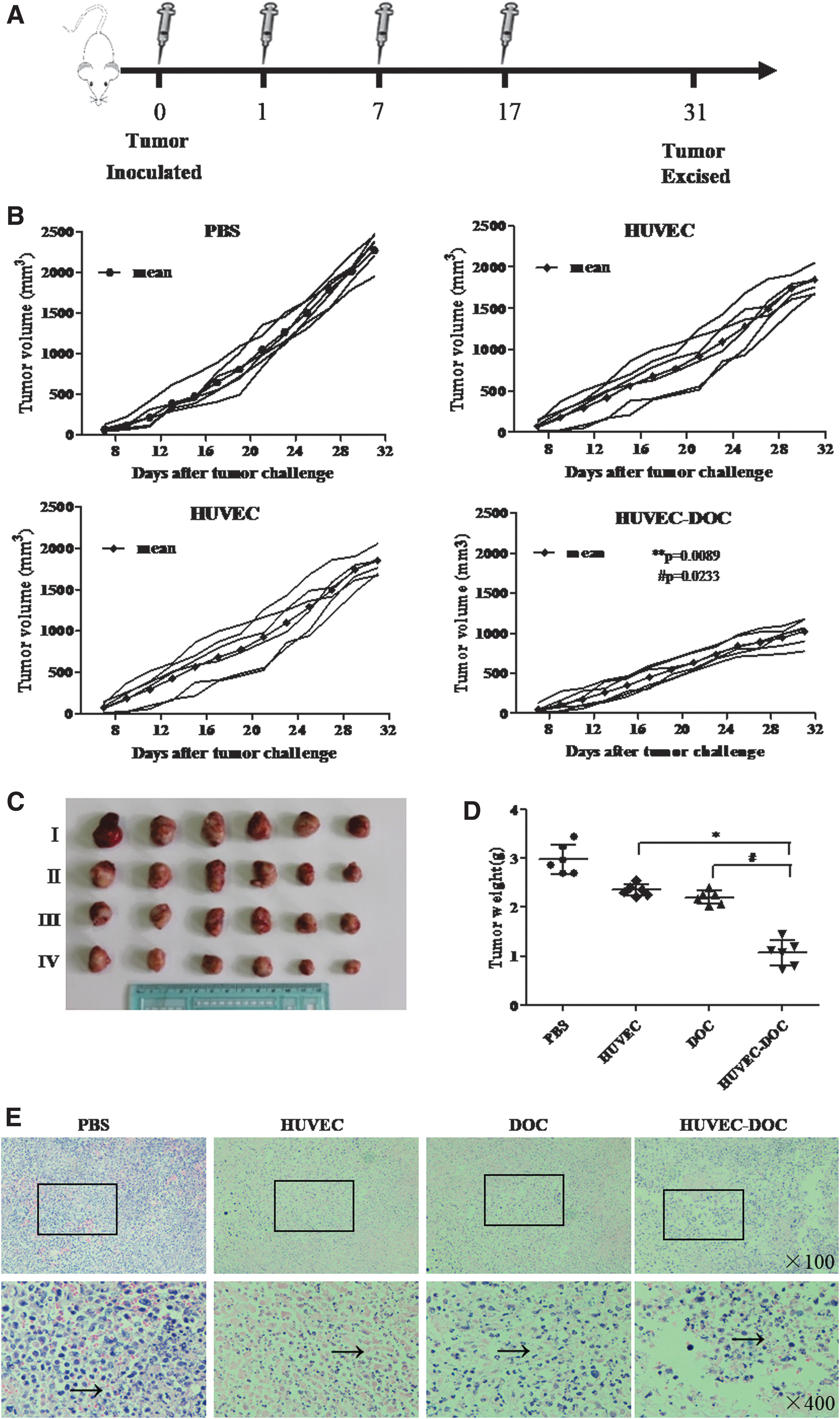
Synergistic antitumor effects elicited by concomitant treatment with HUVEC vaccine and DOC.
HUVEC vaccine and DOC synergistically suppress tumor angiogenesis
To elucidate the antitumor mechanisms of the HUVEC–DOC treatment, the expression of CD31 in tumor sections was detected by immunohistochemical staining. As shown in Figure 2A, although the number of tumor vessels was reduced to some extent after HUVEC or DOC monotherapy, the mean MVD was clearly reduced in tumors from the HUVEC–DOC group (p < 0.05 compared with HUVEC; p < 0.01 compared with DOC). After semiquantitative analysis (Fig. 2B), the mean numbers of blood vessels for the four groups were as follows: HUVEC–DOC (11.5 ± 1.73), HUVEC (21.5 ± 1.91), DOC (25.75 ± 4.86), and PBS (36.5 ± 2.52). Furthermore, HUVEC–DOC treatment resulted in the largest amount of tumor necrosis among the four groups, accompanied by the fewest number of new vessels. The data indicated that HUVEC vaccine combined with DOC could result in synergistic antiangiogenic effects.
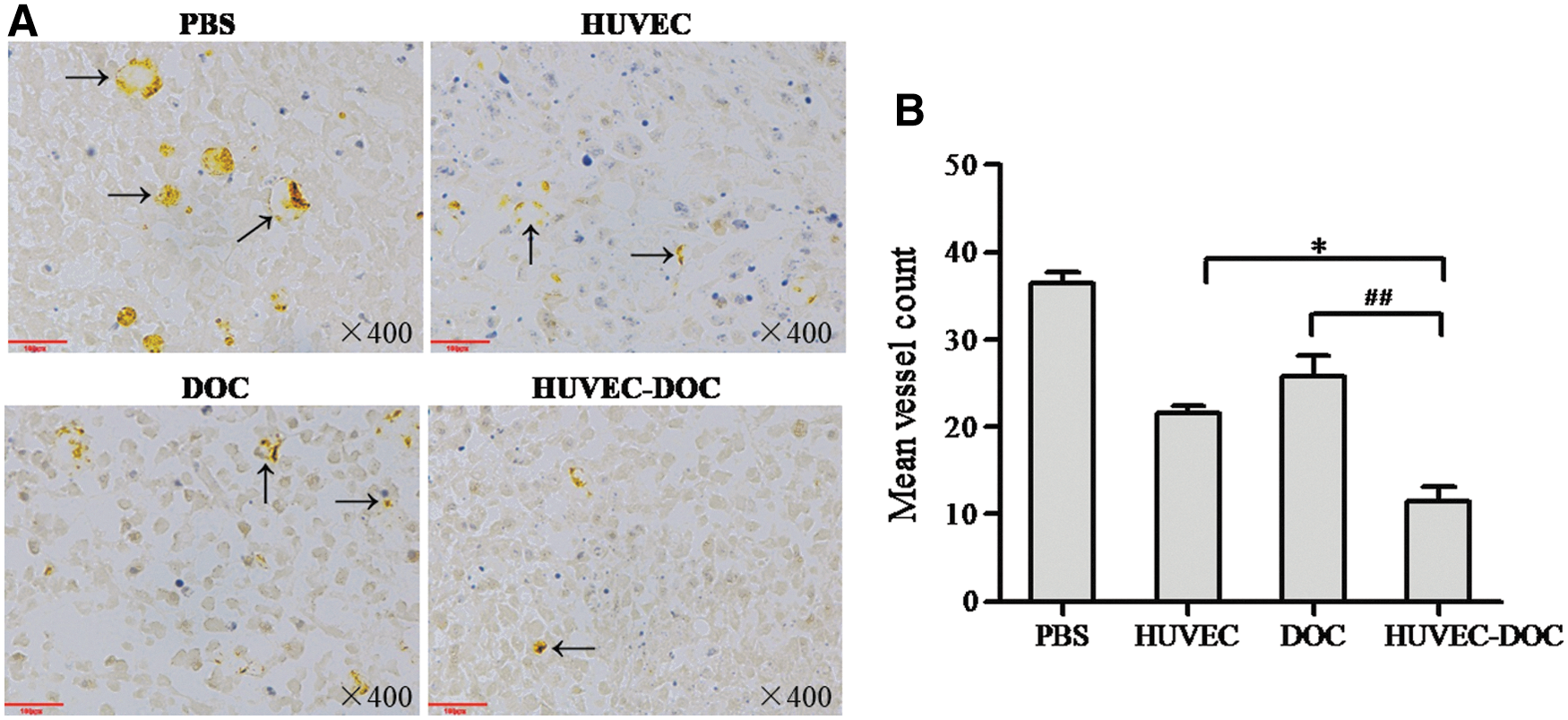
Synergistic antiangiogenic effects elicited by concomitant treatment with HUVEC vaccine and DOC.
HUVEC vaccine and DOC synergistically activate cytotoxic T lymphocyte cells
Next, HUVEC-specific cellular responses were evaluated using a standard LDH release-based cytotoxic T lymphocyte (CTL) assay. Only T lymphocytes isolated from mice treated with a HUVEC vaccine (alone or in combination with DOC) were capable of killing HUVECs dose-dependently in vitro. Furthermore, a significantly stronger killing ability was observed with lymphocytes from HUVEC–DOC-immunized mice compared with those from HUVEC vaccine-immunized mice (p < 0.05; Fig. 3A). However, when the target cells were EMT-6 tumor cells, T lymphocytes from the HUVEC–DOC and HUVEC groups failed to show any cytotoxicity (Fig. 3B), which indicated that the elicited responses were HUVEC-specific.

Synergistic HUVEC-specific cellular responses elicited by concomitant treatment with HUVEC vaccine and DOC.
HUVEC vaccine and DOC synergistically increase IFN-γ concentration
IFN-γ is a relevant mediator in the activation of CTLs, and the concentration of IFN-γ in the supernatant of splenocytes isolated from the mice was further measured by ELISA. The levels of IFN-γ in the HUVEC and HUVEC–DOC groups were both promoted compared with the PBS group, and the amount of IFN-γ in the supernatants of splenocytes from the HUVEC–DOC mice was higher than in mice immunized with HUVEC vaccine alone (Fig. 3C; p < 0.05).
DOC and HUVEC vaccine synergistically increase CD8+ T cells in tumors
The presence of high numbers of CD8+ T cells among tumor-infiltrating lymphocytes was a strong indicator of a favorable clinical outcome; therefore, immunofluorescence was performed to investigate the degree of CD8+ T cell infiltration in the excised tumor tissues. As shown in Figure 3D and E, CD8+ T cell infiltration was observed in tumor xenografts from mice treated with HUVEC vaccine (alone or in combination), and the infiltration of CD8+ T cells was remarkably increased in the HUVEC–DOC group compared with HUVEC vaccine alone (p < 0.05). These results suggested that DOC could assist HUVEC vaccine in eliciting strong HUVEC-specific cellular immune responses, resulting in synergistic antitumor effects.
DOC and HUVEC vaccine synergistically reduce immunosuppressive cells in the tumor microenvironment
Flow cytometry was performed to detect Tregs in digested tumor tissues, as these play an important role in tumor immunosuppression. As shown in Figure 4, the percentage of Tregs in tumors from HUVEC vaccine or HUVEC–DOC groups was decreased to some extent compared with the PBS group, and the number of Tregs in the HUVEC–DOC group was decreased relative to the HUVEC vaccine alone group, suggesting the enhanced antitumor effects observed with HUVEC–DOC.
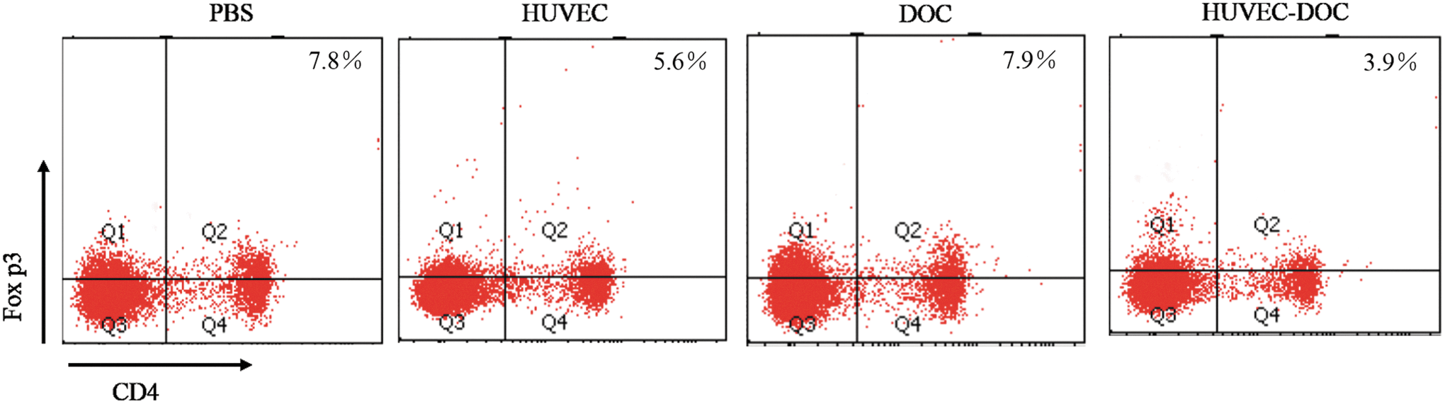
Flow cytometry analysis of Tregs in the tumor microenvironment after different treatments. Treg, regulatory T cells.
Toxicity assessment
Finally, toxicity associated with HUVEC–DOC treatment was evaluated. Body weight was used as an indicator of the overall health of the mice, and there were no significant differences in body weight between the HUVEC–DOC and PBS groups overall (data not shown). Furthermore, no pathological changes were observed in the major organs of mice treated with HUVEC–DOC compared with the PBS group (Fig. 5).
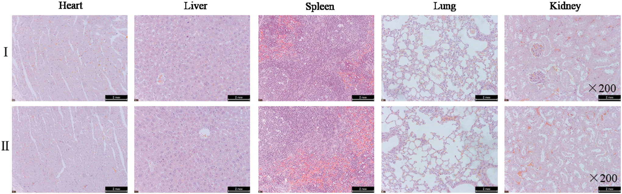
General toxicity observations and H&E staining of the heart, liver, spleen, lung, and kidney tissues (I PBS, II HUVEC–DOC; × 200).
Discussion
Tumor angiogenesis has been proven to be a key hallmark of cancer invasion and metastasis, and antiangiogenic strategies open new paths in breast cancer treatment. 15,16 Accompanied by immunological developments and a more profound understanding of tumorigenesis, cancer vaccines have emerged as efficient approaches for antiangiogenesis-based cancer therapy. 6 –9,17 Additionally, studies have indicated that cancer vaccines are more effective when used in combination therapy. Therefore, it makes sense to design effective combinatorial therapies using antiangiogenic agents. 18,19
In our previous study, the HUVEC vaccine showed an effective antiangiogenic strategy, which could display antitumor efficacy in several animal models. 8 –10 To further optimize the antitumor efficiency of the HUVEC vaccine, DOC was introduced to develop a combination treatment regime. A rapidly growing murine EMT-6 breast cancer model was used to investigate the antitumor efficiency of HUVEC–DOC treatment. It was observed that DOC augmented the antitumor effects of the HUVEC vaccine. Tumor inhibition was associated with the significant activation of HUVEC-specific CTLs, increased IFN-γ-secreting T cells, and increased CD8+ cell infiltration in the tumor microenvironment. Furthermore, no serious adverse effects were observed with HUVEC–DOC treatment.
Chemotherapy has always been considered to be immunosuppressive. However, there is a growing body of evidence showing that certain chemotherapeutics may actually assist immunotherapy by activating the immune system, instead of suppressing it. 20 Chemotherapeutic agents, including cyclophosphamide, paclitaxel, and DOC, are reportedly able to enhance the antitumor activity of cancer vaccines. 21,22 Similarly, in our study, we found that concomitant treatment with DOC and the HUVEC vaccine induced higher HUVEC-specific cellular immune responses relative to HUVEC vaccine alone, resulting in synergistic anti-breast cancer effects. These findings indicate that DOC could be administrated concomitantly with cancer vaccines to induce synergistic antitumor effects, which is attributable to the activation of CTL responses. However, in the present study, only the EMT-6 breast cancer model was adopted to evaluate the antitumor efficacy of the HUVEC–DOC treatment; a greater number and range of tumor models must be studied to more systematically evaluate the antitumor efficiency of this combination treatment regime.
Safety should be considered when DOC is used in combination therapy. Latent toxicity of HUVEC–DOC has also been observed in the present study, though we found the combination treatment to be generally well tolerated. The most common reported side-effects of DOC, such as short-lasting neutropenia and hypersensitivity reactions, were not observed during this study. In addition, body weights of the HUVEC–DOC mice were not significantly affected compared with the PBS control mice. Microscopic examination further proved that there were no pathological changes to the heart, liver, spleen, lung, or kidney after HUVEC–DOC treatment.
Conclusion
We demonstrated that concomitant treatment with HUVEC vaccine and DOC was highly effective in inhibiting murine EMT-6 breast cancer growth without overt toxicity. The antitumor function was accompanied by increased HUVEC-specific CTL responses. Therefore, this rational combination treatment could supplement standard treatments and might provide significant therapeutic benefits to patients with breast cancer.
Footnotes
Acknowledgments
This study was supported by the National Natural Science Foundation of China (grant no. 81541158), the Natural Science Foundation of Shandong Province (grant nos. ZR2018QH005, ZR2018BH046), and the Project of Shandong Province Higher Educational Science and Technology Program (grant nos. J15LM51 and J15LE51).
Authors' Contributions
M.X. and S.Z. conceived the study and developed the criteria. M.L., Q.Y., H.L., W.Z., J.X.G., and C.S. conducted the preliminary search. M.L. and Q.Y. wrote the article. L.Z., S.Z., and M.X. revised the article. All co-authors have reviewed and approved of the article before submission.
Disclosure Statement
No competing financial interests exist.