
Abstract
Objective:
The reverse of chemoresistance and the improvement of sensitivity to chemotherapeutic agents of colorectal cancer cells have great clinical significance and the mechanism underlying the drug resistance is still unclear. REG Iα was reported to be upregulated in colorectal cancer tissues, but the roles of chemoresistance are still unclear.
Materials and Methods:
The expression of REG Iα in colorectal cancer cell lines was assessed by quantitative real-time polymerase chain reaction (Q-PCR). The expression of REG Iα in HCT116 and LOVO cells was knockdown by siRNA. The cell viability and IC50 (half maximal inhibitory concentration) values were analyzed by the CCK8 assay. The proportion of apoptosis and cell cycles were analyzed by flow cytometry. The migration potency of HCT116 and LOVO cells was analyzed by cell migration assay. The protein level of Cyclin D1, CDK4 (cyclin-dependent kinase 4), Bax and Bcl-2 were analyzed by western blot.
Results:
Knockdown of REG Iα enhances the sensitivity to 5-Fu of colorectal cancer cells. REG Iα knockdown promoted the cell apoptosis of HCT116 and LOVO under the 5-Fu treatment. The cell migration and cycle of colorectal cancer cells was also inhibited by REG Iα knockdown. We also found that REG Iα knockdown induced cell cycle arrest and cell apoptosis by Cyclin D1/CDK4 pathway and BAX/BCL-2 pathways.
Conclusions:
Knockdown of REG Iα enhances the sensitivity to 5-Fu of colorectal cancer cells via cyclin D1/CDK4 pathway and BAX/BCL-2 pathways.
Introduction
As one of the most frequent diagnosed cancers worldwide, colorectal cancer has been the fourth leading cause of death globally. 1 Nowadays, the incidence of colorectal cancer has increased significantly due to improved living standards, changed lifestyle, and dietary structure. Despite the declined mortality rate of CRC mainly due to advances of screening tests, surgery, radiation therapy, and adjuvant chemotherapy, >40% of CRC patients almost inevitably die from tumor recurrence and metastasis. 2 The major obstacle for the treatment of colorectal cancer is treatment failure, and the acquired and inherent resistance to chemotherapy represents the major challenge in colorectal cancer treatment. 3 Overcoming intrinsic and acquired drug resistance in treating colorectal cancer patients has great clinical significance and the mechanism underlying the development of drug resistance is poorly understood.
Chemotherapy as one widely used treatment strategy for cancer is also the main treatment methods for colorectal cancer. 4 –6 5-Fluorouracil (5-Fu) and oxaliplatin are the first-line anticancer chemotherapeutic agents and widely used for colorectal cancer treatment. 7 However, chemoresistance for 5-Fu leads to treatment failure of colorectal cancer, and the mechanisms underlying chemoresistance to treatment must be elucidated.
Regenerating gene (Reg), originally isolated as a gene from regenerating pancreatic islets, plays an important role in β cell regeneration and other physiological and pathophysiological processes. 8 –10 REG Iα, as one of the REG family genes, has been found to be involved in the basic biological effects such as cellular proliferation. 11 –14 In gastrointestinal cells, REG Iα is reported to promote the cellular proliferation and highly expressed in the gastrointestinal tissues during tissue injury. 9 For cancer, the REG Iα is found to be overexpressed in lung cancer, 15 squamous esophageal cancer, 16 pancreatic cancer, 17 salivary gland cancer, 18 gastric cancer, 18,19 and colorectal cancers. 20,21 Zheng et al. indicated that aberrant REG expression might be closely linked to the pathogenesis, invasion, or lymph node metastasis of colorectal cancer. 21 Sekikawa et al. found that REG Iα promotes the proliferation and inhibited the H2O2-induced cell apoptosis of colorectal cancer cells. 20 However, the role of REG Iα in chemoresistance of colorectal cancer remains unclear.
In this study, the authors first assessed the relative expression levels of REG Iα in colorectal cancer cell lines and chose the HCT116 and LOVO cells to be used in the subsequent experiment. To explore the potential role of REG Iα in colorectal cancer cells, the expression of REG Iα was knocked down by siRNA. It was found that the knockdown of REG Iα enhances the sensitivity to 5-Fu of colorectal cancer cells. The knockdown of REG Iα significantly inhibited the cell viability and decreased the IC50 values of 5-Fu. In addition, REG Iα knockdown promoted the cell apoptosis of HCT116 and LOVO under 5-Fu treatment. Cell migration and cycle of colorectal cancer cells were also inhibited by REG Iα knockdown. It was also found that REG Iα knockdown induced cell cycle arrest and cell apoptosis by cyclin D1/CDK4 (cyclin-dependent kinase 4) pathway and BAX/BCL-2 pathways.
Materials and Methods
Cell lines
The colorectal cell lines (SW480, HCT116, LOVO, and HT-29) and human small intestinal epithelial cells (HIEC) were obtained from the American Type Cell Culture (Manassas, VA) and cultured in Dulbecco's modified Eagle's medium supplemented with 10% fetal bovine serum (FBS) containing 1% penicillin and streptomycin. The cells were cultured in a humidified incubator under 5% CO2 at 37°C.
Cell transfection
The siRNA oligonucleotides targeting REG Iα and negative control were designed and synthesized by RiboBio Co. Ltd., China. Transfection was performed by Lipofectamine 3000 (Invitrogen, Carlsbad, CA) following the manufacturer's protocol. HCT116 and LOVO cells were transfected by indicated transfection with siRNA targeting REG Iα or siRNA negative control. Then, the cells after transfection were treated with or without 5-Fu for 48 h.
RNA isolation and quantitative real-time PCR
Total RNA was isolated from colorectal cancer cell lines by the Trizol method (Invitrogen). For single-strand cDNA synthesis, random primers, 1 μg RNA template, and Primescript reverse transcriptase (Takara, Japan) were used for reverse transcription by M-MLV (Life Science) according to the manufacturer's instructions. The expression of GAPDH and REG Iα was analyzed by quantitative real-time PCR. Primers used were F: 5′-ACCAGCTCATACTTCATGCTGA-3′ and R: 5′-CCAGGTCTCACGGTCTTCAT-3′ for REG Iα and for GAPDH F: 5′-GGAGCGAGATCCCTCCAAAAT-3′ and R: 5′-GGCTGTTGTCATACTTCTCATGG-3′. Applied Biosystems (ABI) step-one plus sequence detection system (Applied Biosystems, Foster City, CA) was used and comparative cycle threshold method and fold change were calculated as 2−ΔΔCt in gene expression.
Cell viability assay
Cell viability and IC50 values (drug concentration causing 50% inhibition of cell growth) were analyzed by the CCK8 assay. For CCK8 assay, 3000 cells were seeded in 96-well microtiter plates and treated with 5-Fu at various concentrations (0, 0.5, 1, 5, 10, and 50 μM) after indicated transfection. The medium was removed and fresh medium was added to each well, after indicated treatment. Then, 10 μL of CCK-8 solution (CCK-8; Dojin, Japan) was added into each well and incubated for 2 h at 37°C. The absorbance at 450 nm was measured by a microplate reader (Thermo Fisher Scientific, Waltham, MA). The inhibitory and viability rates of each cell line with different treatments were calculated by comparing the OD values of the experimental groups with that of the blank group. Cell viability = (ODDrug − ODBlank)/(ODControl − ODBlank)*100%, cell inhibition = 1 − cell viability. The half-maximal inhibitory concentrations of the different groups were calculated with probit regression analysis by using SPSS version 19.
Apoptosis assay
After treatment with or without 5-Fu, the cells were harvested for apoptosis assay. Forty-eight hours later, the cells (1 × 10⁶) were washed and double stained with Annexin V-FITC/PI (Beyotime, Shanghai, China) following the manufacturer's instructions. FITC fluorescence was detected by a filter with a wavelength of 515 nm, and propidium iodide (PI) fluorescence was detected by a filter with a wavelength of 630 nm. The proportion of apoptosis cells was analyzed by FACS Calibur™ Flow Cytometry (Becton Dickinson, Franklin Lakes, NJ). The apoptosis frequency was calculated as Annexin-FITC(+)/PI(−) and Annexin-FITC(+)/PI(+).
Cell migration assay
For cell migration assay, 1 × 10 5 cells per well in 200 μL cell suspension were added into the upper chamber with 650 μL medium containing 10% FBS in the lower chamber. After 36 h, the unpenetrated cells were removed using a cotton swab and the migrated cells were fixed by 4% formaldehyde and stained with 0.1% crystal violet. Finally, six visual fields were selected for counting number of cells under an optical microscope (Olympus, Germany).
Western blot analysis
Cells were collected and lysed in lysis buffer (50 mM Tris–HCl pH 8.0, 150 mM NaCl, 0.02% NaN3, 0.1% sodium dodecyl sulfate [SDS], 100 μg/mL phenylmethylsufonyl fluoride, 1 μg/mL aprotinin, 1% triton). After centrifugation, cell lysates (100 μg/lane) were collected and subjected to 10% SDS-PAGE (polyacrylamide gel electrophoresis). A BCA assay kit (Beyotime) was used for analyzing the total protein concentration. Then, the protein was transferred onto polyvinylidene difluoride membranes (Millipore). Antibodies against cyclin D1 (1:1500 diluted, No. 2922; CST, Boston, MA), CDK4 (1:1000 diluted; No. sc-70832; Santa Cruz, CA), Bax (1:1000 diluted; No. 2974; CST), GAPDH (1:2000 diluted; No. 5174; CST), Bcl-2 (1:1000 diluted; No. 4223; CST), and horseradish peroxidase-conjugated goat antirabbit secondary antibodies (1:1000; Promab) were used. Protein bands were visualized with ECL-Plus reagent (Millipore) and scanned using a bioimaging analyzer (Bio-Rad, Hercules, CA). Then the integrated optical density of protein bands was analyzed through the software Image-Pro Plus 7.0 (Media Cybernetics, MD) and the relative levels of protein bands were calculated.
5-Ethynyl-2′-deoxyuridine proliferation assays
For 5-ethynyl-2′-deoxyuridine (EdU) assay, cells were sequentially incubated with 10 μM thymidine analog EdU (Beyotime) for 3 h, and fixed with 3.7% formaldehyde. Then, the stained cells were analyzed by FACS Calibur™ Flow Cytometry.
Cell cycle analysis
PI staining was used to analyze the cell cycle distribution. The cells were harvested and fixed in 70% cold ethanol overnight at 4°C. Then the cells were washed twice and pretreated with 50 μg/mL RNase (Beyotime). For PI staining, the cells were treated with 50 μg/mL PI (Sigma) at 37°C for 0.5 h in the dark. FACS Aria Calibur (BD Biosciences) was used to examine the relative proportions of cells in the G0/G1, S, and G2/M phases. Finally, data were analyzed with a software Modfit (Verity Software House).
Statistical analysis
All results are presented as means ± standard error of the mean of at least three independent experiments. Student's t-test was used to assess differences between two groups and one-way analysis of variance was used for multiple comparisons. A value of p < 0.05 was considered to be statistically significant.
Results
Expression levels of REG Iα in colorectal cancer cell lines
First, the authors assessed the relative expression levels of REG Iα in HIEC and colorectal cancer cell lines. As shown in Figure 1A, the expression of REG Iα was significantly higher in HCT116 and LOVO cells than in other cells. Then, the HCT116 and LOVO cells were used in the subsequent experiment. To explore the potential role of REG Iα in colorectal cancer cells, the expression of REG Iα was knocked down by siRNA. The interference efficiency of siRNA is shown in Figure 1B.

Expression levels of REG Iα in colorectal cancer cell lines. The mRNA level of REG Iα in HIEC and colorectal cancer cell lines (SW480, HCT116, LOVO, and HT-29) was analyzed by Q-PCR
Knockdown of REG Iα enhances the sensitivity to 5-Fu of colorectal cancer cells
To determine whether REG Iα confers chemoresistance in colorectal cancer cells, the transfected HCT116 cells were treated with different concentrations of 5-Fu and the cell viability was analyzed (Fig. 2A). The knockdown of REG Iα significantly inhibited the cell viability under the treatment of 5-Fu. IC50 values of HCT116 cells were significantly decreased from 10.77 to 4.89 μM after REG Iα knockdown (Table 1). Similar results were found in LOVO cells (Fig. 2B and Table 1). The results suggest that REG1α may contribute to chemoresistance.

Knockdown of REG Iα enhances the sensitivity to 5-Fu of colorectal cancer cells. The HCT116 and LOVO cells were transfected with siRNA targeting REG Iα or siRNA NC (after 48 h), and treated with different concentrations of 5-Fu (48 h). The cell viability was analyzed by CCK8 assay
IC50 for 5-Fluorouracil in Colorectal Cancer Cell Following Different Treatments
5-Fu, 5-fluorouracil.
Knockdown of REG Iα inhibited cell proliferation and migration of colorectal cancer cells
To further ascertain the role of REG Iα in proliferation and migration of colorectal cancer cells, the authors transfected HCT116 and LOVO cells by siRNA target with REG Iα and siRNA control. The cells were treated with or without 5-Fu and the cell proliferation assay was performed. Cell proliferation of both HCT116 and LOVO cells was inhibited by REG Iα knockdown (Fig. 3A, B). As shown in Figure 3C and D, the migration potency of HCT116 and LOVO cells was markedly inhibited by REG Iα knockdown with or without 5-Fu treatment.
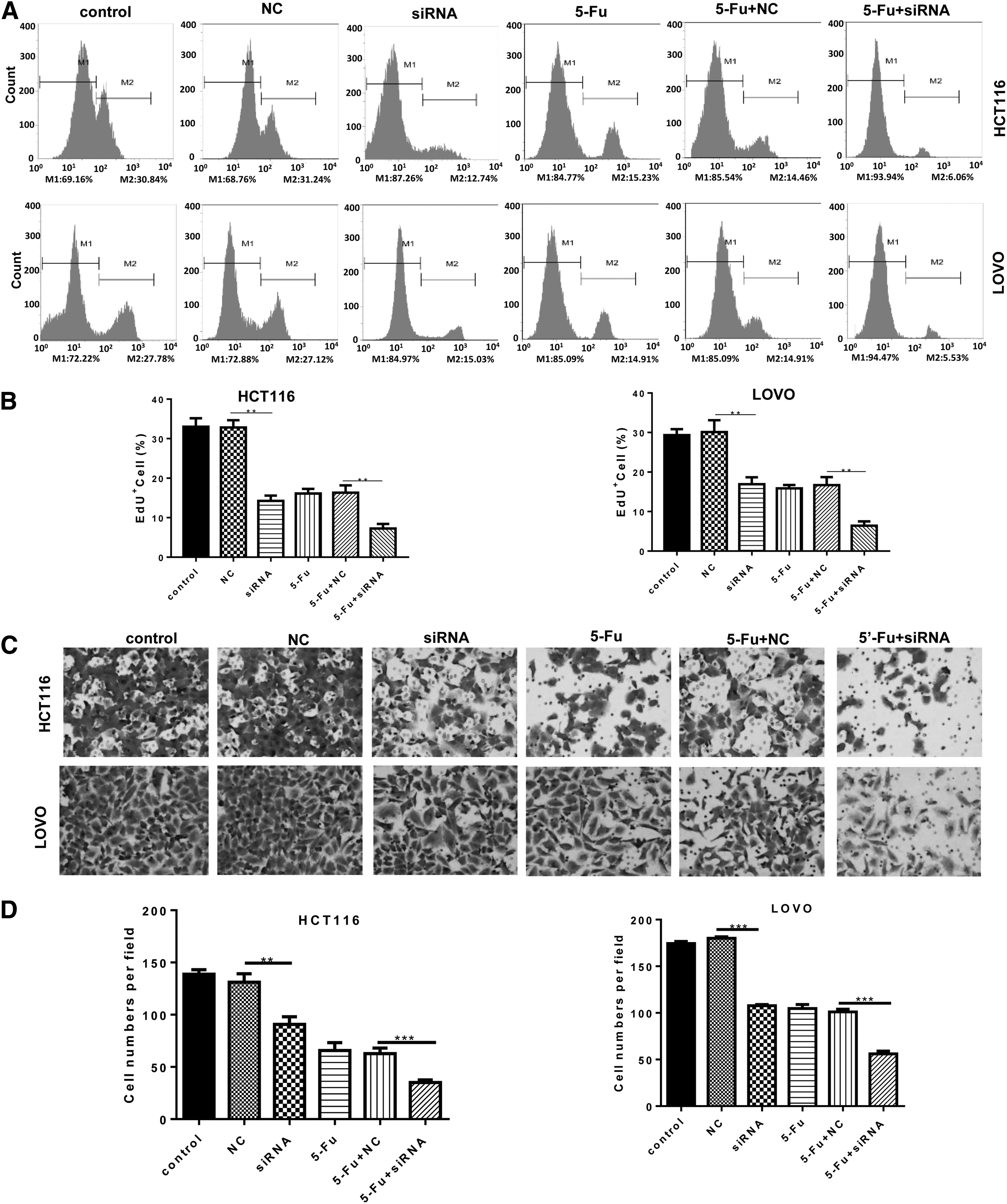
Knockdown of REG Iα inhibited the cell proliferation and migration of colorectal cancer cells. HCT116 and LOVO cells were transfected with siRNA targeting REG Iα or siRNA NC (after 48 h), and subsequently treated with or without 5-Fu (48 h). The EdU proliferation assays of HCT116 and LOVO cells under indicated treatment were performed by flow cytometry. Representative histograms
Knockdown of REG Iα induced cell cycle arrest and apoptosis of colorectal cancer cells
The authors also analyzed the cell cycle of colorectal cancer cells under the indicated treatment. The REG Iα knockdown significantly induced G1 phase arrest of HCT116 and LOVO cells (Fig. 4A, B). Compared with the NC group (negative control: transfected HCT116 and LOVO cells by siRNA control), HCT116 cells transfected with siRNA target with REG Iα displayed markedly decreased proportion of cells in the S phases and increased arrest in G0/G1 phase from 49.00% ± 1.11% to 62.92% ± 2.47% (Fig. 4A, B). The treatment of 5-Fu further increased the arrest in G0/G1 phase in both HCT116 and LOVO cells. In addition, the authors assessed the cell apoptosis of HCT116 and LOVO cells under indicated treatment, and found that the REG Iα knockdown significantly promoted the cell apoptosis of HCT116 (from 3.29 ± 1.40 to 20.74 ± 2.23) and LOVO (from 3.92 ± 1.52 to 29.76 ± 3.47) (Fig. 4C, D). Furthermore, the REG Iα knockdown also promoted the cell apoptosis of HCT116 (from 24.22 ± 2.27 to 36.32 ± 3.65) and LOVO (from 21.95 ± 4.22 to 36.08 ± 1.21) under 5-Fu treatment.
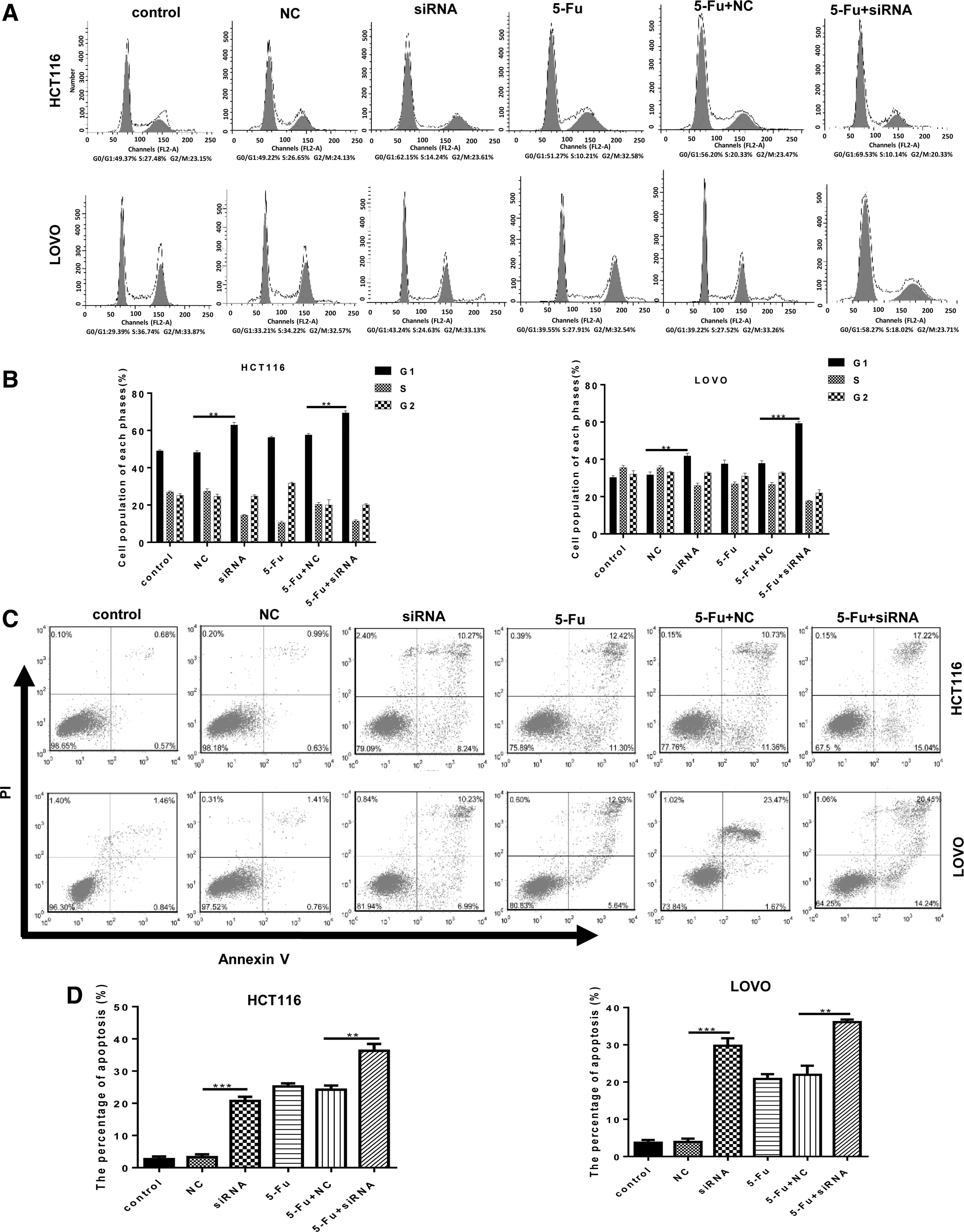
Knockdown of REG Iα induced the cell cycle arrest and apoptosis of colorectal cancer cells. Flow cytometry was used to analyze the cell cycles of HCT116 and LOVO cells after indicated treatment. Representative histograms
Knockdown of REG Iα induced cell cycle arrest and cell apoptosis by cyclin D1/CDK4 pathway and BAX/BCL-2 pathways
Given that the knockdown of REG Iα induced cell cycle arrest and promoted the cell apoptosis of colorectal cancer cells, the authors analyzed the pathway involved. Collecting data showed that the cyclins D and CDK4 drive the cell cycle from G1 to S phase. 22 As shown in Figure 5A and B, the level of cyclin D1 and CDK4 was decreased by REG Iα knockdown in both HCT116 and LOVO cells. In the 5-Fu treatment group, the level of cyclin D1 and CDK4 was further reduced by REG Iα knockdown. In addition, the authors analyzed the apoptosis-related pathway and found that the level of Bax protein is significantly increased and Bcl-2 protein is significantly decreased after treatment of cells with 5-Fu and 5-Fu+siRNA. The 5-Fu treatment further enhanced the protein level of Bax and Bcl-2 in HCT116 and LOVO cells with REG Iα knockdown (Fig. 5C, D).
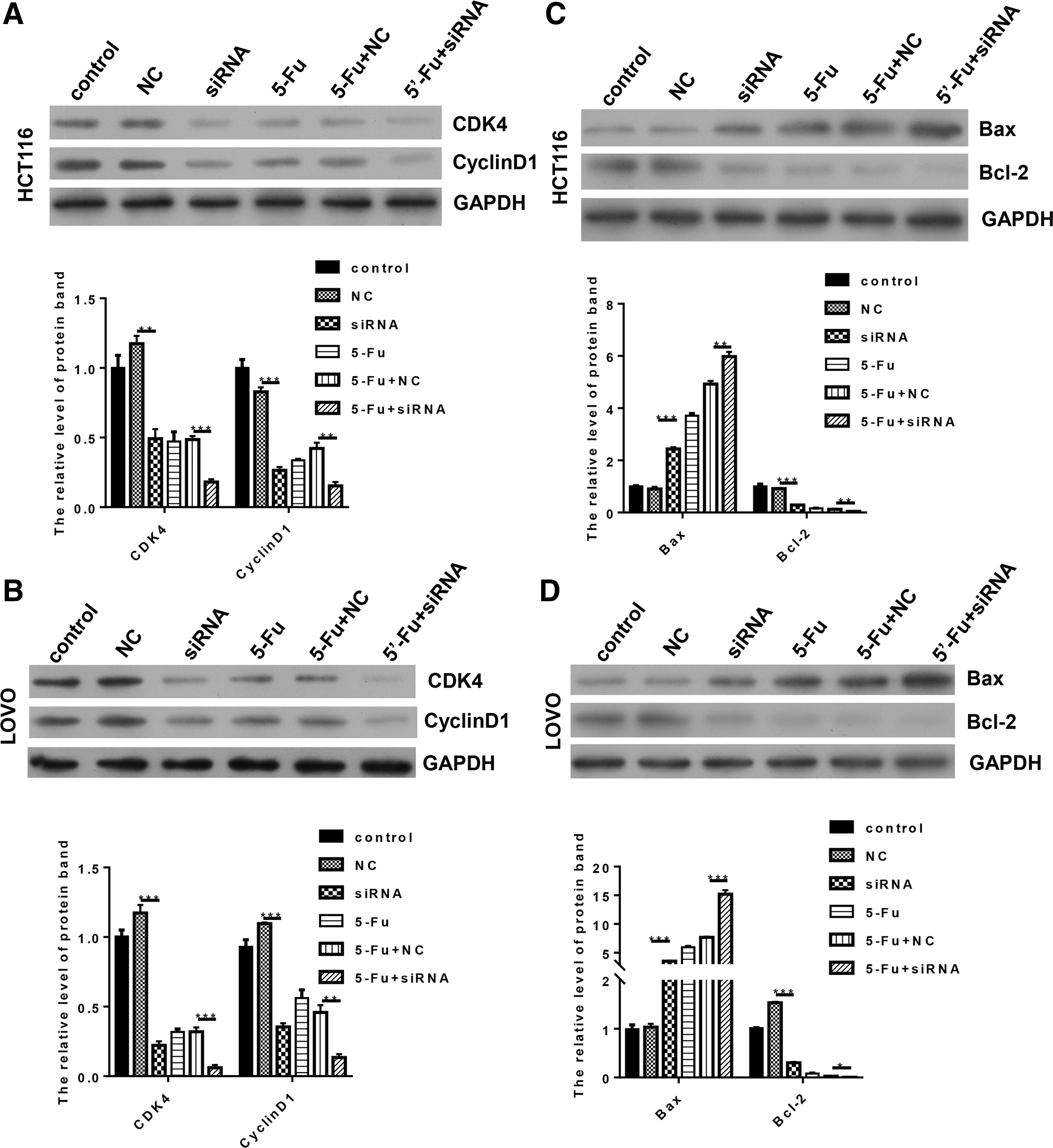
Knockdown of REG Iα induced cell cycle arrest and cell apoptosis by cyclin D1/CDK4 pathway and BAX/BCL-2 pathways. HCT116 and LOVO cells were transfected with siRNA targeting REG Iα or siRNA NC (after 48 h), and subsequently treated with or without 5-Fu (48 h). The protein expression of cyclin D1 and CDK4 in HCT116 and LOVO cells after indicated treatment for 48 h was analyzed by Western blot
Discussion
In recent studies, REG Iα was found to be playing an important role in the pathophysiology of various human inflammatory diseases. REG Iα was expressed not only in the various human inflammatory diseases such as gastritis, 23 pancreatitis, 24 salivary glanditis, 25,26 and colitis, 27 but also in various experimental models of inflammation in animal tissues. 28 In human intestine during Entamoeba histolytica-infected acute colitis and Crohn's disease, the expression of REG Iα was increased. 29 Sekikawa et al. also found the strongly expressed REG Iα in inflamed epithelium, dysplasias, and cancerous lesions of human ulcerative colitis tissues. The expression of REG Iα in human ulcerative colitis tissues was positively correlated with the severity of inflammation and disease duration. 20 In the animal models of inflammation, REG Iα showed a function to protect the intestinal epithelium from parasite-induced apoptosis. Higher probability of spontaneous, and parasite-induced, apoptosis was found in the knockout mice. 30 The collected data indicated that the inflammation might be the main event that triggers REG Iα expression in many tissues including cancer. The results showed the expression of REG Iα in colorectal cancer cells; the key factors in the upstream to trigger REG Iα expression in the colorectal cancer cells need to be further explored in a subsequent research.
It was found that the knockdown of REG Iα enhances the sensitivity to 5-Fu of colorectal cancer cells. The knockdown of REG Iα significantly inhibited the cell viability and decreased the IC50 values of 5-Fu. In squamous cell esophageal carcinoma, REG Iα was considered to be a reliable marker of chemoradiosensitivity. 31 Hayashi et al. also showed that REG I promoted chemoresistance and radioresistance of squamous esophageal cancer cells. 32 Their conclusions are highly consistent with the present findings in colorectal cancer cells. In this study, the knockdown of REG Iα induced cell cycle arrest and cell apoptosis with or without 5-Fu treatment. Cyclin D1 and CDK4 were decreased by REG Iα knockdown in both HCT116 and LOVO cells. In the 5-Fu treatment group, the level of cyclin D1 and CDK4 was further reduced by REG Iα knockdown. In addition, the authors analyzed the apoptosis-related pathway and found that the protein level of Bax protein is significantly increased and Bcl-2 protein is significantly decreased after treatment of cells with 5-Fu and 5-Fu+siRNA. In gastric cancer cells, REG Iα has antiapoptotic effect through STAT3 activation. 19 Sekikawa et al. reported that REG Iα played its antiapoptotic roles by Akt activation and promoting the expression of Bcl-xL and Bcl-2. 33 The results showed the decreased Bcl-2 protein level in HCT116 and LOVO cells by REG Iα knockdown.
In summary, it was shown that REG Iα is expressed in colorectal cancer cell lines and the knockdown of REG Iα enhances the sensitivity to 5-Fu of colorectal cancer cells. The knockdown of REG Iα significantly inhibited the cell viability and promoted the cell apoptosis by cyclin D1/CDK4 pathway and BAX/BCL-2 pathways. The findings provide the idea that therapies targeting REG Iα in colorectal cancer may synergize with chemotherapeutics drugs to increase treatment efficacy and thus improve patient outcomes.
Footnotes
Acknowledgment
This study is supported by grants from Medical Research Program of Chongqing Health and Family Planning Commission (Grant No. 2017MSXM005).
Disclosure Statement
There are no existing financial conflicts.