
Abstract
The Wnt/β-catenin signaling pathway is one of the highly conserved signaling pathway widely reported to play essential roles in the development of various tumors and human cancers, thus serving as a potential target for anticancer therapy. However, the specific effects of the related proteins in the Wnt/β-catenin signaling pathway in nasopharyngeal carcinoma (NPC) still remain elusive. Thus, this study was performed to uncover the correlation between the Wnt/β-catenin signaling pathway-related proteins and the clinical characteristics and prognosis of NPC. NPC tissues were revealed to present high expression of β-catenin and v-myc myelocytomatosis viral oncogene homolog (c-MYC) but low expression of Dickkopf-3 (DKK-3). Immunohistochemical staining revealed that DKK-3 was positively linked to but β-catenin and c-MYC were negatively linked to differentiation, tumor-node-metastasis (TNM) stage and lymph node metastasis of patients with NPC. In addition, c-MYC was identified to be positively correlated to DKK-3 in NPC tissues. The positive expression of β-catenin and c-MYC had negative relations with and that of DKK-3 had positive relations with survival rate of patients with NPC, which was analyzed by Kaplan–Meier method. Moreover, it was shown that later TNM stage and positive expression of β-catenin were risk factors for NPC-related death. These findings provide evidence that the proteins related to the Wnt/β-catenin signaling pathway (DKK-3, β-catenin, and c-MYC) participate in the development of NPC and positive expression of DKK-3 and negative expression of β-catenin, and c-MYC can serve as essential prognostic biomarkers, shedding new light on the prognosis and treatment of NPC.
Introduction
Nasopharyngeal carcinoma (NPC) is identified as an endemic malignancy occurring in the head and neck accompanied by cervical lymph node metastasis (LNM), a main reason for NPC-related death. 1 NPC is usually characterized by various risk factors, pathogenesis, treatment modalities, and clinical presentations. 2 It is comparatively more prevalent in Southern China and even Southeast Asia, with the incidence rate reaching ∼20 in every 100,000 people in endemic areas every year. 3,4 NPC is able to invade adjacent normal tissues and even 70%–80% of NPC patients exhibited metastasis to bone and other organs through lymphatic system or blood at their first diagnosis. 5 As to the management of NPC, radiotherapy is widely recognized to be effective in the early stage because of the radiosensitive features and deep location of NPC. 6 Although many NPC patients could be cured if they were diagnosed and treated at the initial phase, ∼20% patients would be confronted with local recurrence post-treatment. 7 The system of tumor-node-metastasis (TNM) is an essential factor for the prognosis of NPC; however, the current staging system is inadequate to predict the prognosis owing to big variations in the treatment outcomes. 8 Therefore, better understanding of the underlying mechanisms related to NPC prognosis is needed to develop more novel treatment strategies.
The canonical Wnt/β-catenin signaling pathway belongs to one of the most important signaling pathways engaged in many human malignancies and could participate in the development of various cancers and tumors. 9,10 Recent studies indicate that the Wnt/β-catenin signaling pathway is involved in the development of NPC. 11,12 The overactivation of the Wnt/β-catenin pathway was reported in multiple cancers, such as NPC and activation of Wnt/β-catenin signaling pathway may lead to poor prognosis in NPC. 13,14 β-catenin is an essential regulator of the Wnt/β-catenin signaling pathway. 15 Its high expression is closely associated with LNM and low survival of NPC patients, thus serving as a marker for the prognosis of NPC patients. 16 Dickkopf-3 (DKK-3) is recognized as a member belonging to the DKK class of Wnt antagonist with structural and functional divergence. 17 The expression of DKK-3 is found to be reduced in many cancers and could act as a target for poor prognosis. 18 As a negative mediator of the Wnt/β-catenin signaling pathway, DKK-3 depletion has been demonstrated to indicate high survival rate and low metastasis rate in head and neck squamous cell carcinoma (HNSCC). 19 v-myc myelocytomatosis viral oncogene homolog (c-MYC) is an oncogene with deregulated expression in many diseases and acts on critical parts in diverse cellular processes. 20 The Wnt/β-catenin signaling pathway has been found to elevate the expression of c-MYC in teratocarcinoma, thus enhancing cell proliferation. 21 It was revealed that MYC could be taken as a biomarker in NPC. 22 Downregulated c-MYC has been found in NPC cells through enforced microRNA let-7. 23 Based on these findings, the authors hypothesized that these proteins function in the prognosis of NPC. This study is designed to investigate the biological correlation between those proteins and Wnt/β-catenin signaling pathway in NPC.
Materials and Methods
Ethics statement
This study protocol was approved by the Ethics Committee of The Affiliated Hospital of Qingdao University, Qingdao Municipal Hospital. Written informed consent was obtained from all participants or their relatives.
Study subjects
A total of 175 patients (92 men and 83 women, age 22–69 years) who received surgery treatment and were pathologically diagnosed as NPC in the Cancer Center of The Affiliated Hospital of Qingdao University, Qingdao Municipal Hospital from January 2010 to January 2013 were recruited. The obtained specimens were consistent with the NPC histological classification published in 2003 by World Health Organization. 24 The TNM of the specimens was assigned into different stages based on the staging criteria formulated by International Union against cancer (UICC). 25 Inclusion criteria: (1) patients who received surgery treatment in the department of ear-nose-throat in The Affiliated Hospital of Qingdao University, Qingdao Municipal Hospital, and were pathologically diagnosed as NPC and received no other surgical or medical treatment elsewhere before the study; (2) patients with sufficient paraffin-embedded tissue specimens stored in the department of pathology for further experiment, complete clinical records, and accessible contact information. Exclusion criteria: (1) Patients who received tumor resection and biopsy in other hospital before receiving surgery in The Affiliated Hospital of Qingdao University, Qingdao Municipal Hospital; (2) patients who received neoadjuvant chemoradiotherapy, neoadjuvant target treatment, and immunotherapy before surgery.
Immunohistochemical staining
Immunohistochemical (IHC) staining was performed as previously described. 26 In brief, the dissected tumors were paraffin embedded and cut into 4 μm thick sections. These sections were deparaffinized and rehydrated. After antigen retrieval, the sections were incubated with anti-β-catenin, anti-c-MYC, and anti-DKK-3 antibodies (Zsbio Commerce Store, Beijing, China) at 4°C overnight. After incubation with horseradish peroxidase-conjugated secondary antibody (Zsbio Commerce Store, Beijing, China), the sections were counterstained with Mayer's hematoxylin. Ten fields of view at high magnification were randomly selected from each section with 100 cells in each field. Each field was graded according to staining intensity 27 : no staining at all, 0 point; light yellow, 1 point; yellow, 2 points; and brownish yellow, 3 points. The percentage of positive cells was also included: <5%, 0 point; 5%–25%, 1 point; 26%–50%, 2 points; 51%–75%, 3 points; and >75%, 4 points. The product of staining intensity and percentage of positive cells signaled the negative (0–3 points) or positive (≥4 points).
Follow-up
The follow-up started when the patients finished the systematic treatment and were discharged from the hospital and ended in January 2016. A total of 21 patients died for other reasons or were lost to follow-up, or still alive at the end of the study, and these patients were recorded as censored cases. Data of patients lost to follow-up were treated according to the last statistical time. The follow-up was conducted through outpatient, telephone communication, or reference to medical records. The survival time was calculated based on month. Survival analysis was performed by Kaplan–Meier method with the NPC-related death as the terminal indicator.
Statistical analysis
All data were analyzed using SPSS 21.0 statistical software (IBM Corp., Armonk, NY). Enumeration data were presented as percentage and ratio and analyzed by Chi-square test. The interaction among DKK-3, β-catenin, and c-MYC was analyzed with Spearman's rank correlation. The survival rates between positive patients and negative patients were compared by Kaplan–Meier method, and multivariate survival analysis was conducted based on Cox proportional hazards model. A p < 0.05 indicated statistically significant.
Results
β-catenin and c-MYC are highly expressed, whereas DKK3 is poorly expressed in NPC tissues
Initially, IHC staining was performed to detect the positive rate of DKK3, β-catenin, and c-MYC in NPC tissues. It was found that the positive expression of DKK-3 was mainly localized in cytoplasm in brown, whereas that of β-catenin and c-MYC was mainly found in nucleus in brown. Among the tissues from 175 patients with NPC, the positive rates of DKK-3, β-catenin, and c-MYC reached 41.1% (n = 72), 56% (n = 98), and 61.7% (n = 108), respectively. Among the 175 adjacent normal tissues, the positive rates of DKK-3, β-catenin, and c-MYC reached 65.1% (n = 114), 22.9% (n = 40), and 32% (n = 56), respectively. The expression of DKK-3, β-catenin, and c-MYC in NPC tissues were significantly different from that in adjacent normal tissues (all p < 0.001; Fig. 1 and Table 1). These results demonstrate that NPC tissues exhibit high expression of β-catenin and c-MYC but low expression of DKK-3.
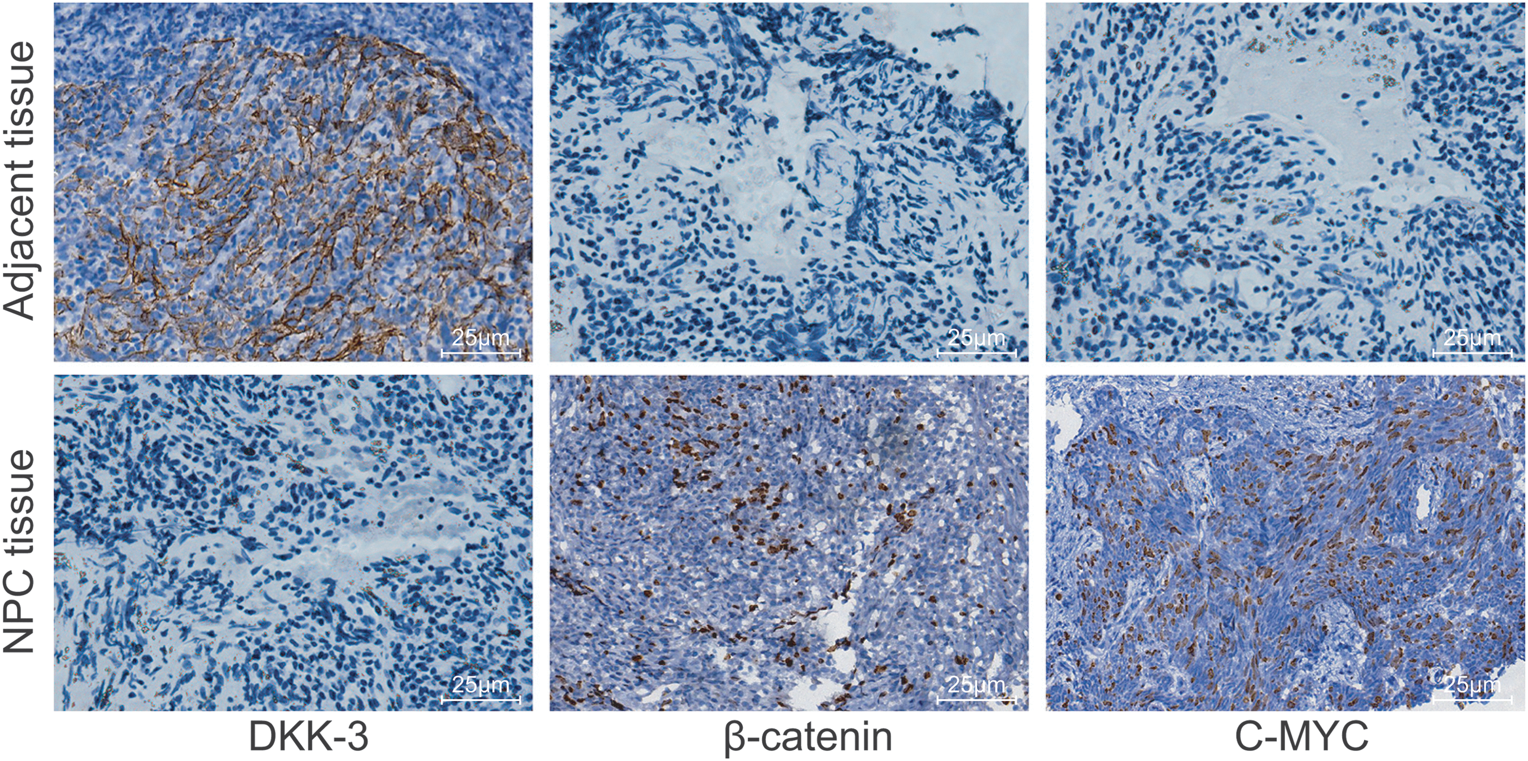
DKK-3 was reduced, whereas β-catenin and c-MYC were elevated in NPC tissues (SP × 400). c-MYC, v-myc myelocytomatosis viral oncogene homolog; DDK-3, Dickkopf-3; NPC, nasopharyngeal carcinoma; SP, streptavidin-peroxidase. Color image is available online.
Expression of Dickkopf-3, β-Catenin, and v-myc Myelocytomatosis Viral Oncogene Homolog in Nasopharyngeal Carcinoma Tissues and Adjacent Normal Tissues
c-MYC, v-myc myelocytomatosis viral oncogene homolog; DDK-3, Dickkopf-3; NPC, nasopharyngeal carcinoma.
Expression of DKK-3 is positively correlated, whereas that of β-catenin and c-MYC is negatively correlated to TNM stage, differentiation, and LNM of patients with NPC
After detecting the expression of DKK-3, β-catenin, and c-MYC in NPC tissues, the focus was shifted to determining the correlation between the expression of DKK-3, β-catenin, and c-MYC and clinical characteristics of patients with NPC. The results showed that the expression of DKK-3, β-catenin, and c-MYC in NPC tissues was all correlated to TNM stage, differentiation, and LNM (p < 0.05). Patients with NPC in later TNM stage, low differentiation, and LNM presented lower DKK-3 positive rates and higher β-catenin and c-MYC positive rates. The expression of DKK-3, β-catenin, and c-MYC had no statistical correlation to the patients' age, gender, tumor location, infiltration depth, tumor size, and tissue type (all p > 0.05; Table 2). According to rank correlation analysis of Spearman nonparametric test, DKK-3 and c-MYC were positively correlated (r = 0.157, p < 0.05), whereas no significant difference was found between β-catenin and c-MYC or between β-catenin and DKK-3 (p > 0.05; Table 3). All in all, DKK-3 is positively associated with, whereas β-catenin and c-MYC are negatively associated with TNM stage, differentiation, and LNM of patients with NPC.
Correlation Between Clinical Characteristics of Nasopharyngeal Carcinoma Patients and the Expression of Dickkopf-3, β-Catenin, and v-myc Myelocytomatosis Viral Oncogene Homolog
TNM, tumor-node-metastasis.
Interaction Among Expression of Dickkopf-3, β-Catenin, and v-myc Myelocytomatosis Viral Oncogene Homolog in Nasopharyngeal Carcinoma Tissues
DKK-3 positive expression is positively related, whereas β-catenin and c-MYC positive expression are negatively related to survival rate of patients with NPC
After all patients received review and follow-up visits, the patients' condition and the survival time were recorded and Kaplan–Meier method and Chi-square test were employed to analyze the relationship between survival rate and the expression of DKK-3, β-catenin, and c-MYC. The follow-up spanned 3–4 years, ending in January 2016. Among the 175 patients with NPC, 84 survived and 91 died with median survival time of 29 months. The median survival time was 29 months for patient with negative DKK-3 expression and 31 months for patients with positive DKK-3 expression. The median survival time was 30 months for patients with negative β-catenin expression and 28 months for patient with positive β-catenin expression. Patients with negative expression of c-MYC had a median survival time of 30 months, whereas patients with positive expression of c-MYC displayed a median survival time of 31 months. The survival time for patients with negative and positive expression of DKK-3, β-catenin, and c-MYC was analyzed with Kaplan–Meier method and the survival curve was drawn accordingly (Fig. 2). The results reveal that the survival rates of patients with DKK-3 positive expression are higher than the patients with DKK-3 negative expression, and that the survival rates of patients with β-catenin and c-MYC positive expression are lower than those with β-catenin and c-MYC negative expression (all p < 0.05).
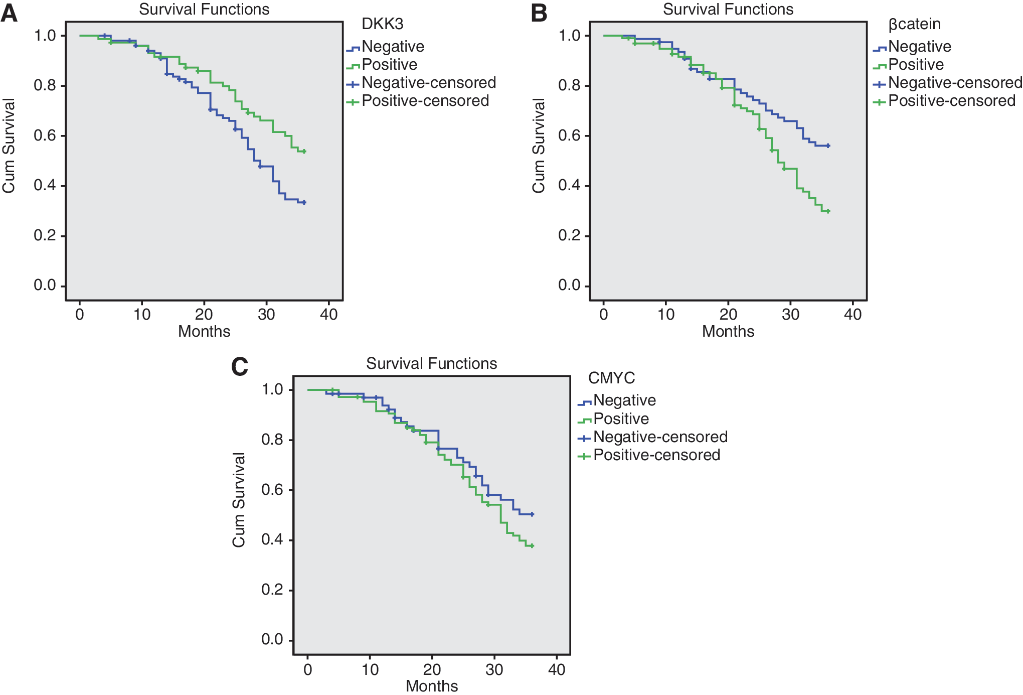
Positive expression of DKK-3 was positively linked, whereas positive expression of β-catenin and c-MYC was negatively linked to survival rates of NPC patients. K-M survival curves of positive and negative expression of DKK-3
Later TNM stage and β-catenin positive expression may lead to NPC-derived death
Cox proportional hazards model was used for multivariate survival analysis. The death of patients with NPC was taken as dependent variables and the expression of DKK-3, β-catenin, and c-MYC in NPC tissues, TNM stage, differentiation, and LNM as independent variables. The findings provide evidence verifying that later TNM stage and β-catenin positive expression are risk factors for NPC-derived death (both p < 0.05; Table 4).
Cox Proportional Hazards Model for Multivariate Survival Analysis in Nasopharyngeal Carcinoma
CI, confidence interval; Exp, exponential; SIG, significance; SE, standard error.
Discussion
NPC as an endemic cancer is a prevalent malignancy with high incidence in Southeast Asia, especially in South China. 28 Thus far, the pathogenesis of NPC has not yet been fully elucidated. Therefore, this study aims to investigate the association between the Wnt/β-catenin signaling pathway and NPC. The findings of these results indicated that the expression of proteins (DKK-3, c-MYC, and β-catenin) related to the Wnt/β-catenin signaling pathway were correlated with the development and progression of NPC, among which β-catenin positive expression could function as one of the detection and evaluation indexes for diagnosis and prognosis of NPC.
First, it was revealed in this experiment that compared with the adjacent normal tissues, DKK-3 positive expression was decreased and β-catenin and c-MYC positive expression were elevated in NPC tissues. DKK-3 is one of the DKK class of DKK-1, DKK-2, DKK-3, and DKK-4, which encode secreted proteins, and DKK-3 transfection has been found to affect the invasive ability of some tumor cells and induce apoptosis, indicating that DKK-3 may exert tumor-suppressive effects. 29 Consistently, DKK-3 was found to be downregulated in numerous human cancers such as bladder cancer and could act as an underlying gene for treatment of cancers. 30,31 Similarly, the depletion of DKK-3, suppressor of the Wnt/β-catenin signaling pathway, was also demonstrated in prostate cancer and restored DKK-3 could suppress the progression of prostate cancer. 32 As a component of the Wnt/β-catenin signaling pathway, β-catenin was upregulated in NPC accompanied by LNM, 33 which was in line with the findings of this study. Besides, its downregulation could greatly repress the development of NPC cells. 3 Moreover, β-catenin plays a crucial role in E-cadherin-mediated cell adhesion. 34,35 Abnormal expression of β-catenin is essential to distal metastasis of various malignant tumors. 36,37 In NPC cells, c-MYC is an oncogene associated with proliferation, and has demonstrated its expression in ∼90% NPC tissues. 23 c-MYC downregulation could inhibit the development of NPC, thus depleted c-MYC could be considered as an underlying target for the NPC treatment. 38
Furthermore, the authors' data indicated that the protein expression of DKK-3, β-catenin, and c-MYC was correlated to the differentiation, TNM stage, and LNM of NPC, and that β-catenin positive expression could function as an important biomarker for NPC prognosis. A previous study confirmed that DKK-3 protein was closely correlated to the metastasis of HNSCC by prolonging disease- and metastasis-free survival and overall survival. 19 The research of Gu et al. demonstrated that DKK-3 was poorly expressed in pancreatic cancer cells, suggesting the role of DKK-3 as a tumor suppressor through downregulating β-catenin expression through the extracellular regulated protein kinase-mediated pathway. 39 Moreover, downregulation of DKK-3 caused by promoter methylation was linked to poor survival of gastric cancer patients, 40 which was consistent with the authors' finding that the positive expression of DKK-3 is a protective factor against NPC-related death. Meanwhile, it has been demonstrated that the methylation of the Wnt signaling regulators (e.g., SFRP1, DKK2, and DKK3) was frequently detected in samples from NPC patients, and these genes are tumor-suppressor genes for NPC based on the functional studies 41 ; thus, it is not surprising that positive expression of DKK-3 is associated with survival rate of NPC. According to the authors' results, negative DKK-3 signals the poor prognosis of patients with NPC. Nevertheless, the regulatory role of DKK-3 in the survival rate of NPC needs to be further studied. Wu et al. found that the expression of β-catenin is associated with tumor metastasis and poor prognosis of colorectal carcinoma. 42 Likewise, in lynch syndrome, the positive expression of β-catenin was found to be correlated to invasive depth of the tumors and LNM. 43 The findings of Song and his colleagues revealed that β-catenin is highly expressed in NPC cell lines and tissues and silenced β-catenin could lead to significantly downregulated expression of downstream genes of the β-catenin signaling pathway, including c-MYC. 44 Evidence also confirmed that the inhibited β-catenin could suppress the proliferation and migration capacities of NPC and thereby β-catenin could serve as a protein to maintain the proliferation and migration capacities of NPC, which may be linked to the network containing both c-MYC and β-catenin. 28 It is also suggested that c-MYC suppression leads to differentiation and inhibit cell proliferation of such cancers as osteogenic sarcoma. 38
Conclusion
To conclude, this study provided evidence that the proteins related to the Wnt/β-catenin signaling pathway were closely correlated to the development and progression of NPC, and that β-catenin positive expression can function as one of the detection and evaluation indexes for diagnosis and prognosis of NPC. The authors' findings contribute to the treatment and management of NPC. However, sample size will be further expanded to explore the correlation between DKK-3 expression and prognosis of patients with NPC, whereas the function of DKK-3 in NPC will also be expected to explore in future. Besides, considering the complex interaction of molecular components, future researches are required for the accurate mechanism underlying NPC.
Footnotes
Acknowledgments
This study was supported by the two topics of Project of Qingdao Municipal Health and Family Planning Commission (nos. 2017-WJZD020 and 016-WJZD013) and Project of Shandong Provincial Health and Family Planning Commission (no. 2005-ZJ006). The authors thank and appreciate their colleagues for their valuable efforts and comments on this article.
Disclosure Statement
No competing financial interests exist.