
Abstract
AHNAK nucleoprotein 2 (AHNAK2) is supposed to participate in calcium signaling and cytoarchitecture by directly interacting with some proteins. Recently, it was identified as a novel candidate oncogene in human tumors. The author's present study aimed to investigate the expression and biological function of AHNAK2 in uveal melanoma (UM). Based on microarray data of 63 UM patients that were downloaded from Gene Expression Omnibus database, the authors found that AHNAK2 expression is higher in UM primary tumor tissues from patients who developed metastases after enucleation than that in UM primary tumor tissues from patients without metastasis after enucleation. On the basis of the data obtained from The Cancer Genome Atlas database, they found that high AHNAK2 expression is closely associated with shorter overall survival time in UM patients. From quantitative reverse transcription polymerase chain reaction analyses, they revealed that the mRNA expression level of AHNAK2 was significantly upregulated in M17 and SP6.5 cell lines compared with that in D78. Functionally, knockdown of AHNAK2 using small interfering RNA in M17 and SP6.5 cells dramatically suppressed cell proliferation, migratory and invasive abilities, as well as inhibited the activation of phosphatidylinositol 3-kinase (PI3K) signaling pathway. Taken together, their results illustrated that AHNAK2 was upregulated in UM and plays a promoting role in the proliferation and migration of UM cells possibly via regulating PI3K signaling pathway.
Introduction
Uveal melanoma (UM), a kind of melanoma that develops in the uveal component, is the most common human intraocular tumor. 1 –3 Almost 50% of the patients diagnosed with UM will develop metastatic UM and die of metastasis. 4,5 Because of the special vasculature of eyes and the distinctive biology of tumor, the areas to which UM metastasizes the most frequently is liver. 6 Radio-plaque, proton-beam, and enucleation are considered as effective treatment strategies to therapy methods for the primary UM in the eye. However, no effective treatment for metastatic UM is available to date. 7 Hence, exploring effective treatment approach is of great significance. Meanwhile, a deeper understanding of the molecular mechanism underlying UM progression will be helpful for us to find out effective therapy methods.
AHNAK nucleoprotein 2 (AHNAK2) is a member of AHNAK family, which are large proteins with molecular mass larger than 600 kDa. 8,9 Since AHNAK2 possesses similar structure and possibly shares some intracellular localizations with AHNAK1, it is supposed to possess similar function of AHNAK1: participation in calcium signaling, cell repair, tumor cell migration, and invasion. 10 –13 Recently, the overexpression of AHNAK2 has been found in pancreatic ductal adenocarcinoma (PDAC) tissues 14 and clear cell renal cell carcinoma (ccRCC) tissues. 15 Also, it was proposed to be a prognostic marker as well as an oncogenic protein in PDAC and ccRCC patients. 14,15 However, the expression of AHNAK2 in UM tissues and its potential function in UM progression remains unclear.
In the present research, the authors revealed the upregulation of AHNAK2 in UM cells and demonstrated that AHNAK2 functioned as a facilitator in the proliferation, migration, and invasion of UM cells for the first time. Mechanistically, we identified that AHNAK2 might exert its influence partially by regulating the activation of phosphatidylinositol 3-kinase (PI3K) signaling pathway.
Methods
Data collection
The gene expression microarrays data of 35 UM primary tumor tissues from patients who developed metastases after enucleation (metastasis group) and 28 UM primary tumor tissues arising from patients without metastases after enucleation (nonmetastasis group) were downloaded from Gene Expression Omnibus (GEO,
Cell lines
UM cell lines M17 and SP6.5 and corneal epithelial cell line D78 were obtained from Shanghai Cell Bank of Chinese Academy of Sciences (Shanghai, China). These cells were cultivated in RPMI-1640 medium with 5% CO2 at 37°C. The culture medium was added with 10% fetal bovine serum, 100 U/mL penicillin, and 100 mg/mL streptomycin.
Real-time quantitative reverse transcription polymerase chain reaction
The authors extracted the total RNA from UM cells using RNAiso Plus (TaKaRa Biotechnology, Dalian, China). Reverse transcription was conducted utilizing HiFiScript cDNA Synthesis Kit (CwBio, Beijing, China). Hereafter, the expression of AHNAK2 was determined by qPCR using Applied Biosystems 7300 Sequence Detection System (Applied Biosystems, Foster, CA). β-Actin and GAPDH were utilized as internal references. The primers used were as follows: AHNAK2 forward: 5′ AGCGTCTGTAGCTTCCTTGT 3′, AHNAK2 reverse: 5′ GGCAGCCTCAGTCGTGTATT 3′; β-actin forward: 5′ TCACCCACACTGTGCCCATCTAC 3′, β-actin reverse: 5′ AGCGGAACCGCTCATTGCCAATG 3′; GAPDH forward: 5′ GGAGCGAGATCCCTCCAAAAT 3′, GAPDH reverse: 5′ GGCTGTTGTCATACTTCTCATGG 3′.
The relative expression of AHNAK2 was calculated using a 2−ΔΔCt formula. 18 All the tests were performed for three independent times.
Cell transfection
Small interfering RNAs (si-RNAs) against AHNAK2 [si-AHNAK2 (1): 5′ GAGCGTCTGTAGCTTCCTT 3′; si-AHNAK2 (2): 5′ GACTCAATGACAAACACAA 3′] and scrambled si-RNA (si-con, 5′ CTTCGATGTCTGGCGAGTTC 3′) were purchased from GenePharma Co. (Shanghai, China) and transfected into M17 and SP6.5 cells using Lipofectamine 2000 (Invitrogen, CA) following the manufacturer's instructions. After 48 h transfection, the knockdown efficiency was determined by quantitative reverse transcription polymerase chain reaction (qRT-PCR) and Western blot.
Cell proliferation assay
Cell Counting Kit-8 (CCK-8; Beyotime Technology, Jiangsu, China) was applied for examining the ability of cell proliferation at 0, 24, 48, and 72 h points in accordance with the instructions provided by the manufacturer. A microplate reader (Bio-Rad, CA) was used to examine the absorbance at 450 nm. The proliferation curve was plotted using GraphPad Prism 5.0 software.
Wound healing assay
The authors performed a wound healing assay after 24 h transfection to investigate the effect of AHNAK2 downregulation on the migration ability of UM cell lines. First, a pipette tip was utilized to make a wound on the cell monolayers. Then they used phosphate-buffered saline buffer to rinse off the wounded cells, and the remaining cells on the plates were continued to be cultivated at 37°C for 24 h. The wound at the beginning of scratching and 24 h after scratching were observed and pictured under an optical microscope (Olympus, Japan) with 100 × magnification. All the experiments were conducted in triplicate.
Transwell assay
To explore the effect of AHNAK2 on the invasive ability of UM cells, transwell invasion assay was conducted using a transwell chamber coated with Matrigel (BD Bioscience). Transfected UM cells were suspended in serum-free medium and seeded into the upper chamber (1 × 10⁵ cells/well). The lower chamber was filled with complete medium. After 48 h routine cultivation, the cells that remained on the upper chamber were removed, and then the cells invaded to the lower chamber were fixed with 4% paraformaldehyde and stained by crystal violet. The results were observed under a microscope and five fields were randomly selected for count.
Western blot
After 48 h transfection, the total proteins were isolated using RIPA lysates (Beyotime). The concentration of the protein was determined using a BCA Protein Assay Kit (Thermo Scientific). Then the protein expression of PI3K, p-PI3K, AKT, and p-AKT were detected by Western blot. The primary antibodies used were the following: PI3K (1:1000, 4249; Cell Signaling Technology [CST]), p-PI3K (1:1000, 4228; CST), AKT (1:1000, 4691; CST), p-AKT (Ser473, 1:1000, 4060; CST), and GAPDH (1:5000, 5174; CST). The membranes were then incubated with appropriate horseradish peroxidase-conjugated anti-mouse/rabbit secondary antibodies (CST). Eventually, the signals were detected via utilizing enhanced chemiluminescence plus detection kit. Quantity One software was used to quantify the protein expression. All the tests were performed for at least three independent times.
Statistical analysis
The experiment data are presented as mean ± standard deviation. Student's t-test was conducted to compare the discrepancy between two groups. For the comparison among three or more groups, one-way analysis of variance combined with a Tukey's post hoc test was applied. SPSS 15.0 software (IBM, Chicago, IL) was applied to carry out the statistical analyses. For the overall survival analysis, Kaplan–Meier method together with log-rank test was utilized. The samples were divided into high and low expression groups according to the median of AHNAK2 expression. Pearson's chi-square (χ 2 ) test was conducted to analyze the correlation between AHNAK2 expression and clinic-pathological characteristics of UM patients. Univariate and multivariate Cox analysis was applied for analysis of prognostic factors. p-Value <0.05 indicated a statistically significant difference.
Results
AHNAK2 is highly expressed in UM cells
On the basis of the microarray data obtained from GEO database (access number: GSE22138), the authors identified that AHNAK2 expression is higher in UM primary tumor tissues from patients who developed metastases after enucleation (metastasis group) than that in UM primary tumor tissues from patients without metastases after enucleation (nonmetastasis group) (Fig. 1A, p < 0.0001). Through qRT-PCR analysis, they discovered that the mRNA expression level of AHNAK2 in M17 and SP6.5 cells was markedly higher than that in D78 normal cells (Fig. 1B, C, p < 0.001). The results of χ 2 test (Table 1, based on the data downloaded from TCGA database) revealed that the expression of AHNAK2 is closely correlated with histological-type (p = 0.034), pathologic-M (p = 0.042), recurrence (p = 0.025), and dead (p < 0.001). Kaplan–Meier analysis indicated that high AHNAK2 expression is dramatically associated with shorter overall survival time (Fig. 1D, p < 0.01). Univariate analysis revealed that AHNAK2 expression, pathologic-M, and recurrence might be considered as prognostic factors for UM (Table 2, p < 0.001). Further multivariate analysis showed that AHNAK2 expression (p = 0.002) and pathologic-M (p < 0.001) were potential independent risk factors for overall survival of patients with UM. Then they analyzed the correlation between AHNAK2 and HTR2B (a prognostic gene) by Pearson's correlation analysis on GEPIA website. A positive correlation was observed between AHNAK2 and HTR2B (R = 0.65, p = 1.2E-10, Fig. 1E), which further supports the results of multivariate analysis that AHNAK2 was a risk factor for overall survival of patients with UM.

AHNAK2 expression is higher in UM cells.
The Association Between AHNAK2 Expression and Clinical Features of Uveal Melanoma Patients
p < 0.05.
Univariate and Multivariate Analysis of Clinic Pathologic Features for Overall Survival in Uveal Melanoma Cells
HR, hazards ratio; CI, confidence interval. * p < 0.05.
Knockdown of AHNAK2 restrained UM cell proliferation
On the purpose of investigating the specific role of AHNAK2 in UM progression, they constructed SP6.5 and M17 cell lines with silenced AHNAK2 using RNA interference. From Figure 2A–D, the authors can easily observe that the level of AHNAK2 in SP6.5 cells decreased obviously both at mRNA and protein levels after transfected with si-AHNAK2 (1) or si-AHNAK2 (2). Moreover, when using si-AHNAK2 (2), the knockdown efficiency is higher than 80%. Hence, the authors chose si-AHNAK2 (2) to perform the following experiment. Afterward, they carried out CCK8 assay to determine the effect of depletion of AHNAK2 on UM cell proliferation. As shown in Figure 3A and B, the proliferation ability of M17 and SP6.5 cells was significantly declined in si-AHNAK2 group compared with that in their corresponding si-con group (p < 0.01).
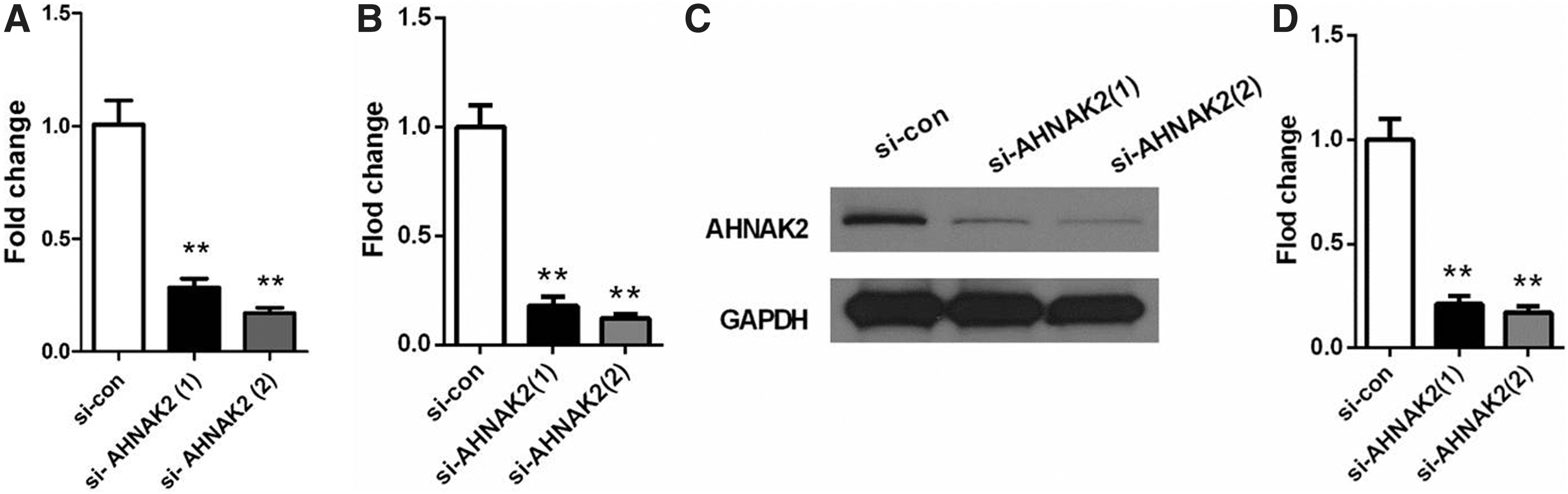
The expression level of AHNAK2 is downregulated in SP6.5 cells after transfected with si-AHNAK2.
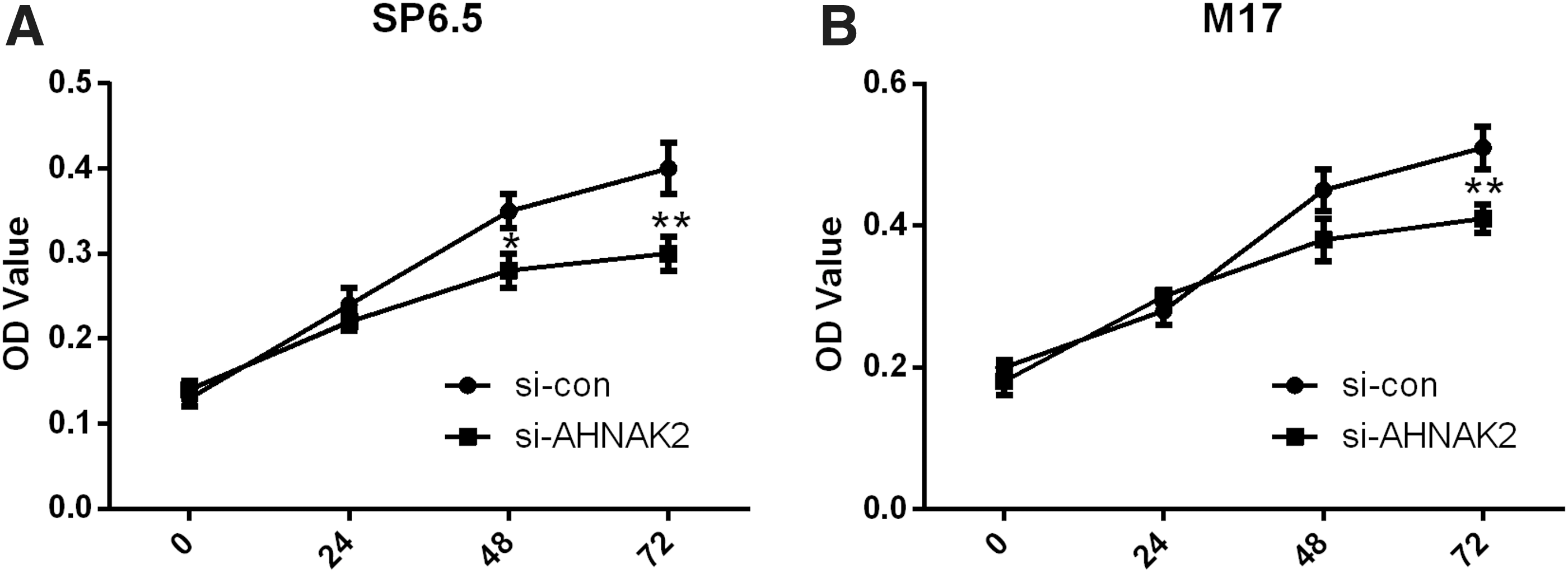
The OD value was lower in si-AHNAK2 group than that in si-con group.
Knockdown of AHNAK2 restrained UM cell migration and invasion
Hereafter, they examined the migration ability of M17 and SP6.5 cells using a wound healing assay. The authors found that the relative migration distance of M17 cells reduced significantly in si-AHNAK2 group compared with that in si-con group (p < 0.01, Fig. 4A, B). The similar results were observed in SP6.5 cells transfected with si-AHNAK2 (Fig. 4C, D, p < 0.01).
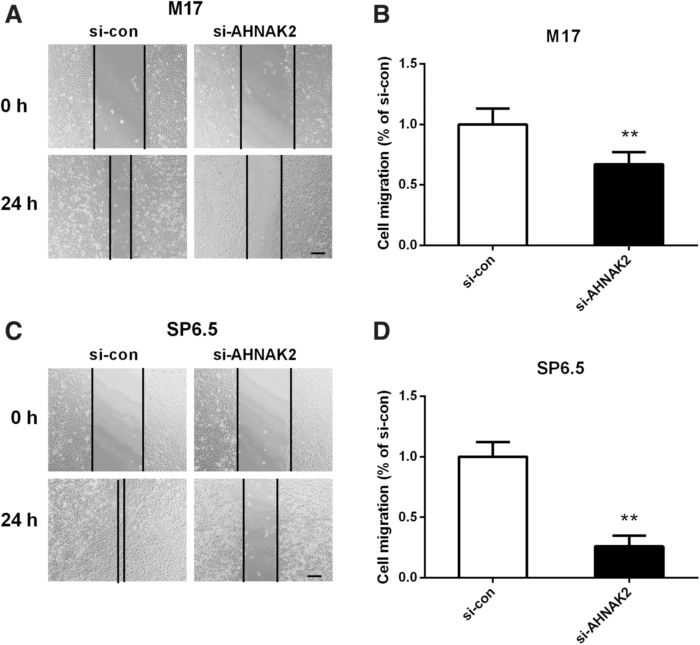
The migration distance was reduced in si-AHNAK2 group than that in si-con group.
On the purpose of investigating the effect of AHNAK2 on the invasive ability of UM cells, they then conducted transwell invasion assays. The results revealed that the number of invaded M17 cells in si-AHNAK2 group (65 ± 16) was notably less than that in the si-con group (136 ± 19, p < 0.01, Fig. 5A, B). Consistently, the number of invaded SP6.5 cells in si-AHNAK2 group (58 ± 19) was also smaller than that in of the si-con group (249 ± 27, p < 0.01, Fig. 5C, D).

The invasive ability was decreased in si-AHNAK2 group than that in si-con group.
These data implied that knockdown of AHNAK2 significantly inhibited UM cell migration and invasion, suggesting a promoting role of AHNAK2 in UM motility.
Knockdown of AHNAK2 restrained the activation of PI3K signaling pathway
The expression level changes of PI3K, AKT, p-PI3K, and p-AKT after silencing AHNAK2 were then determined by Western blot. As shown in Figure 6A, the authors found that the levels of PI3K and AKT were basically unchanged in SP6.5 cells after transfected with si-AHNAK2, while the expression levels of p-PI3K and p-AKT reduced obviously in the si-AHNAK2 group. Quantification of the protein expression levels found that the relative expression ratio of p-PI3K/PI3K and p-AKT/AKT in the si-AHNAK2 group decreased to 39.1 ± 4.1% and 51.5 ± 6.2% of the si-con group correspondingly (p < 0.01, Fig. 6B). This result indicated that knockdown of AHNAK2 dramatically suppressed the activation of PI3K/AKT signaling pathway.

PI3K signaling pathway was inhibited after knockdown of AHNAK2.
Discussion
To date, research concerning the specific function of AHNAK2 is still limited. AHNAK2 has been speculated to be a complementary protein of AHNAK1, since depletion of AHNAK1 caused the upregulation of AHNAK2, with no obvious phenotypes observed in the mouse. 11,14 Furthermore, AHNAK2 was identified to participate in the stress-induced FGF1 secretion pathway. 19 In 2016, Bhasin et al. first found that AHNAK2, combined with another four proteins, could help sensitive and specific diagnosis of PDAC by meta-analysis of transcriptome data. 20 Hereafter, Di et al. further confirmed the prognostic value of AHNAK2 in PDAC by bioinformatics analysis and tissue-based evidences. 14 Recently, AHNAK2 is supposed to be an oncogenic protein and a novel prognostic marker in ccRCC. 15 In the present study, by analyzing the AHNAK2 expression based on several sets of database and determination of its level in UM cell lines, the authors illustrated that AHNAK2 is overexpressed in UM, and high expression of AHNAK2 was identified to be positively associated with histological-type, pathologic-M, recurrence, and dead. Moreover, they found that AHNAK2 was expressed at higher levels in high risk UM. In addition, they revealed that AHNAK2 was positively correlated with HTR2B. The expression of HTR2B was increased the metastasizing UM. 21 The detection of high HTR2B mRNA expression is a discriminating mark to identify the primary UM tumors at risk of developing toward the metastatic disease. 21 –24 Their data suggested that the high expression of AHNAK2 was associated with the high risk of UM. However, more efforts are urgently needed to prove the clinical prognostic value of AHNAK2 in UM.
M17 and SP6.5 cell lines with depleted AHNAK2 were developed in the study to evaluate the effect of AHNAK2 on UM cell proliferation and migration. The results first demonstrated that knockdown of AHNAK2 significantly suppressed the proliferation and migration of M17 and SP6.5 cells in vitro, indicating that AHNAK2 acted as a promoter in the growth and migration of UM. To date, the knowledge on the function of AHNAK2 in tumor growth and migration is limited. 25 Studies on ccRCC have revealed that silencing AHNAK2 hindered ccRCC cell proliferation and migration both in vitro and in vivo in nude mice. 15 Collectively, these phenomena intimated the oncogenic function of AHNAK2.
Hereafter, the authors explored the expression level changes of PI3K signaling pathway-related proteins by Western blot after silencing AHNAK2. Their results exhibited that p-PI3K and p-AKT expression levels were dramatically decreased in SP6.5 cells with silenced AHNAK2, indicating that knockdown of AHNAK2 present an inhibitory effect on PI3K signaling pathway. PI3K signaling pathway is widely known to participate in many cellular processes such as epithelial-mesenchymal transition, metabolism, growth, survival, and motility, which are critical for cancer progression. 26,27 This pathway has attracted a wide range of interests as a potential therapeutic target for metastatic tumors. 28,29 Excessive activation of PI3K signaling pathway has been observed in UM. 30 Hepatocyte growth factor (HGF)-induced UM cell migration has been reported to depend on the activation of PI3K signaling pathway. And PI3K signaling pathway activated by HGF/c-Met Axis was implicated in the decease of cell adhesion molecules (E-cadherin and β-catenin). 31 Hence, the authors supposed that AHNAK2 might affect UM cell proliferation and migration partially by regulating PI3K signaling pathway.
In conclusion, the authors revealed that AHNAK2 is expressed at higher levels in high-risk UM. Moreover, their results demonstrated that AHNAK2 might play a promoting role in UM proliferation and migration, which possibly via regulating PI3K signaling pathway. Animal experiments are urgently needed to further confirm the results the authors obtained in vitro. Furthermore, how AHNAK2 talks with PI3K signaling pathway is also worthy for further study.
Author Contributions
M.L. and Y.Z. designed this work. M.L., Y.L., and Y.M. performed the experiments, analyzed, and explained the data. M.L. drafted the article. Y.Z. revised the article critically for important intellectual content.
Footnotes
Disclosure Statement
There are no existing financial conflicts.
Funding Information
No funding was received for this article.