
Abstract
Introduction:
The advent of the Germanium-68 (Ge-68)/Gallium-68 (Ga-68) generator has contributed enormously to a plethora of molecular imaging approaches for in vivo identification of tumor characteristics. The present study compares the effect of 1,4,7,10-tetraazacyclododecane-1,4,7,10-tetraacetic acid (DOTA) and 1,4,7-triazacyclononane,1-gluteric acid-4,7-acetic acid (NODAGA) bifunctional chelators on radiolabeling of arginine–glycine–aspartic acid (RGD) dimer, an antagonist of integrin αvβ3 with Ga-68 and their biodistribution in C57BL/6 mice bearing melanoma and in patients with breast carcinoma.
Methods:
Radiolabeling parameters for DOTA-(RGD)2 and NODAGA-(RGD)2 with Ga-68 were optimized in-house. After quality control procedures, preclinical studies were done in C57BL/6 mice bearing melanoma. The percent radioactivity associated with per gram of various organs and tumor (% ID/g) was analyzed. Positron emission tomography–computed tomography patient imaging was performed in clinically diagnosed locally advanced breast carcinoma patients (n = 30). The uptake of various organs and lesions for both radiotracers was compared.
Results:
Radiolabeling yield >95% was obtained by heating 15–20 μg of peptide at 95°C for 5–10 min and 3.5–4.0 reaction pH. NODAGA-(RGD)2 could also be radiolabeled at room temperature, but 40–50 μg peptide was required. Animal biodistribution study revealed the kidney as the major excretory organ for both the radiotracers. Maximum counts were observed in tumor at 45 min. During the clinical study, liver, spleen, bilateral brain ventricles, salivary glands, and intestines were the organs with physiological uptake of both Ga-68-DOTA-(RGD)2 and Ga-68 NODAGA-(RGD)2. The major excretory route was through kidneys. All primary lesions were picked by both the radiotracers. Additionally, in 5 patients, metastatic lesions were also picked up.
Conclusion:
DOTA- and NODAGA-chelated RGD2 were successfully radiolabeled with Ga-68. Good tumor to background contrast exhibited by Ga-68-DOTA-(RGD)2 and Ga-68 NODAGA-(RGD)2 in both preclinical and clinical studies suggested that both radiotracers can be used as potential molecular tools for imaging angiogenesis.
Introduction
Identifying and developing biomarkers as a potential radiopharmaceutical has emerged as a great area of interest in the field of nuclear medicine as it will help in an earlier detection of tumor response. Fluorine-18 (F-18)-based 6-flourodeoxy glucose (F-18 FDG) is the most commonly used positron emission tomography (PET) radiopharmaceutical in clinical setup. However, with the advent of a small transportable Germanium-68 (Ge-68)/Gallium-68 (Ga-68) generator, various biomolecules (predominantly peptides) have been labeled with Ga-68. Ga-68 DOTATATE/NOC and Ga-68 PSMA-11 are widely used for neuroendocrine tumors and prostate cancer imaging, respectively. Many other receptor-targeting Ga-68-based radiopharmaceuticals such as exendin-4, CPCR4 trifluoroacetate, and ubiquicidin have been explored for various oncological and non-oncological applications. 1 –4 The radiolabeling chemistry for F-18-based tracers is complicated. Also, on-site cyclotron availability and necessity of dedicated radiochemistry module for radiolabeling are the limiting factors for the universal accessibility of F-18 tracers. In contrast, Ga-68 being a generator-produced radioisotope can easily be explored for radiolabeling of various biomolecules in a hospital-based radiopharmacy, as Ga-68 chemistry is simpler and is also compatible with the pharmacokinetics of many peptides. Furthermore, frequent elutions (2–3 times a day) are possible due to shorter half-life of Ga-68 (T1/2 = 68 min, max e+ energy 1.92 MeV, mean 0.89 MeV). 5 –8
For radiolabeling a biomolecule with Ga-68, it must be chelated by a suitable bifunctional chelator (BFC). Because of increasing use of Ga-68-based radiotracers a variety of compatible BFC ligands have been investigated. Out of the various non-macrocyclic and cyclic chelating ligands, 1,4,7,10-tetraazacyclododecane-1,4,7,10-tetraacetic acid (DOTA) was once considered the best bifunctional chelating ligand for Ga-68-based tracers due to its high kinetic stability. However, due to the prerequisites of high temperature and long incubation time, compatibility with heat-sensitive biomolecules such as proteins is challenging. Chelating ligands such as N′N-bis(2-hydroxybenzyl)ethylendiamin-N,N′-diaceticacid (HBED), N,N′,N″-(1,4,7 triazacyclononane-1,4,7-triyl) triacetic acid (NOTA), and its derivatives such as 1,4,7-triazacyclononane-1-succinic acid-4,7-diacetic acid (NODASA) and 1,4,7-triazacyclononane,1-gluteric acid-4,7-acetic acid (NODAGA), 3,3′,3″-(((1,4,7-triazonane-1,4,7-triyl)tris(methylene))tris(hydroxy-phosphoryl)) tripropanoic acid (TRAP), 3-hydroxy-4-pyridinone (HOPO), and so on, have been developed for radiolabeling with Ga-68. 9,10
Integrins are one of the various biomarkers predominantly overexpressed on endothelial cells of neovasculature and tumor cells compared with normal endothelial cells. Out of the 24 integrins known till date, integrin αvβ3 is identified to regulate angiogenesis more strongly. The overexpression of integrin αvβ3 can be seen in numerous malignancies, ranging from carcinoma, sarcoma, to neuroendocrine tumors. 11 –13 The accurate determination of tumor stage is necessary in deciding the most effective treatment strategy, which usually leads to a better prognosis resulting in long-term survival. Repeated motifs are present on integrin αvβ3 that can recognize arginine–glycine–aspartic acid (RGD) sequence. Various single-photon emission computed tomography and PET radioisotopes have been used to radiolabel an RGD peptide sequence, which acts as an antagonist for integrin αvβ3. 14 –20 Integrin αvβ3 overexpression has been studied clinically using different radionuclides such as Technetium-99m (Tc-99m)-RGD, F-18-RGD, Ga-68-RGD, and Copper-64 (Cu-64)-RGD with encouraging results and good tolerance in patients. 5,6,21 –25 The role of integrin αvβ3 has also been studied recently in non-oncological diseases such as Moya-Moya, myocardial ischemia, and rheumatoid arthritis. 26 –28 In the present study, the effect of macrocyclic BFCs DOTA and NODAGA was observed on radiolabeling and biodistribution of cyclic RGD dimer peptide in mice bearing melanoma and clinically diagnosed locally advanced breast cancer (LABC) patients. The outline of the study is represented in Figure 1.
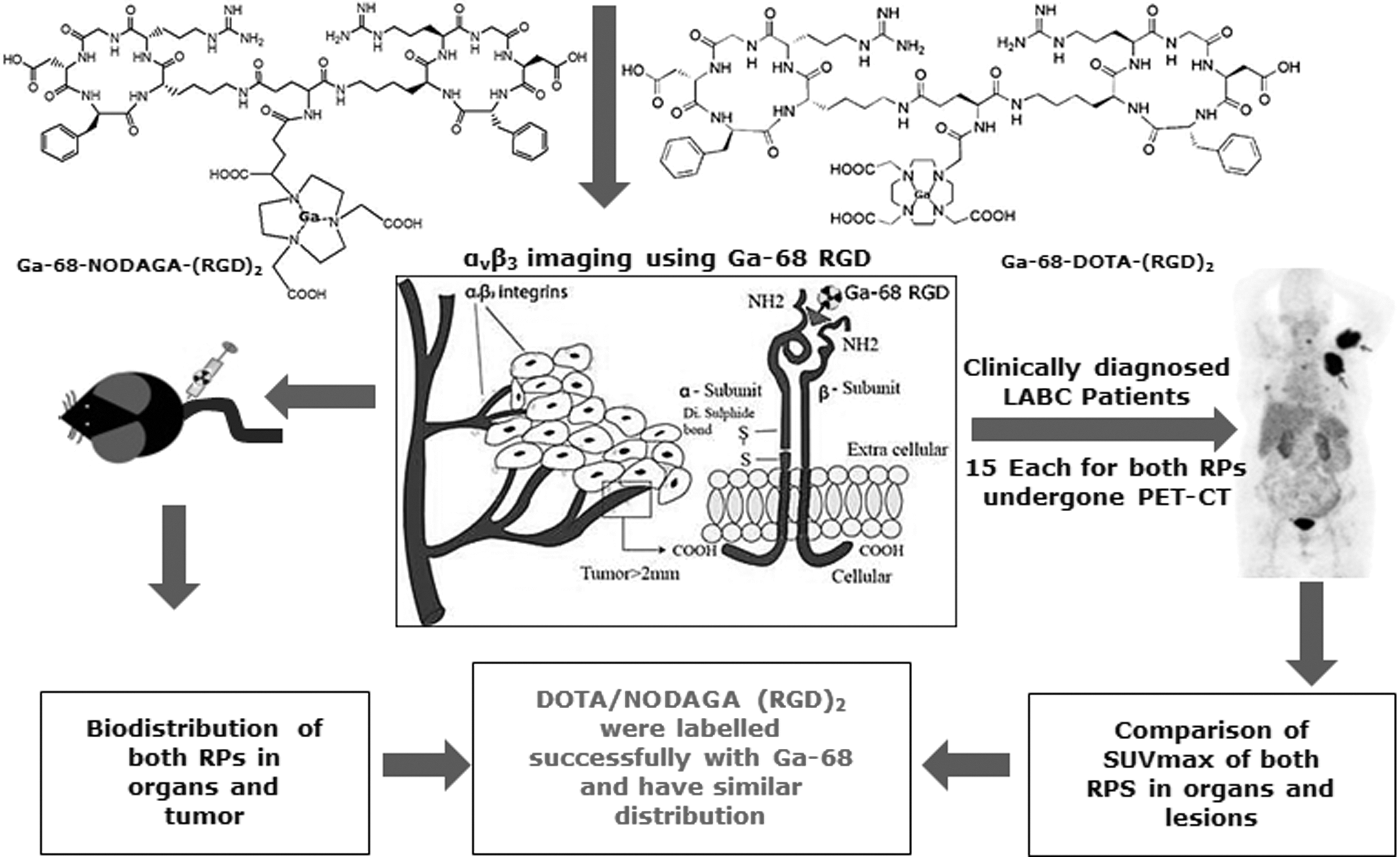
Schematic diagram representing the outline of the study protocol.
Materials and Methods
Ga-68 was obtained from a commercially available Ge-68/Ga-68 generator (ITG, Germany) for optimization of radiotracers and clinical studies thereof. Preclinical studies were performed using an in-house-developed Ge-68/Ga-68 generator with CeO2-PAN as column matrix. 29 Radiolabeling was done in a semi-automated fluidic labeling module (ITG). DOTA-E[c(RGDfK)]2—that is, DOTA-(RGD)2 (E = glutamic acid, f = phenylalanine, K = lysine, R = arginine, G = glycine, D = aspartic acid)—and NODAGA-E-[c(RGDfK)]2—that is, NODAGA-(RGD)2—were purchased from ABX Advanced Biochemical Compounds, Germany. The other chemicals and reagents used were also of analytical grade.
Radiolabeling of DOTA- and NODAGA-conjugated cyclic RGD2
DOTA- and NODAGA-conjugated dimeric cyclic RGD peptides were dissolved in water (1 mg/mL) and stored at −20°C. Ga-68 was eluted from a 999 MBq (27 mCi) Ge-68/Ga-68 generator. Purification of the elute before radiolabeling was not required. Various reaction parameters studied during the optimization include elution volume (2–4 mL), peptide amount (10–50 μg), reaction pH (3–5), and incubation time (5–20 min). The radiolabeling of DOTA-(RGD)2 and NODAGA-(RGD)2 with Ga-68 was performed at 95°C as well as at room temperature.
Purification and characterization
Purification of both the radiolabeled products was done with a preconditioned C-18 Sep-pak cartridge (Waters). The characterization of purified radiolabeled samples was done by matrix-assisted laser desorption ionization-time of flight (MALDI-TOF) mass spectrometry (Ultraflextrem; Brukers Daltonics, Germany) and high-performance liquid chromatography (HPLC). For MALDI-TOF, 3–5 μL of the samples dissolved in 0.01% trifluoroacetic acid (TFA) was spotted onto the ground steel plate using cyano-4-hydroxycinnamic acid as a matrix. Nitrogen laser was used to irradiate the samples. For HPLC, water (A) and acetonitrile (B) with 0.1% TFA were used as the mobile phase. Separation was done using the gradient elution technique (0–4 min 95% A, 4–15 min 95% A to 5% A, 15–20 min 5% A, 20–25 min 5% A to 95% A, 25–30 min 95% A). A dual-pump HPLC unit equipped with a C-18 reversed phase HiQ-Sil (5 μM, 25 × 0.46 cm) column was used to maintain the flow rate of 1 mL/min. Signals corresponding to UV 270 nm and radioactivity using the NaI(TI) detector were monitored to record the elution time of the product from the column.
Quality control
Quality control tests were performed according to the procedure described earlier. 24 Briefly, radiolabeling efficiency and radiochemical purity of the labeled product was determined by instant thin-layer chromatography (TLC) using 0.5 M sodium citrate solution (pH 5.5) and NH4OAc:MeOH (1:1) solution as the mobile phases. The silica gel-coated strips were read with a TLC scanner (EZ-SCAN TLC Scanner; Berkeley). Sterility testing was done by incubating the radiolabeled sample volume equivalent to the injection volume (2 mL) in tryptic soya broth medium (10 mL) at 37°C. Turbidity in broth was observed up to 7 d for the presence of any growth. Pyrogen testing was done using a point-of-use portable test system (PTS; Charles River). To determine the endotoxin contents, 25 μL of the sample was added to each well in the PTS cassette and was incubated at 38°C for 15–20 min. The observations were noted with 50%–200% spike recovery with sensitivity of 0.01 endotoxin unit (EU) per milliliter. Gas chromatography (Varian 3500) was used to measure the residual ethanol content in the product. On the basis of area under the curve of ethanol standard (4000 ppm), the concentration of ethanol in the product was calculated. The stability of both the compounds was checked by incubating the radiolabeled samples with phosphate-buffered saline (PBS) at room temperature for up to 6 h.
Biodistribution studies
The avidity of RGD peptide for αvβ3 integrins was evaluated in C57BL/6 mice bearing melanoma. All the animal experiments were carried out in strict compliance with the national laws relating to the conduct of animal experimentation after taking ethical clearance from the Institutional Animal Ethics Committee. Melanoma cells (ATCC® CRL-6475™) were cultured in Dulbecco's modified Eagle's medium, supplemented with 10% fetal bovine serum. The culture was incubated at 37°C in 5% CO2 environment. Melanoma cells were suspended in 200 μL PBS and were used for the tumor build-up. Tumor cells (∼1 × 10⁶) were injected into the right thigh of each C57BL/6 mice (weighing 20–25 g) subcutaneously. Animals were allowed to grow till 0.2–0.4 g of tumor mass was attained (2–3 weeks). Through the tail vein, 3.7–5.5 MBq (100–150 μCi) of Ga-68-DOTA-(RGD)2 and Ga-68-NODAGA-(RGD)2 was injected. Animals were sacrificed post anesthesia by cervical death at 15, 30, and 60 min post intravenous (i.v.) administration. Three animals were scarified at each point of time. The percent radioactivity associated with per gram of various organs and tumor (% ID/g) was computed.
To confirm that the uptake of RGD peptide in tumor cells was receptor-specific, blocking studies were performed by administering 500 μg (20–25 mg/kg of body weight) of cold DOTA-(RGD)2 and NODAGA-(RGD)2 along with 100 μCi of Ga-68-DOTA-(RGD)2 and Ga-68-NODAGA-(RGD)2 in the C57BL/6 mice (n = 3, each radiotracer) bearing melanoma. All animals were sacrificed at 30 min post injection (p.i.) of the radiotracers. % ID/g associated with the tumor and various tissues in the absence and presence of excess DOTA-(RGD)2 and NODAGA-(RGD)2 peptides was compared.
Patient imaging
Patient investigations were duly approved by the institutional ethics committee (Ref. No. Histopath/13/NK/2421, dated December 8, 2013). Written informed consent was also obtained from all the patients included in this study. A total of 30 female patients [n = 15, each for Ga-68-DOTA-(RGD)2 and Ga-68-NODAGA-(RGD)2] were enrolled in the study. All the included patients were clinically diagnosed to have LABC. Imaging protocol optimized in the previous study was used. 24 Whole-body images from base of skull to mid-thigh were acquired with a dedicated PET-computed tomography (CT) system (Discovery 710; GE Health Care, Milwaukee) 45 min post i.v. administration of 111–185 MBq (3–5 mCi) of Ga-68 DOTA-(RGD)2 and Ga-68 NODAGA-(RGD)2. After reconstruction, all scans were independently reviewed by a certified nuclear medicine physician who was blinded to the patient's history, diagnosis, and findings on other imaging studies. The uptake of Ga-68-DOTA-(RGD)2 and Ga-68-NODAGA-(RGD)2 was assessed from a circular region of interest over the entire lesion/lesions and expressed as the maximum standardized uptake value (SUVmax). The mean SUVmax values were calculated for blood pool, brain ventricles, thyroid, lung, liver, spleen, kidneys, intestine, and the lesion for Ga-68-DOTA-(RGD)2 and Ga-68-NODAGA-(RGD)2. Primary tumor to liver and blood pool ratios were calculated by dividing the primary tumor SUVmax with liver SUVmax and blood pool SUV max values, respectively, and then the mean of these values was calculated.
Results
Radiolabeling of DOTA- and NODAGA-conjugated cyclic RGD2
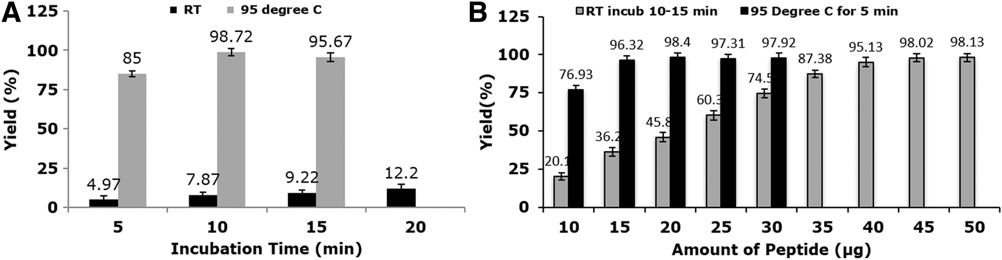
Both DOTA-(RGD)2 and NODAGA-(RGD)2 peptides were labeled with Ga-68 with >95% radiolabeling yield and >99% radiochemical purity. The optimal peptide amount, pH, and elution volume for both were 15–20 μg, 3.5–4.0, and 3 mL, respectively. Heating of the reaction mixture at 95°C for 10 min was required for radiolabeling of DOTA-(RGD)2 and 5 min for NODAGA-(RGD)2 as shown in Figure 2 . Under these conditions, the maximum radiolabeling yield observed was >95% with both the chelators. NODAGA-(RGD)2 had also been radiolabeled with Ga-68 at room temperature. However, for achieving radiolabeling yield >95% at room temperature, 40–50 μg of peptide [NODAGA-(RGD)2] amount was required (Fig. 2B).
Purification and characterization
MALDI-TOF mass spectra of Ga-68-DOTA-(RGD)2 and Ga-68-NODAGA-(RGD)2 showed an increased mass near the weight of Ga-68 (1771 and 1776, respectively) compared with the pure compound mass (1704 and 1708, respectively), conforming good attachment as shown in Figure 3A and B. The radio-HPLC of both radiolabeled products showed a retention time of 15.6 min. Free Ga-68, if present, was eluted in void volume (Fig. 3C, D).
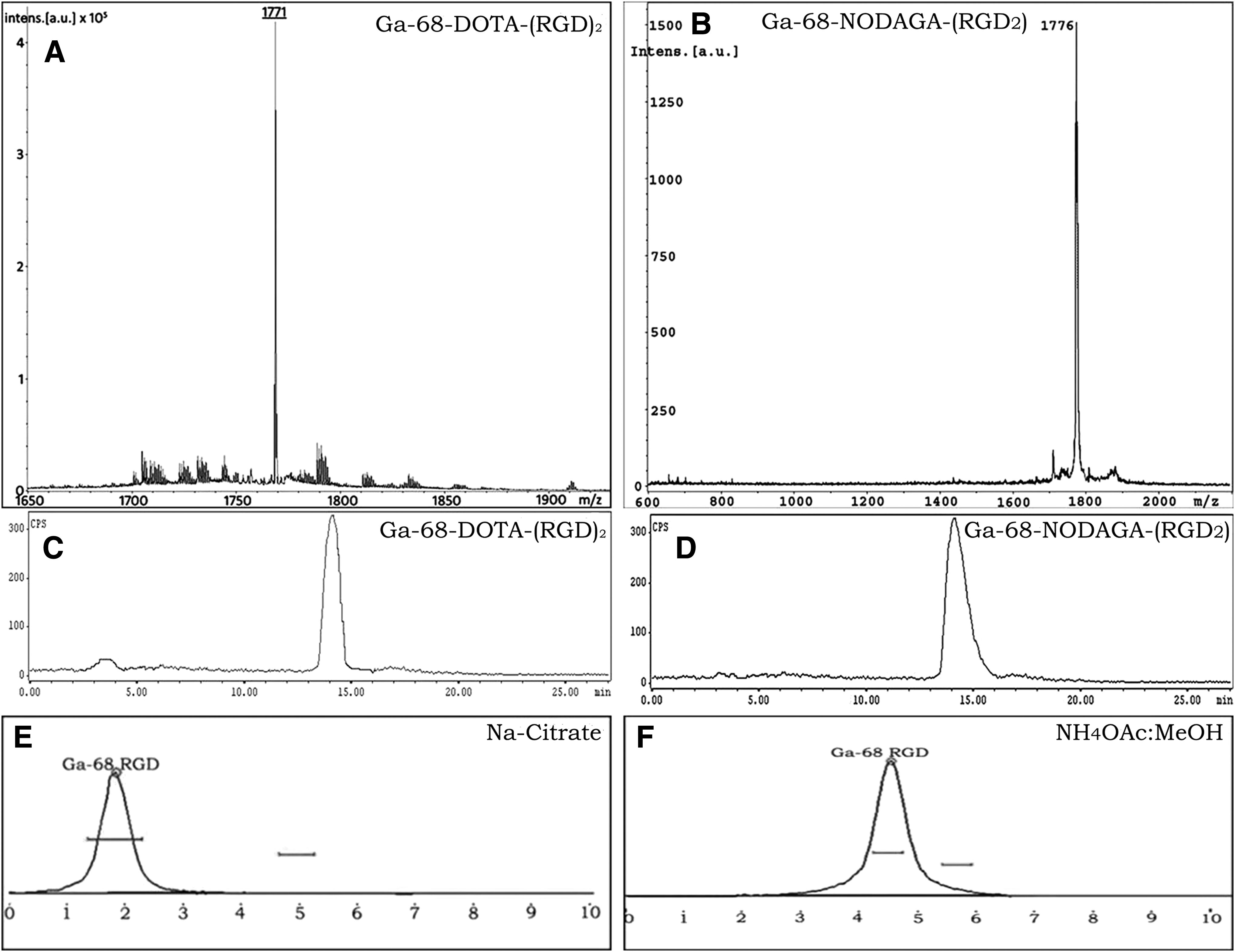
Matrix-assisted laser desorption ionization-time of flight mass spectra
Quality control
The radionuclide and radiochemical purity of Ga-68-DOTA-(RGD)2 and Ga-68-NODAGA-(RGD)2 was >99% after C-18 cartridge purification (Rf = 0.1–0.2 in sodium citrate and 0.4–0.5 in NH4OAc:MeOH as shown in Fig. 3E, F). The samples were sterile as no growth was observed for up to 7 d of incubation. The endotoxin content was <4 EU/mL (1.2–3.7 EU/mL; normal level 175 EU/mL). The residual ethanol content was between 1993 and 2490 ppm in all samples. Both compounds were found to be stable at room temperature for up to 6 h.
Biodistribution studies
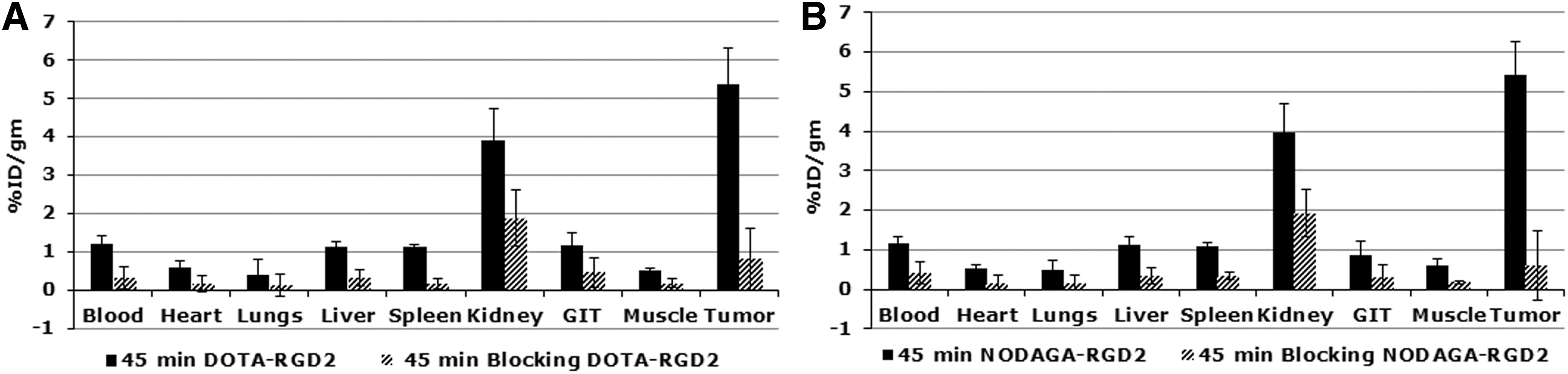
Biodistribution studies were done at 15, 30, and 60 min post i.v. administration of 3.7 MBq (100 μCi) of Ga-68-DOTA-(RGD)2 and Ga-68-NODAGA-(RGD)2. Significant tumor counts were observed at 15 min p.i., which increased with time as noted at 30 min. However, at 60 min p.i. of the radiotracer, the counts were observed to have decreased. Both radiotracers exhibited a rapid blood pool clearance. Among the non-targeted organs, maximum counts were observed in the liver and spleen. A small amount of radioactivity was also noted in the intestine and lungs (Table 1).
Percent Radioactivity Associated with Per Gram (% ID/g) of Various Organs and Tumor Post Administration of 3.7 MBq (100 μCi) Ga-68-DOTA-(RGD)2 and Ga-68-NODAGA-(RGD)2, Respectively, at Different Time Intervals
Ga-68, Gallium-68; DOTA, 1,4,7,10-tetraazacyclododecane-1,4,7,10-tetraacetic acid; NODAGA, 1,4,7-triazacyclononane,1-gluteric acid-4,7-acetic acid; RGD, arginine–glycine–aspartic acid.
Both Ga-68-DOTA-(RGD)2 and Ga-68-NODAGA-(RGD)2 were predominately excreted through the kidneys. The uptake of Ga-68-DOTA-(RGD)2 and Ga-68-NODAGA-(RGD)2 in the presence and absence of excess cold RGD peptide was also studied. A significant reduction in tumor counts was noted for both radiotracers after co-injection of excess cold RGD peptide at 30 min as shown in Figure 4.
Effect of co-injection of cold
Patient imaging
A total of 30 female patients with a mean age of 50 years (range 24–73 years) were included in the study. Out of these patients, the estrogen receptors (ERs) were found to be positive in 4 patients; 8 patients each were Her2-positive and triple positive (ER/progesterone receptor [PR]/Her 2); and 7 patients were triple negative. Receptor status could not be determined in 3 patients. The physiological uptake of both the tracers was noted in bilateral brain ventricles, thyroid, liver, spleen, and salivary glands. The chief organ for the excretion of these tracers was the kidneys. Variable tracer uptake was noted in the small intestine at 45 min post i.v. tracer administration. Good tumor to background ratio was seen with both the tracers along the primary lesions (Figs. 5 and 6). Out of 30 patients, 5 patients [n = 3 and 2 for Ga-68-DOTA-(RGD)2 and Ga-68-NODAGA-(RGD)2, respectively], who were clinically diagnosed with LABC, were found to be metastatic on RGD PET-CT scan (Fig. 5).
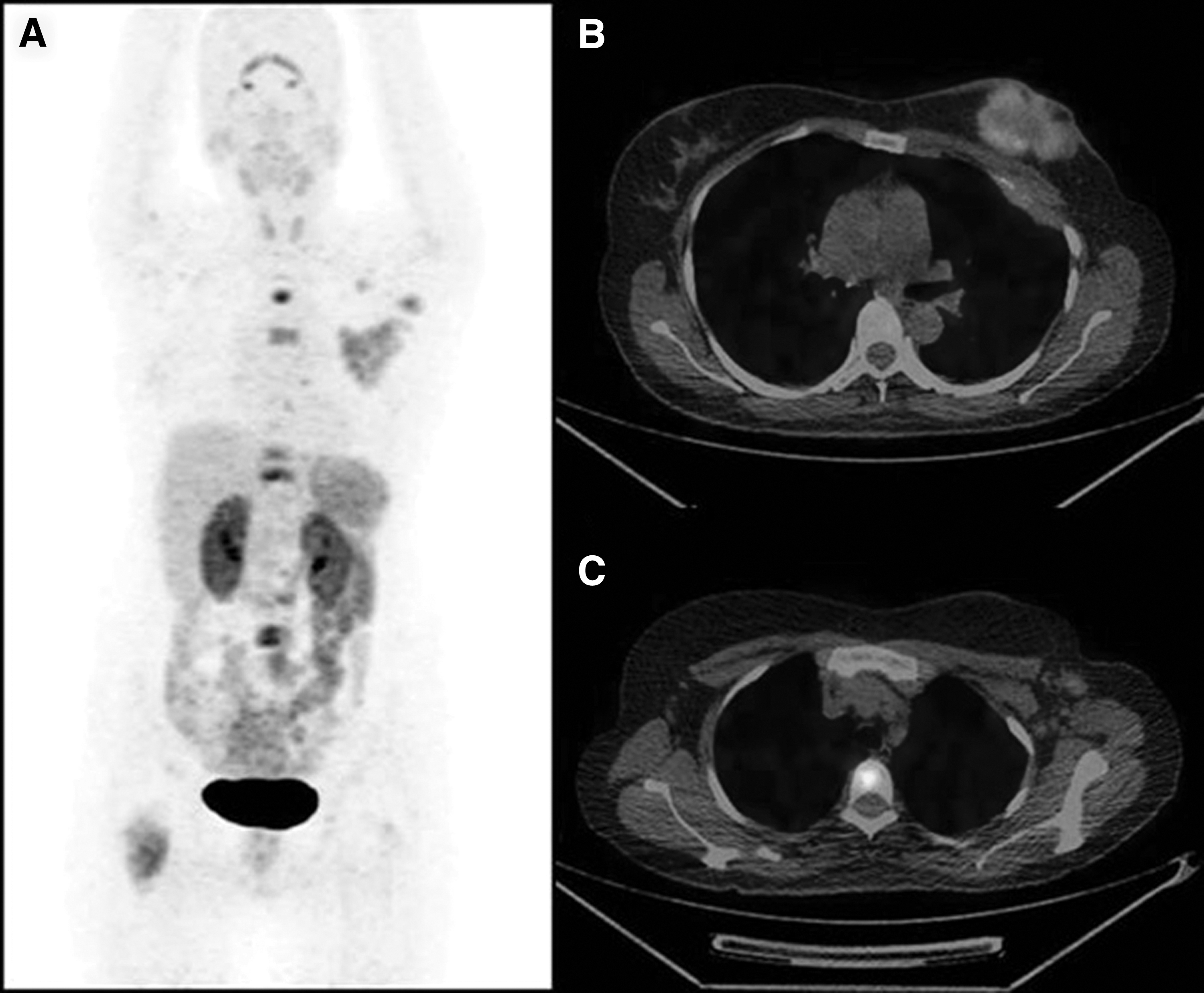
MIP PET image

MIP PET image
Both the tracers picked the primary lesion in 100% of the patients with a mean SUVmax of 6.15 (1.54–19.34) for Ga-68-DOTA-(RGD)2 and 5.9 (2.78–10.76) for Ga-68-NODAGA-(RGD)2. The mean SUVmax values for various organs and lesions are shown in Figure 5. The mean value of SUVmax ratio for the primary tumor to the liver for Ga-68-DOTA-(RGD)2 versus Ga-68-NODAGA-(RGD)2 was 1.73 versus 1.49, and for the primary tumor to the blood pool, the SUVmax ratio was 3.93 versus 5.00. No adverse reactions were noted in any of the patients with either radiotracer.
Discussion
BFCs play an important role in the labeling chemistry of Ga-68. The present study compared the effect of macrocyclic chelators DOTA and NODAGA attached to the cyclic RGD dimer on radiolabeling and biodistribution in mice and in patients' breast carcinoma.
In contrast to pre-concentration, Ga-68 was used directly for radiolabeling as eluted from the generator. 6,30 –33 Only 6–11 nmol (10–20 μg) of DOTA-(RGD)2 and NODAGA-(RGD)2 peptides was used for labeling with 8–25 mCi of Ga-68. A maximum of 98% radiolabeling yield was obtained under the mentioned optimum conditions. However, in earlier studies, 1–50 nmol of the peptide reportedly gave a 74%–97% yield. 16 –18,30 –32 Also, the amount of Ga-68 that was eluted decreased as the generator became older, and the same was also experienced by us. Depending upon the activity eluted, the peptide amount can be optimized. Along with averting receptor saturation, this attuned approach can reduce the cost of production of labeled RGD.
Depending upon the nature of BFCs, the RGD peptide can be radiolabeled with Ga-68 at varied temperatures. 15 –18,30,31,34 In this study, by incubating the reaction mixture at room temperature, the maximum yield achieved for Ga-68-DOTA-(RGD)2 was only 12%. However, >95% radiolabeling yield was achieved by heating the reaction mixture at 95°C for 10 min. In a similar study done by Dijkgraaf et al., 7 incubation of 20 min at 95°C was recommended for obtaining 93%–95% radiolabeling yield for Ga-68-DOTA-(RGD)2. 6 Whereas, for NODAGA-(RGD)2 to achieve a radiolabeling yield >95%, an increased amount of peptide (40–45 μg, 23–26 nmol) was used where the reaction mixture was incubated at room temperature for 10–15 min. However, a reduced amount of peptide (15–20 μg, 8–11 nmol) was observed with heating of the reaction mixture at 95°C for 5 min. Oxboel et al., 18 in another study, recommended 20 nmol of the peptide for a radiolabeling yield >95% at room temperature. 7 In the present study, the heating protocol was found to be better than incubation at room temperature as it not only decreased the preparation time but also increased the labeling yield even with a low peptide concentration (8–11 nmol).
In the present study biodistribution studies were performed on C57BL/6 mice bearing melanoma. Counts were observed in the blood, lungs, kidneys, liver, and gastrointestinal tract, indicating that physiological uptake decreased gradually with time at 30 and 60 min. The results were found to be in concordance with other biodistribution studies reported earlier in animals bearing glioma, melanoma, breast, and lung tumor xenografts. 16,18,30 –32,34,35 % ID/g associated with the tumor was 5.13 at 30 min, which increased to 5.37 at 45 min. The size of the tumor varied from 0.2 to 0.4 g, but no significant difference was noted in the activity (% ID/g) of tumors. However, a reduced uptake was reported in earlier studies due to the presence of necrotic or hypoxic areas in large tumors. 33,36 Sharp decrease of radiotracers counts in tumor during the blocking study revealed that the uptake of Ga-68-DOTA/NODAGA-(RGD)2 was receptor-specific. No significant difference was observed in the biodistribution characteristics of Ga-68-labeled DOTA and NODAGA-chelated (RGD)2 peptide due to the specificity of RGD to integrin αvβ3 irrespective of the BFC attached. Cold RGD was given ∼25 times (w/w) of radiolabeled peptide; hence there was a competition between radiolabeled and cold RGD. The bowmen capsule, proximal, and distal tubules were reported to express αvβ3. 37 The reduced counts of both radiotracers were observed after co-injection of cold RGD peptide. Low counts in the kidneys may probably be because of saturation of these receptors.
In patient studies, the uptake of both the tracers was independent of ER/PR/Her2 receptor expression. Ga-68 DOTA complexes showed a low stability constant compared with NOTA and its derivatives (Kd 21.3 vs. 31.0); however, as is evident from Figure 7, no significant difference was observed between the mean SUVmax values of various organs. Our results showed that both these tracers can be used in angiogenesis imaging with equal efficacy to achieve good-quality images that may be helpful in further staging and management of patients. In future, these tracers may play a vital role in the selection of patients and response assessment to anti-angiogenesis therapy and may bring a ray of hope to patients who did not respond to available treatment options, by replacing Ga-68 with therapeutic Lutetium-177 or Yttrium-90. 38 However, further evaluation is needed with more prospective studies and a larger patient population.
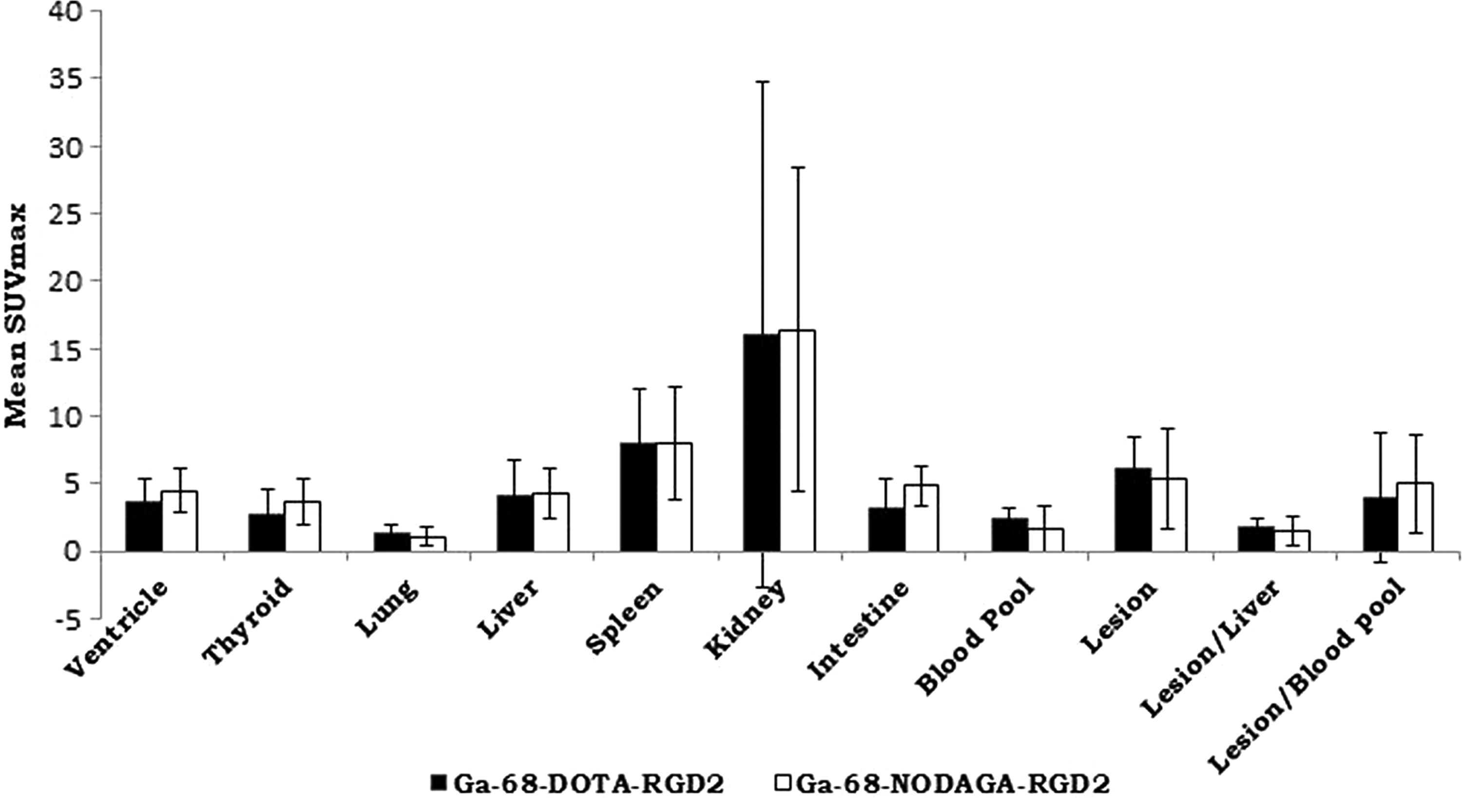
Mean SUVmax values of various organs and primary lesions in LABC patients at 45 min post injection of 111–185 MBq (3–5 mCi) Ga-68-DOTA-(RGD)2 and Ga-68-NODAGA-(RGD2), respectively.
Conclusion
Both DOTA- and NODAGA-conjugated RGD peptides were efficaciously radiolabeled with Ga-68. Preclinical studies with C57BL/6 mice and preliminary clinical investigations in patients with LABC, suggested that Ga-68-labeled DOTA and NODAGA-conjugated RGD2 peptides have the potential to be a promising agent for non-invasive molecular imaging of neo-angiogenesis in tumors that overexpress αvβ3 integrins.
Footnotes
Acknowledgement
The authors S.C. and A.D. sincerely acknowledge the valuable contribution of Dr. H.D. Sarma, Radiation Biology and Health Sciences Division, Bhabha Atomic Research Centre, Mumbai, in conducting animal studies.
Authors Contributions
The role of each author was as follows: R.V.: radiolabeling optimization, QC and preparation of radiopharmaceuticals, and article writing; J.S.: design of the study and article revision; S.K.: conduction of PET studies and reporting; S.C.: study design, radiochemistry and animal experimentation; A.D.: study design, radiochemistry and animal experimentation; G.S.: surgical oncologist providing primary treatment and patient referrals; B.R.M.: design of the study, patient reporting, and article editing. All co-authors have reviewed and approved the article before submission.
Disclosure Statement
There are no existing financial conflicts.