
Abstract
Background:
Long noncoding RNA (lncRNA) GATA6-AS regulates the growth of endothelial cells, indicating its involvement in human diseases. The present study aimed to investigate the roles of GATA6-AS in glioma.
Results:
GATA6-AS was significantly upregulated in glioma. GATA6-AS levels in plasma was positively and significantly correlated with its expression levels in glioma tissues. TUG1 was significantly downregulated in glioma and was inversely correlated with GATA6-AS. GATA6-AS overexpression led to TUG1 downregulation, whereas TUG1 overexpression failed to significantly affect GATA6-AS. GATA6-AS overexpression promoted glioma cell proliferation and inhibited apoptosis, TUG1 overexpression inhibited cell proliferation and attenuated the effects of GATA6-AS overexpression.
Conclusions:
Therefore, lncRNA GATA6-AS affects cell proliferation and apoptosis and regulates TUG1 expression in glioma.
Introduction
Glioma is the most common type of malignancy developed from the central nervous system. 1 Although the incidence of glioma is low and only 5 out of 100,000 will be affected by this disease during their life time, glioma poses a severe threat to public health due to its extremely aggressive nature and unacceptable high mortality rate. 2 At present, the median survival time of glioma patients after conventional treatments is still below 15 months. 3 It is believed that the development and progression of glioma require the involvement of a complex molecular modulation and gene interaction network. 4,5 However, molecular mechanism of glioma is still largely unknown, resulting in difficulties in clinical treatment and prevention.
Genome-wide gene expression analysis has shown that the protein-coding transcripts only account for less than 2% of all transcriptions. 6,7 It is suggested that most human genes actually transcribe noncoding RNAs (ncRNAs), which should be included to explain the complexity of human gene expression in evolutional and physiological aspects. 8 Long (>200 nt) ncRNAs (lncRNAs) have been recognized as critical determinants in human diseases, 9 such as different types of cancer. 10 LncRNA GATA6-AS regulates the growth of endothelial cells, 11 and the abnormal growth of endothelial cells is involved in many types of diseases, including glioma. 12 This study therefore was carried out to investigate the involvement of GATA6-AS in glioma.
Materials and Methods
Research subjects
This study included 58 patients who were diagnosed in PLA Rocket Force General Hospital from May 2015 to April 2018. All patients were subjected to pathological examinations, which were performed by three experienced pathologists. Inclusion criteria: (1) glioma patients with the first diagnosis; (2) patients with no treatment performed before admission; (3) patients willing to participate in this study and signed informed consent. Exclusion criteria: (1) patients who received any treatments before admission; (2) patients complicated with other diseases. There are 18 cases of grade I, 16 cases of grade II, 14 cases of grade III, and 10 cases of grade IV. The 58 patients included 32 males and 26 females, and age ranged from 36 to 68 years, with a mean age of 50.5 ± 5.6 years. This study was approved by the Ethics Committee of the aforementioned hospital before the admission of patients.
Specimens and cell lines
Fasting blood (5 mL) was extracted from each participant before treatments. Blood was transferred to ethylenediamine tetraacetic acid (EDTA) tubes and centrifuged at 1200 g for 10 min to collect plasma samples.
Tumor tissues and tumor adjacent normal tissues were collected from each participant through biopsy. All tissues were confirmed by three experienced pathologists.
Our study included 2 human glioma cell lines, Hs 683 and CCD-25Lu (ATCC). Eagle's Minimum Essential Medium (10% fetal bovine serum [FBS]) was used as cell culture medium and cell culture conditions were 37°C and 5% CO2.
RT-quantitative polymerase chain reaction
TRIzol reagent (Invitrogen) was mixed with tissue powders (ground in liquid nitrogen), plasma, and in vitro cultivated cells to extract total RNAs. Following DNase I digestion, SuperScript IV Reverse Transcriptase (Thermo Fisher Scientific, Inc.) was used to synthesize cDNA through reverse transcription. To detect the expression of GATA6-AS and TUG1, polymerase chain reaction (PCR) systems were prepared using the SYBR® Green Quantitative RT-qPCR Kit (Sigma-Aldrich, St. Louis, MO), and all PCRs were performed with 18S as endogenous control. All data normalizations were performed according to 2−ΔΔCT method.
Cell transfection
GATA6-AS and TUG1 expression pcDNA3.1 vectors were constructed by Sangon (Shanghai, China). Cells of Hs 683 and CCD-25Lu cell lines were cultivated at confluence of 70%–80%, followed by cell transfections performed using Lipofectamine 2000 Transfection Reagent (Thermo Fisher Scientific, Inc.) with vectors at a concentration of 10 nM. Control and negative control cells were untransfected cells and cells transfected with empty vectors, respectively. Cells were harvested at 24 h after transfection for subsequent experiments (see Figure 1).

GATA6-AS was significantly upregulated in glioma tissues. RT-quantitative polymerase chain reaction results showed that GATA6-AS expression was significantly upregulated in glioma tissues than in normal healthy tissues (*p < 0.05).
In vitro cell proliferation assay
Cells were harvested at 24 h after transfection. Cells were mixed with Eagle's Minimum Essential Medium (10% FBS) at a ratio of 4 × 10⁴ cells per 1 mL cell culture medium to prepare a single cell suspension, and each well of a 96-well plate was filled with 100 μL cell suspension. Cells were cultivated in an incubator (37°C, 5% CO2), and CCK-8 solution (10 μL) was added into each well every 24 h until 94 h. After that, cell culture was performed for additional 4 h, followed by the addition of 10 μL dimethyl sulfoxide (DMSO). OD values at 450 nM were measured.
Cell apoptosis assay
Cells were harvested at 24 h after transfection, and 6 × 10⁴ cells were mixed with Eagle's Minimum Essential Medium (1% FBS) to prepare single cell suspensions. Each well of a six-well plate was filled with 2 mL cell suspension, followed by cell culture for 48 h. After that, cells were subjected to 0.25% trypsin digestion. Finally, propidium iodide (Dojindo, Japan) and Annexin V-FITC (Dojindo) staining was performed and flow cytometry was carried out to detect apoptotic cells.
Statistical analyses
Mean ± standard deviation was calculated using data from three biological replicates. All statistical analyses were performed using GraphPad Prism 6 software. Comparisons between normal and tumor tissues were performed by paired t-test. Differences among multiple cell treatment groups were explored by analysis of variance (ANOVA) (one-way) and Tukey's test. Correlation analysis was performed using linear regression. Univariate analysis was performed to analyze the correlations between clinical factors and GATA6 expression. p < 0.05 indicated a difference with statistical significance.
Results
GATA6-AS was significantly upregulated in glioma tissues
RT-quantitative PCR (RT-qPCR) was performed to detect the differential expression of GATA6-AS in glioma and adjacent normal tissues of 58 glioma patients. Compared with normal tissues, significantly upregulated expression of GATA6-AS was observed in glioma tissues, indicating the involvement of this lncRNA in glioma. Univariate analysis found that age (> or ≤50 years), gender, and tumor stages have no significant effects on GATA6-AS expression. Risk ratios were 0.812, 1.428, and 1.232, respectively; 95% confidence intervals were 0.555–1.768, 0.711–2.020, and 0.508–2.545, respectively; p-values were 0.343, 0.888, and 0.596, respectively.
Plasma levels of GATA6-AS were correlated with its expression levels in glioma tissues
RT-qPCR was also performed to detect GATA6-AS in plasma. Linear regression was performed to investigate the correlations between plasma levels of GATA6-AS and its expression in glioma or normal tissues. It was observed that plasma levels of GATA6-AS were significantly and positively correlated with its expression levels in glioma tissues (Fig. 2A), but not in adjacent normal tissues (Fig. 2B).
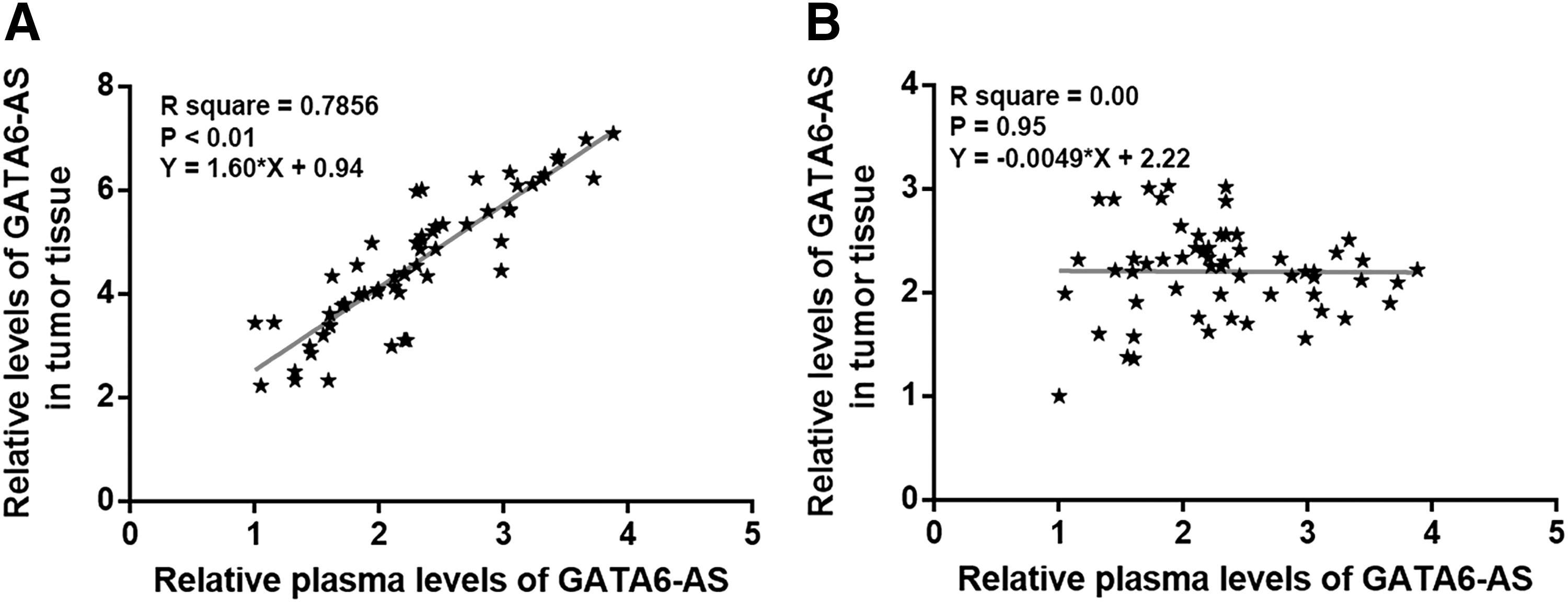
Plasma levels of GATA6-AS were correlated with its expression levels in glioma tissues. Linear regression analysis showed that plasma levels of GATA6-AS were significantly and positively correlated with its expression levels in glioma tissues
TUG1 was downregulated in glioma and inversely correlated with GATA6-AS
Differential expression of TUG1 in glioma and adjacent normal tissues of 58 glioma patients was detected by RT-qPCR. Compared with normal tissues, TUG1 was significantly downregulated in glioma tissues (Fig. 3A, p < 0.05). Linear regression was performed to investigate the correlations between expression levels of GATA6-AS and TUG1. It was observed that expression levels of TUG1 were inversely correlated with expression levels of GATA6-AS only in glioma tissues (Fig. 3B), but not in adjacent healthy tissues (Fig. 3C).
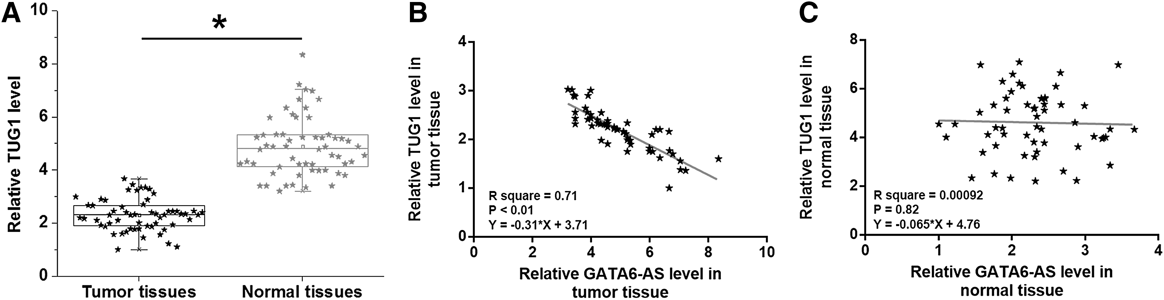
TUG1 was downregulated in glioma and inversely correlated with GATA6-AS. TUG1 was significantly downregulated in glioma tissues than in adjacent normal tissues
GATA6-AS is an upstream inhibitor of TUG1 in glioma cells
Cells of Hs 683 and CCD-25Lu cell lines were transfected with GATA6-AS and TUG1 expression vectors to explore the possible interactions between GATA6-AS and TUG1. Overexpression of GATA6-AS and TUG1 was reached in cells of both cell lines at 24 h after transfection (Fig. 4A, p < 0.05). Compared with untransfection (control [C]) cells and cell transfected with empty vectors (negative control [NC]), GATA6-AS overexpression led to the downregulation of TUG1 (Fig. 4B, p < 0.05), whereas TUG1 overexpression failed to significantly affect GATA6-AS (Fig. 4C).
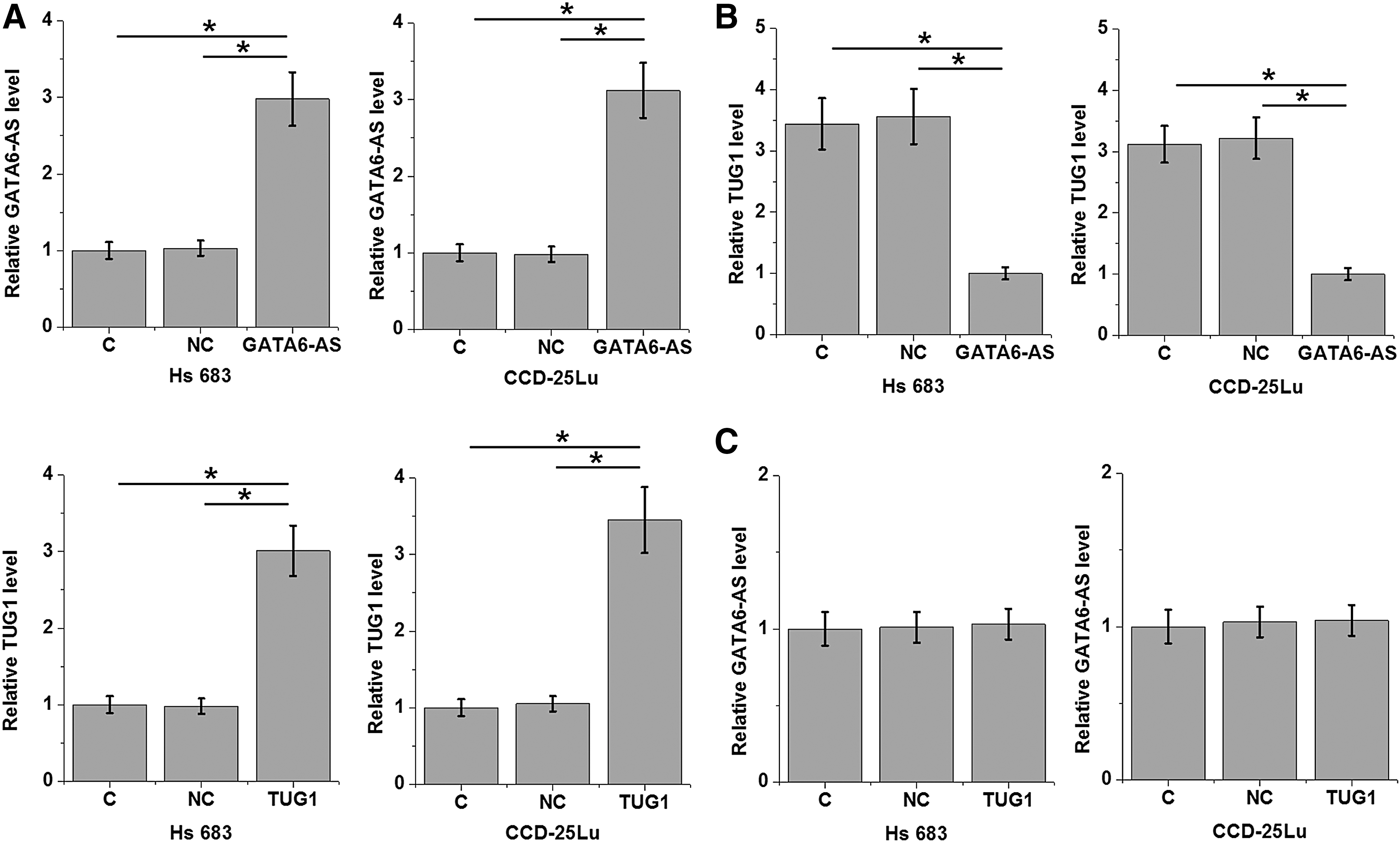
GATA6-AS is an upstream inhibitor of TUG1 in glioma cells. Overexpression of GATA6-AS and TUG1 was reached in cells of both cell lines at 24 h after transfection
GATA6-AS regulated glioma cell proliferation and apoptosis through TUG1
Results of in vitro cell proliferation and apoptosis assay showed that, compared with control (C) and negative control (NC) groups, GATA6-AS overexpression led to promoted proliferation (Fig. 5A) and inhibited apoptosis (Fig. 5B) of cells of both Hs 683 and CCD-25Lu cell lines (p < 0.05), TUG1 overexpression played an opposite role, and attenuated the effects of GATA6-AS overexpression.
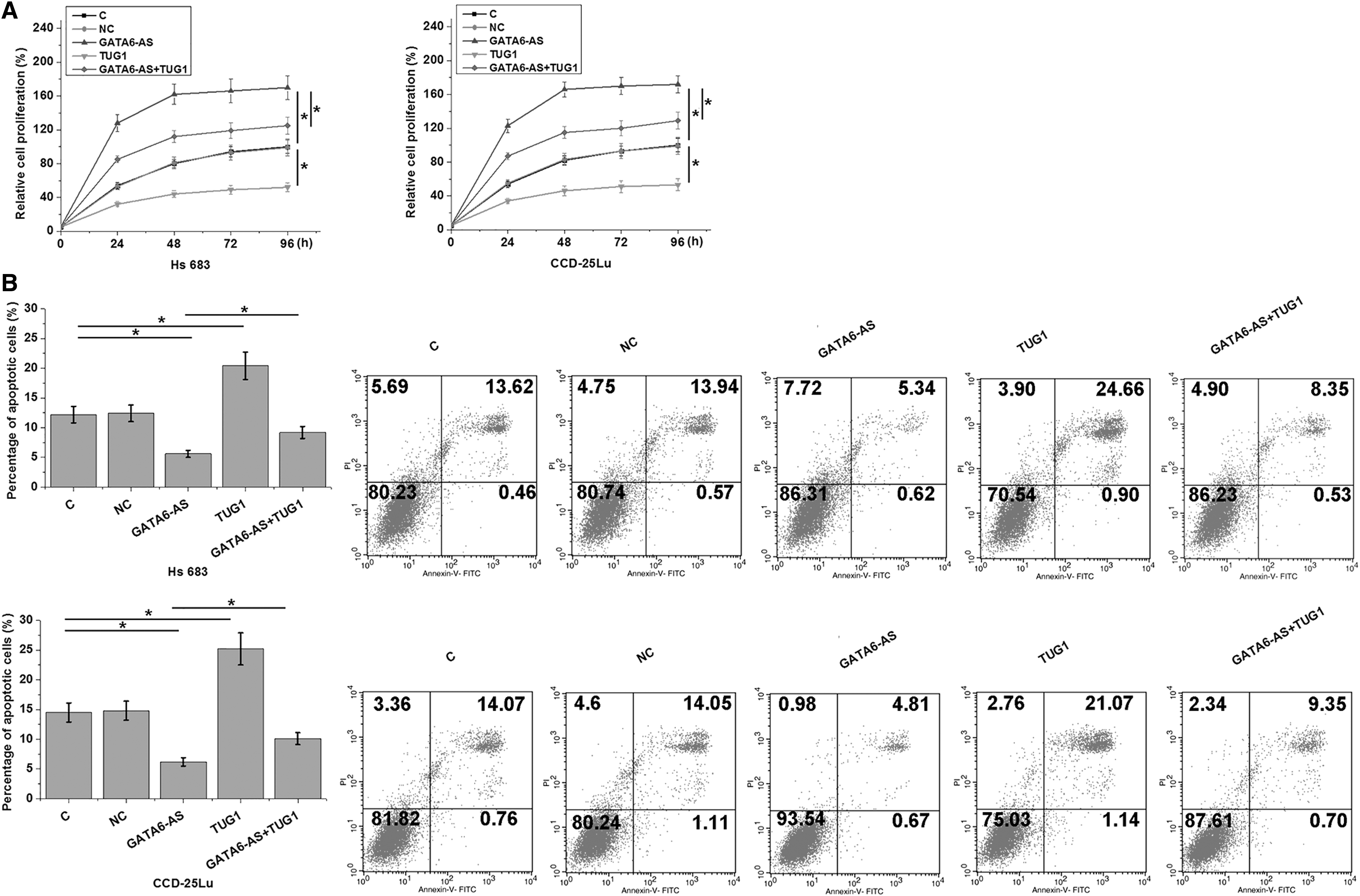
GATA6-AS regulated glioma cell proliferation and apoptosis through TUG1. GATA6-AS overexpression led to promoted proliferation
Discussion
GATA6-AS regulates the growth of endothelial cells, 11 which play critical roles in the development of different types of human diseases. However, the involvement of GATA6-AS in glioma is still unknown. This study first reported the oncogenic function of GATA6-AS in glioma.
The preliminary RNA-seq data have showed that lncRNA TUG1 was downregulated in glioma and is inversely correlated with GATA6-AS (data). Therefore, the present study explored the interactions between GATA6-AS and TUG1 in glioma. TUG1 plays different roles in different types of cancer. In the development of intrahepatic cholangiocarcinoma, TUG1 was upregulated and the overexpression of TUG1 promoted cancer development by regulating glutamine metabolism through Sirt3/GDH axis. 13 In contrast, TUG1 was downregulated in glioma and promotes the apoptosis of glioma cells, resulting in inhibited cancer progression. 14 This study also observed the downregulated TUG1 in glioma tissues compared with adjacent normal tissues. In addition, TUG1 not only promoted the apoptosis of glioma cell apoptosis but also inhibited cancer cell proliferation. This study further confirmed the tumor suppression role of TUG1 in glioma and enriched the authors' understanding on the role of TUG1 in glioma.
This study first reported the upregulation of GATA6-AS in glioma. LncRNAs are usually expressed in specific types of cells or tissues to regulate the expression of downstream genes at multiple levels, indicating the involvement of lncRNAs in specific cellular processes. 15,16 However, certain lncRNAs may be released into the circulating system to achieve systemic regulation of gene expression. 17 This study detected the GATA6-AS in all glioma patients. In addition, plasma levels of GATA6-AS were significantly and positively correlated with its expression levels in glioma tissues, but not in adjacent normal tissues. Therefore, GATA6-AS synthesized in tumor tissues can enter blood, indicating that GATA6-AS may be a signaling molecular mechanism for systemic gene expression regulation.
This study proved that GATA6-AS is likely an upstream inhibitor of TUG1 in glioma cells. The interactions between different lncRNAs have not been well studied in cancer biology. Therefore, this study provided new insights to cancer development. However, molecular mechanism of the interactions between GATA6-AS and TUG1 is still unknown.
In conclusion, GATA6-AS is upregulated in glioma and affects cell proliferation and apoptosis and regulates TUG1 expression in glioma.
Footnotes
Disclosure Statement
There are no existing financial conflicts.
Funding Information
No funding was received for this article.