
Abstract
Background:
Trans-splicing ribozymes (TSR) are useful anticancer agents targeting cancer-specific transcripts and replacing the RNA to induce anticancer gene expression specifically and selectively in cancer cells. Similar to other gene therapy methods, it is also important to evaluate the transgene expression for target specificity and ribozyme activity.
Materials and Methods:
In this study, the authors performed in vivo small animal positron emission tomography (PET) imaging and biodistribution assay to evaluate human telomerase reverse transcriptase (hTERT) RNA-targeting-specific TSR, which directs the expression of herpes simplex virus type 1 thymidine kinase (HSV1-tk) gene selectively in hTERT-positive tumors through targeted RNA replacement of the hTERT transcript.
Results:
The hTERT RNA-targeted HSV1-tk expression with TSR was monitored by PET imaging with 124I labeled 2′-fluoro-2′-deoxy-1-β-D-arabinofuranosyl-5-iodouracil, which is one of the thymidine derivatives acting as substrates for HSV1-tk, in hTERT-positive tumor-bearing mice.
Conclusions:
Imaging of hTERT RNA-targeted HSV1-tk expression by TSR could be used in the development of advanced gene therapy using tumor-specific TSR.
Introduction
Gene therapy has been studied as an alternative therapeutic approach in cancer by three main strategies, including the replacement of the mutant gene with a normal gene, silencing of a mutated gene, and genetic approaches to directly kill the cancer cells. Specific and safe gene targeting are essential to achieve these strategies effectively without adverse effects. 1 Trans-splicing ribozymes (TSR) based on Tetrahymena group I intron mediate the cleavage of target RNA and trans-ligation of an exon attached at their 3′ end to the downstream U nucleotide of the cleaved target RNA. 2 Therefore, TSR has been reported as a useful anticancer agent by cancer-specific transcript targeting and RNA replacement to induce anticancer gene activities specifically and selectively in cancer cells. 3 –6
Even though TSR are stable and specific for in vivo applications, it is still important to evaluate the transgene expression to verify target specificity and activity of ribozyme. Molecular imaging methods using reporter gene are most appropriate for the evaluation of TSR because target specificity and activity of ribozyme are directly correlated with the expression of transgene, reporter gene. Reporter gene imaging provides noninvasive evaluation of endogenous biological processes in living subjects and is performed using various imaging modalities. 7 –10
A few studies reported that endogenous mRNAs can be imaged by TSR and optical imaging reporter genes in cultured cells or tumor-bearing mouse models. 11 –13 However, although optical imaging using fluorescence or bioluminescence reporter gene is associated with increased sensitivity or resolution, its clinical application is limited due to attenuation, scattering, and differential absorption of light by tissues resulting in poor quantification. 14 Conversely, radionuclide imaging based on accumulated radioactivity that reflects the expression of the reporter genes facilitates accurate quantification in human subjects as well as in small animals.
The authors used an adenovirus carrying human telomerase reverse transcriptase (hTERT) RNA-specific trans-splicing ribozyme that contains the herpes simplex virus 1 thymidine kinase (HSV1-tk) gene previously reported. 6 HSV1-tk can be used as a reporter gene as well as a suicide therapeutic gene, and when coupled to 2′-fluoro-2′-deoxy-1-β-D-arabinofuranosyl-5-[124I]iodouracil ([124I]FIAU) or 9-(4-[ 18 F]fluoro-3-(hydroxymethyl)butyl)guanine ([ 18 F]FHBG), it is the first and most widely used radionuclide-based reporter system. 15 –18 Radiolabeled FIAU is highly resistant to metabolic degradation because of the protective group in 2′-fluoro, which acts against enzymatic cleavage of the N-glycosyl linkage by nucleoside phosphorylases. 19 Furthermore, Chou et al. reported that 2′-fluoro substituent resists catabolism and this property increases metabolic stability in vivo. 20
In this study, the authors performed an in vivo small animal positron emission tomography (PET) imaging and biodistribution assay with [124I]FIAU to evaluate the hTERT RNA-targeting TSR, which directs the expression of HSV1-tk gene selectively in hTERT-positive tumor by targeted RNA replacement of the hTERT transcript.
Materials and Methods
Recombinant adenoviruses
Ad-Rz-HSV1tk containing hTERT-specific TSR and HSV1-tk, and Ad-HSV1tk, as a control, containing HSV1-tk driven by the cytomegalovirus promoter were constructed and generated as described (Fig. 1). 4,6
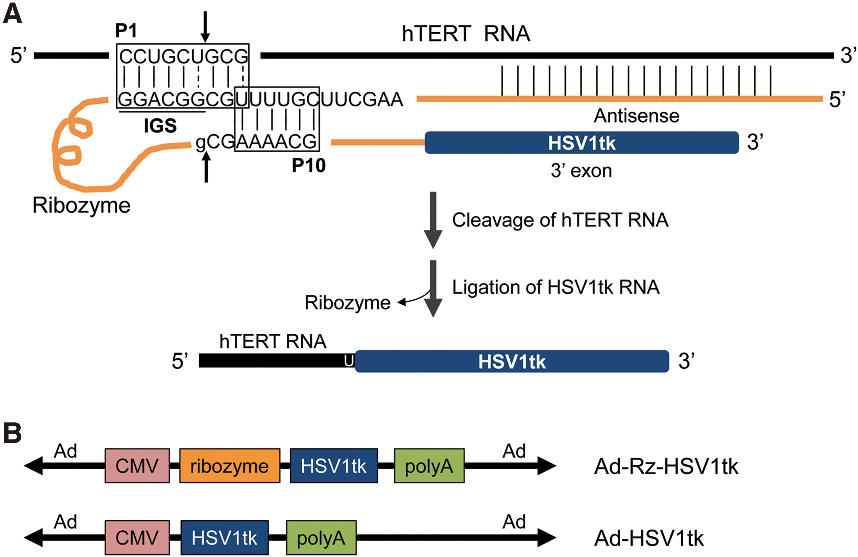
Schematic diagram of hTERT-dependent selective expression of HSV1-tk RNA.
Cell culture
The hTERT-positive human colon cancer cells (HT-29) were cultured in Dulbecco's modified Eagle's medium (DMEM) and hTERT-negative normal human lung fibroblast cells (IMR-90) were cultured in minimum essential medium supplemented with 10% fetal bovine serum, 100 U/mL penicillin, 100 g/mL streptomycin, and 250 ng/mL amphotericin B in an atmosphere containing 5% CO2 at 37°C.
[124/125I]FIAU synthesis
Radiolabeling was performed as previously described with a few modifications. 21 Na124I (MC-50 cyclotron; Korea Institute of Radiological and Medical Sciences [KIRAMS], Seoul, Korea) or Na125I (Perkin Elmer, Inc., Waltham, MA) was added to a solution containing stannylated precursor, 2′-fluoro-2′-deoxy-1-β-D-arabinofuranosyl-5-(tri-n-butyltin)-uracil (25 μg; FutureChem Co., Seoul, Korea), followed by the addition of 30% hydrogen peroxide/acetic acid (1:3, v/v). After quenching with aqueous saturated sodium metabisulfite, the radioiodinated FIAU was isolated on a C18 Sep-Pak cartridge (Waters Corp., Milford, MA), and eluted with ethanol. The ethanol was evaporated and the radioiodinated FIAU was formulated with a cell culture medium or saline.
The overall radiochemical yields were 40% ± 2% for [124I]FIAU and 70% ± 3% for [125I]FIAU. Radiochemical purities measured by radio-high-performance liquid chromatography, as previously described, 22 were over 98% and molar activities about 700 MBq/μmol for [124I]FIAU and 34 MBq/μmol for [125I]FIAU.
In vitro analysis of transgene activity by TSR
To validate HSV1-tk gene expression by a trans-splicing reaction, HT-29 and IMR-90 cells were seeded in six-well plates at densities of 5 × 10⁵ and 2 × 10⁵, respectively, per well. After overnight incubation at 37°C, cells were infected with adenovirus at various multiplicity of infection and maintained for 24 h in serum-free media. The transfection efficiency of adenoviruses was established by reverse transcription-polymerase chain reaction (RT-PCR) analysis of HSV1-tk gene expression in all adenovirus-infected cells.
PCR analysis was conducted with total RNA prepared using the Easy-Spin™ RNA extraction kit (iNtRON Biotechnology, Seongnam, Korea). Complementary DNA (cDNA) synthesis was performed with Superscript III reverse transcriptase (Invitrogen, Carlsbad, CA). PCR amplifications of individual cDNA were performed (95°C for 5 min; 30 cycles at 95°C for 30 s and 57°C for 30 s; and 72°C for 30 s and 72°C for 5 min) using Maxime PCR PreMix Kit (i-Star Taq) (iNtRON Biotechnology) and specific primers. The oligonucleotide primer sequences were as follows: β-actin; sense (5′-GTGGGGCGCCCCAGGCACCAG-3′), antisense (5′-CTCCTTAATGTCACGCACGAT-3′), HSV1-tk; sense (5′-GCGAACATCTACACCACCCA-3′), antisense (5′-AGTGAGCCTCCCCCATCTC-3′), and hTERT; sense (5′-CGGAAGAGTGTCTGGAGCAA-3′), antisense (5′-GGATGAAGCGGAGTCTGGA-3′).
To detect trans-spliced molecules (TSM), the first PCR was performed with a 5′ primer specific for the trans-splicing junction (5′-GGGGAATTCA GCGCTGCGTCC-3′) and with HSV1-tk (5′-GTTATCTGGGCGCTTGTCAA-3′). A second PCR was conducted with inward primers: 5′-GCTGCGTCCTGCTAAAAC-3′ and 5′-CAGTAGCGTGGGC ATTTTCT-3′. 23 Amplified products were analyzed by ethidium bromide–stained agarose gel electrophoresis. Results were interpreted using Multi Gauge V3.0 (Fujifilm, Tokyo, Japan). Expression values of hTERT, HSV1-tk, and TSM genes were normalized with β-actin.
The uptake assay of [125I]FIAU was performed by incubating infected cells at 37°C for 10, 60, and 120 min with [125I]FIAU (74 kBq per well) in the culture medium. After incubation, the medium was removed, and the cells were washed twice with ice-cold phosphate buffered saline. The cells were harvested by treatment with 0.5% trypsin and resuspended in culture medium to neutralize the trypsin. The cellular accumulation of radioactivity was measured using a gamma counter (1480 WIZARD; PerkinElmer, Inc.) and normalized to the number of viable cells. Data represent the mean ± standard deviation of three wells.
Animal study
Animal care, maintenance, and treatment in these studies followed the protocols approved by the Institutional Animal Care and Use Committee of the Korea Institute of Radiological and Medical Sciences. Housing conditions for mice were controlled with 12 h light/12 h dark cycle (lights on at 8:00
Xenografts were produced in 6-week-old female Balb/c-nu/nu mice (n = 15) (Nara Biotech., Seoul, Korea) through subcutaneous injection of 1 × 10⁷ tumor cells in 100 μL of serum-free cell culture medium. Adenoviruses at a concentration of 1 × 10⁹ pfu in 100 μL of saline were injected into the tumor or muscle when the tumors reached a diameter of 8–11 mm, and PET images were acquired at 48 h postinjection. At this time, the average body weight of the mice was 23.85 ± 1.26 g. Potassium perchlorate (100 mg/kg) was administered orally 2 h before [124I]FIAU injection and intraperitoneally just before imaging. At the end of the experiment, the authors performed CO2 euthanasia with flow rate of 10%–30% of the cage volume per minute.
In vivo PET imaging and biodistribution
The mice were anesthetized with 2% isoflurane in oxygen and injected with 7.4 MBq of [124I]FIAU into the tail vein and maintained under normal conditions for 2 h, to obtain an acceptable PET image. PET images were acquired using an Inveon small animal PET/SPECT/CT system (Siemens Medical Solutions) and MicroPETR4 scanner (Concorde Microsystems, Knoxville, TN). PET images were scanned under a 1,200 s acquisition time and a 511 KeV energy level (low level = 350 KeV and upper level = 650 or 750 KeV, respectively). During acquisition of the PET images, mice were anesthetized using 2% isoflurane. The level of radioactivity in tumor was measured as % injected dose per gram by region of interest (ROI), which normalizes the activity for body weight and injected activity. Reconstruction of data set was accomplished using Inveon Research Workplace software (Siemens Medical Solutions).
The biodistribution study was conducted by euthanizing the mice and collecting tissues (blood, heart, liver, lung, spleen, kidney, stomach, small intestine, large intestine, muscle, and tumor) at 48 h postinjection of [124I]FIAU. All tissues were weighed and their radioactivity was measured with a γ-counter (1480 WIZARD; Perkin Elmer, Inc.).
Statistical analysis
Data were expressed as the mean ± standard error of the mean. Unpaired two-tailed Student's t-test was used to compare the two groups. A value of p < 0.05 was considered statistically significant.
Results
Selective induction of transgene expression by Ad-Rz-HSV1tk in hTERT-positive cancer cells
HT-29 and IMR-90 cells were infected with Ad-HSV1tk or Ad-Rz-HSV1tk adenovirus. At 24 h postinfection, the authors performed RT-PCR to investigate the gene expression of hTERT, HSV1-tk, and TSM in HT-29 and IMR-90 cells (Fig. 2). In HT-29 cells, the hTERT-positive cells, hTERT gene expression was observed in the control group as well as the groups infected with Ad-HSV1tk and Ad-Rz-HSV1tk. Conversely, in hTERT-negative IMR-90 cells, hTERT gene expression was not observed in any groups.

The hTERT RNA-dependent HSV1-tk expression in hTERT-negative (IMR-90) and hTERT-positive (HT-29) cells.
The expression level of HSV1-tk RNA fused with the ribozyme in all Ad-Rz-HSV1tk-infected cells was consistent with that of HSV1-tk RNA in all Ad-HSV1tk-infected cells, suggesting that the ribozyme expression level in both cells did not affect the induction of functional HSV1-tk RNA. Otherwise, hTERT-dependent TSM expression by ribozyme was observed in Ad-Rz-HSV1tk-infected HT-29 cells alone. These results demonstrate that Ad-Rz-HSV1tk-induced TSM expression occurred specifically in hTERT-positive cells.
Evaluation of HSV1-tk activity induced by Ad-Rz-HSV1tk in HT-29 and IMR-90 cells
The authors investigated whether ribozyme HSV1-tk specifically induces the accumulation of FIAU in hTERT-expressing cancer cells through [125I]FIAU uptake study.
Figure 3 shows HSV1-tk activity detected by [125I]FIAU uptake in HT-29 and IMR-90 cells. Adenovirus-infected cells were incubated for 10, 60, and 120 min in [125I]FIAU-containing media, followed by measurement of [125I]FIAU uptake using a γ-counter. In HT-29, HSV1-tk activities of Ad-HSV1tk- and Ad-Rz-HSV1tk-infected groups increased time dependently, whereas in IMR-90, the HSV1-tk activity increased in the Ad-HSV1tk-infected group alone. These data indicate that [125I]FIAU uptake driven by HSV1-tk activation depends on the presence of hTERT in Ad-Rz-HSV1tk-infected cells.
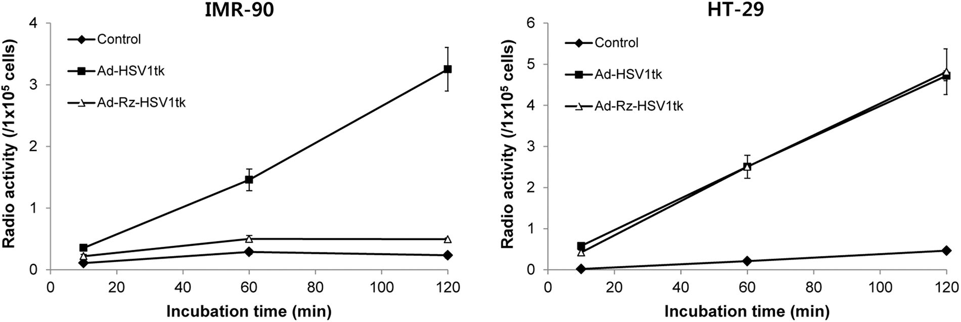
The hTERT RNA-dependent HSV1-tk activity. Time-dependent selective induction of HSV1-tk activity by hTERT ribozyme was detected by [125I]FIAU uptake in HT-29 and IMR-90 cells.
Imaging hTERT mRNA expression using ribozyme HSV1-tk in HT-29 tumor-bearing mice
To investigate the hTERT-positive cancer targeting by ribozyme HSV1-tk through [124I]FIAU PET imaging, the authors generated tumor-bearing BALB/c nude mice by subcutaneous injection of HT-29 cells dorsally into the right shoulders. After 2 weeks, they divided the mice into two groups and intratumorally injected saline as control (n = 5) and Ad-Rz-HSV1tk (n = 5), respectively. At 48 h after adenovirus injection, [124I]FIAU (7.4 MBq/head) was administered to tail vein of the mouse and imaged using PET at 2 h postinjection (Fig. 4A). According to ROI analysis, the [124I]FIAU uptake in the Ad-Rz-HSV1tk-infected HT-29 group was significantly higher (1.54 ± 0.34 %ID/g) than in the control group (1.06 ± 0.17 %ID/g) (p = 0.02), although it was lower than in the Ad-HSV1tk group (data now shown) (Fig. 4B).
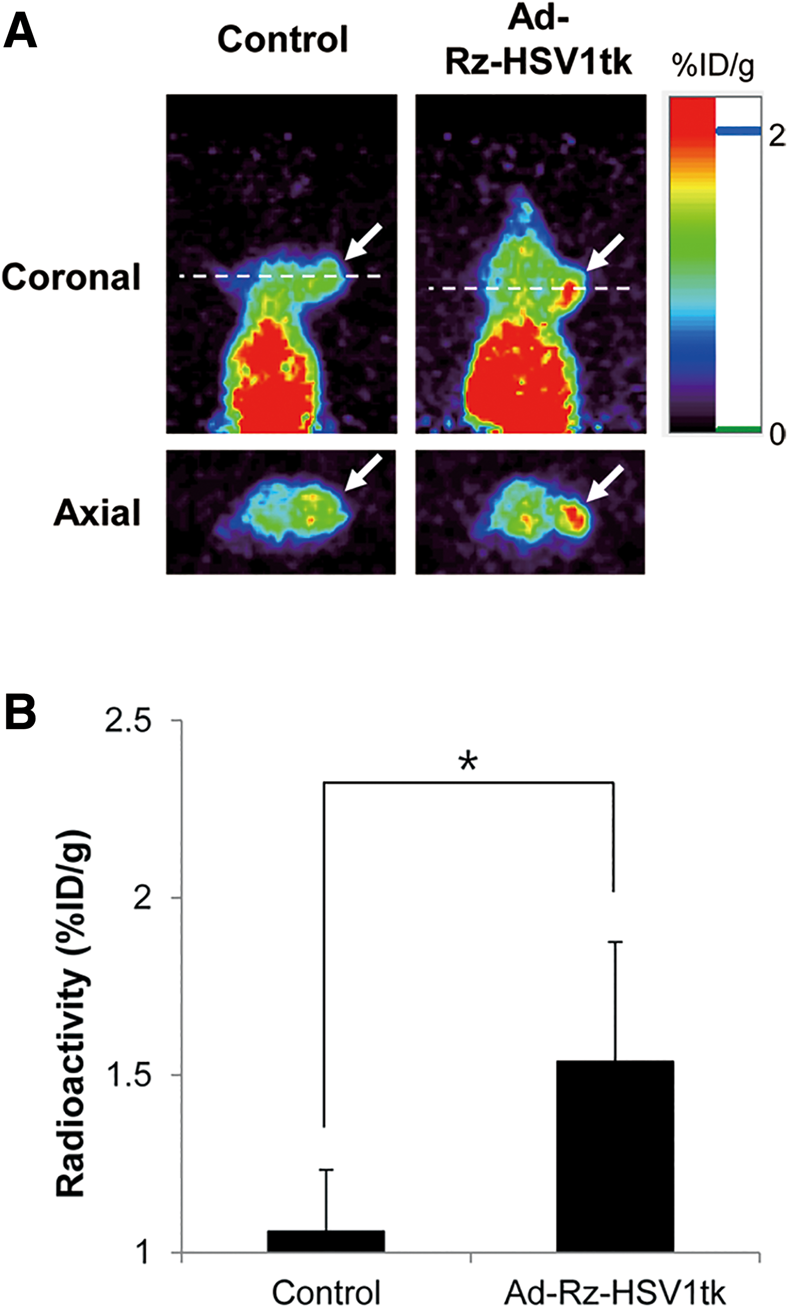
[124I]FIAU uptake by efficient targeting of Ad-Rz-HSV1tk in HT-29 tumor-bearing mice.
Figure 5A illustrates the comparative [124I]FIAU uptake of HT-29 tumor and normal muscle with or without Ad-Rz-HSV1tk infection. In control mice, muscle and HT-29 tumors showed no [124I]FIAU uptake (n = 2). However, Ad-Rz-HSV1tk-infected HT-29 tumor showed higher levels of [124I]FIAU uptake than Ad-Rz-HSV1tk-infected muscle (n = 3). These data indicate that Ad-Rz-HSV1tk-infected hTERT-positive tumor can be noninvasively detected by PET imaging with [124I]FIAU.
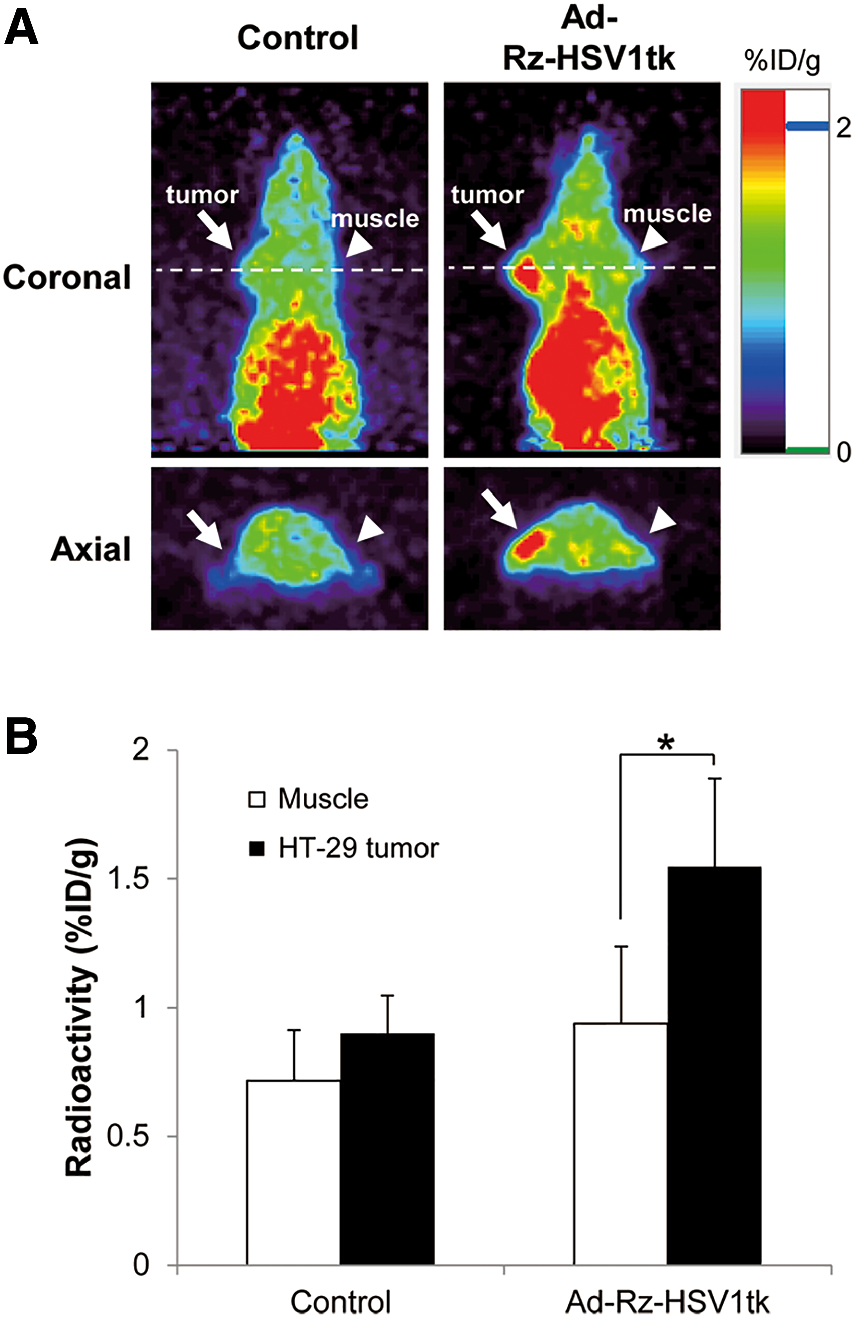
Comparison of [124I]FIAU uptake by muscle and tumor in HT-29 tumor-bearing mice.
Biodistribution of [124I]FIAU in HT-29 tumor-bearing mice
To investigate hTERT-positive cancer targeting by hTERT-specific TSR, the authors performed biodistribution analysis with a γ-counter after a 48 h injection with [124I]FIAU. Biodistribution results showed also high levels of [124I]FIAU uptake in the Ad-Rz-HSV1tk-injected HT-29 tumor along with PET/computed tomography (CT) images acquired at 24 h after adenovirus injection in the same mice (Fig. 6). Furthermore, it has high specificity based on tumor-to-muscle (18.38-fold) and tumor-to-blood (11.5-fold) ratios obtained from the biodistribution data (Table 1). Furthermore, Ad-Rz-HSV1tk-injected tumor uptake was 6.27-fold higher compared with the control tumor. In this study, the highest uptake of [124I]FIAU was detected in the small intestine, which was consistent with a previous study, suggesting urinary excretion of FIAU through the small intestine. 24
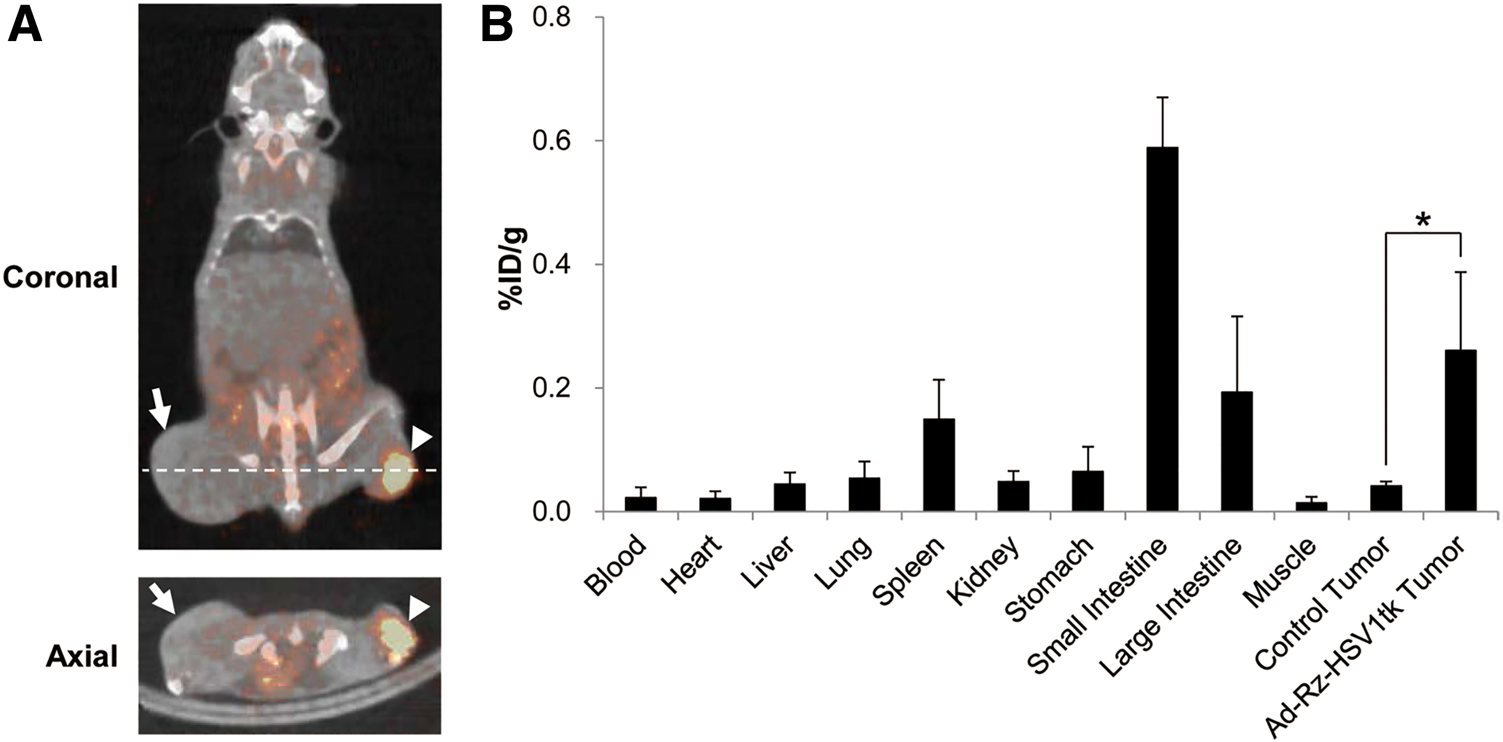
[124I]FIAU PET/CT image and biodistribution.
Biodistribution Results of [124I]FIAU at 24 h After PET/CT Imaging in the Same Tumor-Bearing Mice (n = 3)
Data are presented as mean ± standard deviation for 3 animals.
CT, computed tomography; [124I]FIAU, 124I labeled 2′-fluoro-2′-deoxy-1-β-D-arabinofuranosyl-5-iodouracil; %ID/g, % injected dose per gram of tissue; PET, positron emission tomography.
Discussion
The viral tk has broader substrate specificity than mammalian tk and phosphorylates the pyrimidine and purine nucleoside derivatives, as well as deoxythymidine. 25 The thymidine derivative, FIAU, has been used as a reporter probe for HSV1-tk gene expression monitoring by radioiodination. 26 It is transported into a cell and is phosphorylated by activated HSV1-tk, and is incorporated into the DNA. 27 The authors detected [125I]FIAU only in the hTERT-positive cells and not in the hTERT-negative cells, even though ribozymes (represented as HSV1-tk RNA production) were present in the hTERT-negative cells (Figs. 2A and 3) due to the absence of hTERT-dependent splicing by ribozyme in hTERT-negative cells. These data suggest that the methods using Ad-Rz-HSV1tk and [125I]FIAU might represent a highly effective diagnostic and therapeutic strategy in hTERT-positive cancer.
FIAU is labeled with the positron-emitting radionuclide 124I (T1/2 = 4.08 d). Tjuvajev et al. demonstrated that analysis using [124I]FIAU reporter probe is a highly specific noninvasive imaging modality to identify and quantitate HSV1-tk expression. 28 Also, Kim et al. reported that [125I]FIAU single-photon emission computed tomography (SPECT) and [ 18 F]FHBG PET contributed to evaluation of successfully targeted gene expression of pancreatic adenocarcinoma upregulated factor-targeting TSR and liver-specific hTERT-targeting TSR in pancreatic cancer and hepatocellular carcinoma mice model, respectively. 29,30 Also, another similar research for hTERT targeting had been done about combining the HSV1-tk with retrovirus vector in the therapy of hepatocellular carcinoma, development of the multimodal imaging probe using hTERT antibody, and the [ 18 F]FDG PET/CT imaging and serum hTERT mRNA quantification in cancer diagnosis. 31 –33
However, in this study, the authors used adenoviral vector system for cancer-specific HSV1-tk expression by ribozyme and they conducted hTERT-specific TSR-targeted imaging using PET, which showed higher sensitivity and spatial resolution than SPECT. Moreover, they showed whole-body PET imaging with [124I]FIAU, which has greater sensitivity, dynamic range, and lower abdominal background radioactivity than [ 18 F]FHBG. 34 Figures 4 and 5 indicate that [124I]FIAU PET imaging can be effectively used to verify HSV1-tk RNA replacement by TSR. These data demonstrate that [124I]FIAU can be utilized to quantitatively evaluate HSV1-tk gene expression by hTERT-specific TSR.
Conclusions
In summary, the authors demonstrated that the HSV1-tk trans-splicing activity with TSR depends on the presence of hTERT. The trans-splicing activity is attributed to selective detection of the hTERT-positive tumor. Furthermore, they demonstrated that hTERT-specific uptake of [124I]FIAU reporter probe provides PET imaging and quantification of therapeutic gene expression. These results contribute to diagnostic assessment and gene therapy by hTERT-specific TSR in hTERT-positive cancer.
Footnotes
Authors' Contributions
M.J.S.: conception (constructing the idea for research and the article); data collection (execution of the experiments); analysis and interpretation (interpretation and presentation of the results); data processing (taking responsibility in data management and reporting); literature review; and writer (taking responsibility in the construction of the article). J.H.P.: data collection (execution of the experiments) and literature review. K.C.L.: data collection (execution of the experiments) and critical review (reviewing the article before submission). Y.J.L. and T.S.L.: conception (constructing the idea for research) and critical review (reviewing the article before submission). T.H.C.: data collection (execution of the experiments) and analysis and interpretation (interpretation and presentation of the results). S.W.L.: data collection (execution of the experiments); conception (constructing the idea for research); and critical review (reviewing the article before submission). K.I.K.: conception (constructing the idea for research); design (planning methodology to reach the conclusion); data collection (taking responsibility in execution of the experiments); analysis and interpretation (interpretation and presentation of the results); and critical review (reviewing the article before submission). J.H.K.: conception (constructing the idea for research); design (planning methodology to reach the conclusion); analysis and interpretation (interpretation and presentation of the results); and critical review (reviewing the article before submission).
Disclosure Statement
There are no existing financial conflicts.
Funding Information
This work was supported by the National Research Foundation of Korea (NRF) grant funded by the Ministry of Science and ICT (NRF-2015R1C1A1A02036885, NRF-2017M2A2A02070985, and NRF-2017R1A2B2003368).