
Abstract
Background:
E2F-mediated cell proliferation enhancing long noncoding RNA (EPEL) is an oncogenic long noncoding RNA (lncRNA) in lung cancer, but its involvement in other malignancies is unknown. We aimed at exploring the potential role of EPEL in ovarian endometrioid adenocarcinoma.
Results:
Plasma levels of EPEL and p53 were significantly upregulated in ovarian endometrioid adenocarcinoma patients. In effect, EPEL overexpression distinguished ovarian endometrioid adenocarcinoma patients from the control group. Plasma levels of EPEL and p53 were reversely correlated in ovarian endometrioid adenocarcinoma patients. EPEL overexpression led to downregulated p53 expression in cells of human ovarian endometrioid adenocarcinoma cell line SNU-251, whereas no significant changes in the expression level of EPEL were observed after p53 expression. EPEL overexpression increased, whereas small interfering RNA (siRNA) silencing reduced the viability of SNU-251 cells under carboplatin treatment. In addition, p53 overexpression attenuated the effects of EPEL overexpression on cancer cell viability under carboplatin treatment.
Conclusions:
Therefore, the overexpression of EPEL may participate in the development of chemoresistance of cancer cells to carboplatin in ovarian endometrioid adenocarcinoma by downregulating p53.
Introduction
As the major type of endometrial cancer, ovarian endometrioid adenocarcinoma is the third most common type of genital tract among women. 1 Surgical resection is a promising strategy for the treatment of ovarian endometrioid adenocarcinoma at early stages. 2,3 However, the application of surgery is limited by its invasive nature. 4 The development and popularization of chemotherapy significantly prolong the survival time of ovarian endometrioid adenocarcinoma patients at advanced stages. 5,6 But long-term use of chemical drugs causes the development of resistance in cancer cells, leading to reduced chemosensitivity. 7 Therefore, how to overcome chemoresistance is a major task in the clinical treatment of ovarian endometrioid adenocarcinoma by using different chemical drugs.
p53 is a well-characterized tumor suppressor gene that participates in cancer biology by inhibiting the proliferation, migration, and invasion, and promoting the apoptosis of cancer cells. 8,9 Besides that, downregulation of p53 expression and promoted p53 protein degradation and aggregation have also been proven to play pivotal roles in the development of chemoresistance in cancer cells to chemical drugs, such as carboplatin. 10 –12 It is known that p53 may interact with different long noncoding RNAs (lncRNAs) to achieve its signal transduction in cancer development and progression. 13 E2F-mediated cell proliferation enhancing lncRNA (EPEL) is a recently identified lncRNA in lung cancer. 14 This study revealed that EPEL may participate in the development of chemoresistance of cancer cells to carboplatin in ovarian endometrioid adenocarcinoma by downregulating p53.
Patients and Methods
Cell line and specimens
Human SNU-251 (ATCC) ovarian endometrioid adenocarcinoma cell line was used in this study. Cells were cultured with Dulbecco's modified Eagle's medium (DMEM; C0003-01) + 10% fetal bovine serum (15%) at 37°C in a 5% CO2 incubator.
Plasma samples used in this study were derived from the blood of 42 ovarian endometrioid adenocarcinoma patients and 33 healthy controls who visited Dushanzi People's Hospital from May 2015 to January 2018. Tumor tissue specimens were also obtained from the 42 ovarian endometrioid adenocarcinoma patients. Patient inclusion criteria were: (1) new cases diagnosed by pathological examinations; (2) patients willing to donate blood and tumor samples. Patients' exclusion criteria were: (1) patients who were suffering for other diseases; (2) patients who received any treatment within 100 d before admission. Age of the patient group ranged from 48 to 67 years, with a mean age of 55.55 ± 4.71 years. There were 6, 12, 9, and 15 cases at clinical stage I–IV, respectively. The 33 healthy controls were enrolled in the physiological health examination center of Dushanzi People's Hospital. All those healthy controls showed normal physiological conditions. Age of the control group ranged from 51 to 66 years, with a mean age of 56.43 ± 3.66 years. Patient and control groups have similar age distributions. All participants understood the whole experimental protocol. Ethics committee of Dushanzi People's Hospital approved this study. All participants signed informed consent.
Reverse transcription quantitative real-time PCR
Following instructions of RNAzol RT kit (Sigma-Aldrich), total RNA was extracted and subjected to reverse transcription to prepare complementary DNA (cDNA) samples. Polymerase chain reaction (PCR) mixtures were prepared by using SuperScript III Platinum One-Step qRT-PCR Kit (Thermo Fisher Scientific, Inc.). PCRs were carried out through the following conditions: 95°C for 56 s, and then 95°C for 12 s and 60°C for 35 s for 40 cycles. Primer sequences were: 5′-GAGGCAGACCACGTGAGAG-3′ (forward) and 5′-CAGATTTAAACCCCGCACTG-3′ (reverse) for human EPEL; 5′-GAGCTGAATGAGGCCTTGGA-3′ (forward) and 5′-CTGAGTCAGGCCCTTCTGTCTT-3′ (reverse) for p53; and 5′-GACCTCTATGCCAACACAGT-3′ (forward) and 5′-AGTACTTGCGCTCAGGAGGA-3′ (reverse) for endogenous control β-actin. All data were processed by using the 2−ΔΔCT method. The one with the lowest expression level was set to a relative level of 1.
Enzyme-linked immunosorbent assay
Levels of p53 in plasma were measured by using a human p53 enzyme-linked immunosorbent assay (ELISA) Kit (ab46067) from Abcam. All operations were done following the details described in the manufacturer's instructions.
Cell transfection
EPEL and p53 expression vectors as well as EPEL small interfering RNA (siRNA) 5′-UACAAAACUCUGGAACCUC(dTdT)-3′ and negative control siRNA 5′-CCUACGCCACCAAUUUCGU(dTdT)-3′ were synthesized by GenePharma (Shanghai, P.R. China). According to the manufacturer's instructions, transection was performed by using lipofectamine 2000 reagent (11668-019; Invitrogen, Carlsbad, CA) with vectors and siRNA as dosages of 15 and 50 nM, respectively. The control group (C) was cells without transfection. The negative control group (NC) was cells transfected with empty vectors or control siRNA. Overexpression rate above 200% and knockdown rate below 50% (at 24 h after transfection) were reached before subsequent experiments.
MTT assay
After transfection, cells were harvested and single-cell suspensions (4 × 10⁴ cells/mL) were made. A 96-well plate was used to cultivate cells (100 μL cell per well), and carboplatin was added at dosages of 100, 200, and 300 μM. Cells were cultured under normal conditions (37°C, 5% CO2) for 6 h, followed by adding 10 μL MTT. Cells were cultured for an additional 4 h, and optical density (OD) values at 570 nm were measured.
Western blot analysis
Total protein was extracted by using Total Protein Extraction Kit (2140; Merck Millipore). After that, protein was denatured in boiling water for 5 min. Electrophoresis was then performed by using 12% sodium dodecyl sulfate-polyacrylamide gel electrophoresis (SDS-PAGE) gel. Gel transfer to PVDF membranes (Thermo Fisher Scientific, Inc.) was performed, and membranes were blocked (2 h in 5% skimmed milk). The membranes were first incubated with primary antibodies of p53 (1:1300, rabbit antihuman; ab131442; Abcam) and GAPDH (1:1300, rabbit antihuman, ab181602; Abcam) at 4°C overnight, followed by incubation with IgG-HRP secondary antibody (1:1000, goat antirabbit, MBS435036; MyBioSource) at room temperature for 2 h. Signals were developed by using the chemiluminescence method, and signals were processed by using Image J 1.48 software (NIH, Bethesda, MD).
Statistical analysis
Mean values of three biological replicates were used to express all data. Student's t test was used to explore differences between two groups. One-way analysis of variance and Tukey test were used to explore differences among multiple groups. Correlations were analyzed by Pearson correlation coefficient. p < 0.05 was the cutoff of statistical significance.
Results
Dysregulation of EPEL and p53 were observed in ovarian endometrioid adenocarcinoma patients
qRT-PCR and ELISA were performed to measure the levels of EPEL and p53 in plasma of both ovarian endometrioid adenocarcinoma patients (EA group) and healthy controls (Control group). Compared with the control group, plasma levels of EPEL were significantly higher in the EA group (Fig. 1A, p < 0.05). In contrast, plasma levels of p53 were significantly lower in the EA group than in the control group (Fig. 1B, p < 0.05). It is worth noting that the expression level of EPEL increased, whereas the expression level of p53 decreased slightly but not significantly with the increase in clinical stage (p > 0.05, data not shown).

Dysregulation of lncRNA EPEL and p53 were observed in endometrioid adenocarcinoma patients. Compared with healthy controls, plasma levels of lncRNA EPEL were significantly higher
Upregulated plasma levels of EPEL distinguished ovarian endometrioid adenocarcinoma patients from healthy controls
With healthy controls as true negative cases and ovarian endometrioid adenocarcinoma patients as true positive cases, receiver operating characteristic (ROC) curve analysis was performed to evaluate the potential of plasma EPEL in the diagnosis of ovarian endometrioid adenocarcinoma. Results showed that the area under the curve was 0.8700, with a standard error of 0.04036 and a 95% confidence interval of 0.7909–0.9492 (Fig. 2, p < 0.0001).
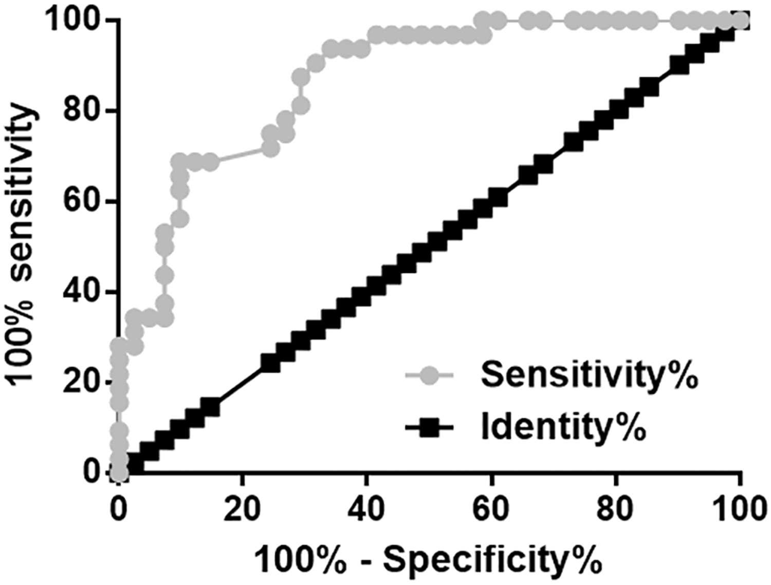
Upregulated plasma levels of EPEL distinguished ovarian endometrioid adenocarcinoma patients from healthy controls. ROC curve was used to analyze the diagnostic vale of EPEL for ovarian endometrioid adenocarcinoma. In ROC curve analysis, true positive cases were ovarian endometrioid adenocarcinoma patients and true negative cases were healthy controls. Black line is the line of identity, and light green line is the diagnostic line. ROC, receiver operating characteristic.
A reverse correlation between plasma levels of EPEL and p53 was found in ovarian endometrioid adenocarcinoma patients
Pearson correlation coefficient analysis revealed that plasma levels of EPEL and p53 were significantly and inversely correlated in ovarian endometrioid adenocarcinoma patients (Fig. 3A). In contrast, no significant correlation between plasma levels of EPEL and p53 was found in healthy controls (Fig. 3B). An attempt was made to further explore the correlation between EPEL and p53 in tumor tissues. The expression of EPEL and p53 messenger RNA (mRNA) in tumor tissues was detected by qRT-PCR. Pearson correlation coefficient analysis revealed that expression levels of EPEL and p53 in tumor tissue were significantly and inversely correlated (Fig. 3C).

A reverse correlation between plasma levels of EPEL and p53 was found in ovarian endometrioid adenocarcinoma patients. EPEL and p53 were significantly and reversely correlated in ovarian endometrioid adenocarcinoma patients
Overexpression of EPEL mediated the downregulation of p53 in cells of SNU-251 ovarian endometrioid adenocarcinoma cell line
The expression of p53 in cells of SNU-251 ovarian endometrioid adenocarcinoma cell line was detected by western blot after transfection of EPEL expression vectors. Compared with C and NC groups, significantly downregulated p53 expression was observed in SNU-251 cells with EPEL overexpression (Fig. 4A, p < 0.03). In contrast, p53 overexpression showed no significant effects on the expression level of EPEL in SNU-251 cells (Fig. 4B).

Overexpression of EPEL mediated the downregulation of p53 in cells of SNU-251 ovarian endometrioid adenocarcinoma cell line. Overexpression of EPEL downregulated p53 in cells of SNU-251 ovarian endometrioid adenocarcinoma cell line
EPEL overexpression induced chemoresistance in SNU-251 cells to carboplatin
As shown in Figure 5A, altered expression of EPEL and p53 mRNA was observed after transfection (p < 0.05). Compared with C and NC groups, overexpression of EPEL significantly increased, whereas EPEL siRNA silencing and p53 overexpression significantly reduced the viability of SNU-251 cells under treatment of different concentrations of carboplatin (Fig. 5B, p < 0.05). In addition, compared with SNU-251 cells transfected with EPEL expression vectors alone, SNU-251 cells transfected with both EPEL and p53 expression vectors had significantly reduced viability.
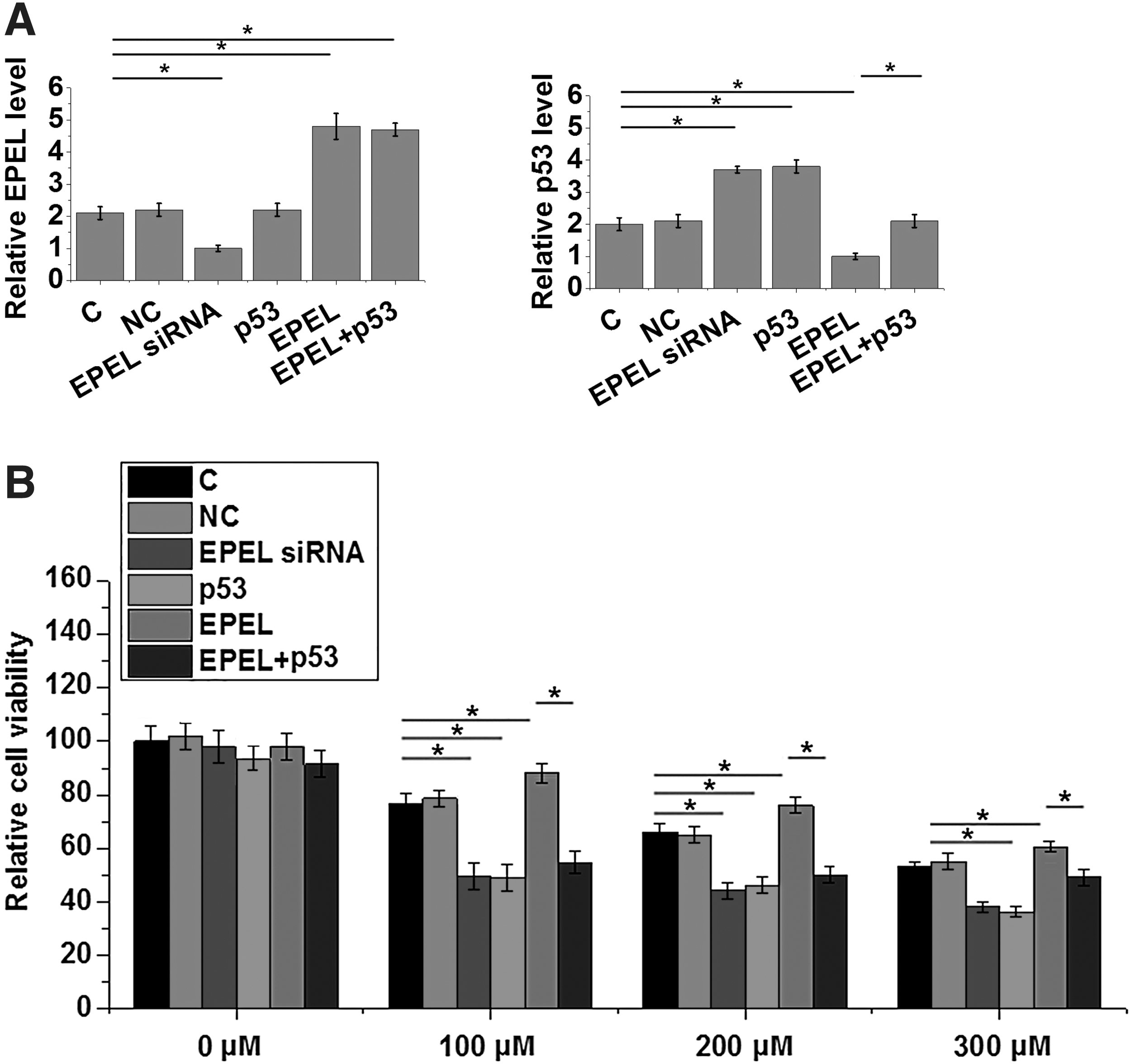
EPEL overexpression induced chemoresistance of SNU-251 cells to carboplatin. Altered expression of EPEL and p53 mRNA was observed after transfection
Discussion
EPEL is a recently reported lncRNA with oncogenic function characterized only in lung cancer. 14 Our study first reported that EPEL is also likely an oncogenic lncRNA in ovarian endometrioid adenocarcinoma. EPEL overexpression in ovarian endometrioid adenocarcinoma may promote the development of chemoresistance of cancer cells to carboplatin in ovarian endometrioid adenocarcinoma by downregulating p53.
Development of ovarian endometrioid adenocarcinoma not only leads to altered expression of a large set of proteins 15 but also globally affects the expression of lncRNAs, 16 which encode no protein and participate in human disease with RNA as the functional form. 17 In our study, plasma levels of EPEL were found to be significantly higher in ovarian endometrioid adenocarcinoma patients than in healthy controls. In effect, ROC curve analysis showed that overexpression of EPEL effectively distinguished patients with ovarian endometrioid adenocarcinoma from healthy controls. Therefore, EPEL is also likely an oncogenic lncRNA in ovarian endometrioid adenocarcinoma and may possess diagnostic potential for this disease.
p53 as a tumor suppressor gene that is usually downregulated in human malignancies, including ovarian endometrioid adenocarcinoma. 18 It has been proved that detection of the degree of the decrease of expression level of p53 in tumor tissues provides guidance for the prognosis of ovarian endometrioid adenocarcinoma. 19 Consistent with previous studies, in this study we also observed significantly downregulated p53 in plasma of ovarian endometrioid adenocarcinoma patients compared with healthy controls, further confirming its role as a tumor suppressor gene in this disease.
Our preliminary microarray data revealed a potential reverse correlation between the expression levels of p53 mRNA and EPEL in tumor tissues across plasma specimens derived from ovarian endometrioid adenocarcinoma patients. In this study, we further confirmed the reverse correlation between plasma levels of p53 mRNA and EPEL in ovarian endometrioid adenocarcinoma patients. Our in vitro cell experiments revealed that EPEL is likely an upstream inhibitor of p53 in the regulation of the development of chemoresistance of cancer cells to carboplatin in ovarian endometrioid adenocarcinoma. This conclusion is proposed based on the following observations: (1) EPEL led to downregulated p53 expression in ovarian endometrioid adenocarcinoma cells, whereas p53 overexpression failed to significantly affect the expression of EPEL; (2) p53 overexpression significantly attenuated the enhancing effects of EPEL overexpression on the viability of ovarian endometrioid adenocarcinoma cells under carboplatin treatment. However, p53 overexpression only partially attenuated, but failed to totally reverse this effect, indicating that EPEL may interact with multiple pathways to participate in this process. We also speculate that the inhibitory effect of EPEL overexpression on p53 expression in ovarian endometrioid adenocarcinoma cells is indirect, because there is no significant correlation between plasma levels of EPEL and p53 in healthy controls.
Chemoresistance is a major challenge in the treatment of almost types of gynecological cancers, such as epithelial ovarian cancer, 20 and novel therapeutics are needed to overcome chemoresistance. 21 It has been reported that pleiotropic interaction between drug-resistant cells and reduced immunosurveillance may participate in the development of chemoresistance in epithelial ovarian cancer. 20 Our future studies will investigate the involvement of this factor in ovarian endometrioid adenocarcinoma. In addition, patients' conditions should be considered in the management of endometrial cancer. 22,23 For instance, elderly female patients are more fragile and personalized therapies should be development for these patients.
This article is the first to report the regulation of p53 by EPEL in the regulation of chemoresistance in ovarian endometrioid adenocarcinoma. Therefore, the regulation of EPEL may assist the treatment of this disease by chemotherapies. However, this study is challenged by the small sample size and the lack of in vivo animal model experiments. Our future studies will try to solve these problems.
In conclusion, the development of ovarian endometrioid adenocarcinoma is accompanied by the upregulated expression of EPEL. Overexpression of EPEL may participate in the development of chemoresistance in cancer cells to carboplatin in ovarian endometrioid adenocarcinoma by downregulating p53.
Availability of Data and Materials
The analyzed data sets generated during the study are available from the corresponding author on reasonable request.
Ethics Approval and Consent to Participate
This study was approved by the Ethics Committee of Dushanzi People's Hospital. The research has been carried out in accordance with the World Medical Association Declaration of Helsinki. All patients and healthy volunteers provided written informed consent before their inclusion within the study.
Consent for Publication
All authors have read and approved the final article.
Footnotes
Authors' Contributions
L.Z., J.Y., Y.D., and C.W. conceived and designed the experiments, and they did the article preparation; L.Z., J.Y., X.L., and X.F. performed the experiments; and L.Z., J.Y., X.L., X.F., Y.D., and C.W. analyzed data.
Disclosure Statement
The authors declare that they have no competing interests.
Funding Information
No funding was received for this article.