
Abstract
Background:
Human epidermal growth factor receptor 2 (HER2) is found to be amplified in ∼15%–20% of breast cancers. In this study, the authors report the synthesis and comparative in vitro therapeutic efficacy of 177Lu-CHX-A″-DTPA-trastuzumab and 177Lu-CHX-A″-DTPA-F(ab′)2-trastuzumab to determine their potential as theranostic agents for patients with breast cancer.
Materials and Methods:
Bivalent F(ab′)2-trastuzumab was produced by enzymatic digestion of trastuzumab, conjugated with p-SCN-Bn-CHX-A″-DTPA and subsequently radiolabeled with 177Lu. Cell viability, membrane toxicity assays, and apoptosis analysis were carried out with 177Lu-CHX-A″-DTPA-trastuzumab and 177Lu-CHX-A″-DTPA-F(ab′)2-trastuzumab in HER2-positive ovarian (SK-OV-3) and breast cancer (SK-BR-3 and MDA-MB-453) cells.
Results:
In vitro cell binding studies showed ∼20%–25% binding of 177Lu-CHX-A″-DTPA-trastuzumab and 177Lu-CHX-A″-DTPA-F(ab′)2-trastuzumab to SK-OV-3, SK-BR-3, and MDA-MB-453 cells. The cells exhibited similar degree of membrane integrity and cellular toxicity when treated with same amount (activity) of 177Lu-CHX-A″-DTPA-F(ab′)2-trastuzumab and 177Lu-CHX-A″-DTPA-trastuzumab, and the toxicity was dose dependent. The mode of cell death was predominantly by apoptosis and necrosis with both the radioimmunoconjugates.
Conclusions:
The results indicated that the efficacy of both the radioimmunoconjugates, in terms of inducing cell death, was similar thereby ascertaining their potential as good therapeutic agents for patients with breast cancer.
Introduction
Breast cancer is one among all cancers with increased incidence, mortality rate, and high economic and social costs. Human epidermal growth factor receptor 2 (HER2) positivity accounts for about 15%–20% of breast and epithelial ovarian cancers along with its corresponding encoded protein at abnormally high levels. 1,2 HER2 or ErbB2 (Neu) belongs to a receptor tyrosine-specific protein kinase family, the EGFR (epidermal growth factor receptor) family consisting of four EGF receptors, EGFR (ErbB1), ErbB2 (Neu), ErbB3, and ErbB4. Members of the EGFR family contain a cytoplasmic tyrosine kinase domain, a single transmembrane domain, and an extracellular domain that is involved in ligand binding and receptor dimerization. 1 –3 HER2 overexpression gives rise to signals that potentiate dysregulated proliferation, oncogenesis, metastasis, and perhaps resistance against apoptosis-inducing therapeutic agents. 4
Trastuzumab (Herceptin®), a humanized IgG1 monoclonal antibody, is used in immunotherapy for targeting the extracellular domain of the HER2 in tumors overexpressing this receptor. 5 Although immunotherapy with trastuzumab is associated with survival benefits, these tumors often develop resistance, 6 and new approaches for targeting them are being actively investigated. 7,8 Radioimmunotherapy, using radiolabeled monoclonal antibodies, is one such strategy widely used for treatment of cancer in recent times.
Several studies have utilized radiolabeled intact trastuzumab for targeting HER2 in the preclinical or clinical settings for diagnosis and therapy. 9 –13 The use of intact trastuzumab directly for theranostic applications in cancer still has many limitations, due to its large size, slow pharmacokinetics, and poor tumor penetration. 14 –17 The slow pharmacokinetics results in slow elimination of the radioimmunoconjugates from the blood and normal tissues yielding low tumor/blood (T/B) and tumor/normal tissue (T/NT) ratios. 18 However, in case of intact antibody such as trastuzumab, which gets internalized within the cell rapidly, the slow pharmacokinetics could have beneficial effect as it can produce a sustained effect in patients with solid tumors. The limitations associated with intact antibody can be overcome by fragmentation of the intact antibody by enzyme digestion using pepsin to yield bivalent F(ab′)2 fragments. Radiolabeled F(ab′)2 fragments are being used in clinical and preclinical studies for theranostic applications due to their improved pharmacokinetics and tumor penetration characteristics. 19 –22 Although the mechanism of action of trastuzumab has been well studied for immunotherapy, 6 very few reports on radiolabeled trastuzumab and radiolabeled trastuzumab F(ab′)2 fragments are available. 23 F(ab′)2-trastuzumab does not mediate antibody-dependent cellular cytotoxicity or complement-dependent cytotoxicity in vitro due to lack of Fc portion, and hence, the in vitro antiproliferative and pro-apoptotic effects of both intact trastuzumab and F(ab′)2 fragments are similar. 24 The efficacy and the mechanism of action in case of radiolabeled trastuzumab and F(ab′)2-trastuzumab are likely to be similar under in vitro conditions; however, it is possible that under in vivo conditions the effects may be different. 24 Understanding the mechanism of action, particularly its mode of cellular toxicity, would offer information for improving the efficacy of these radioimmunoconjugates in clinical settings.
Lu-177 decays with a half-life of 6.73 d by emission of β− particles with maximum energies of 497 keV (78.6%), 384 keV (9.1%), and 176 keV (12.2%) to stable 177Hf. 25 The emission of suitable energy γ photons of 113 keV (6.4%) and 208 keV (11%) enables simultaneous scintigraphic studies. 25 The low energy of its γ and β− is suitable for imaging and therapeutic purposes. 23 Moreover, simple and easy complexation of Lu-177 with bifunctional chelators (BFCAs), such as DTPA and DOTA under appropriate reaction conditions, makes it an attractive radioisotope for theranostic applications. 15,26
The aim of this study was preparation of 177Lu-CHX-A″-DTPA-F(ab′)2-trastuzumab and 177Lu-CHX-A″-DTPA-trastuzumab and perform comparative studies of their cellular toxicity and mechanism of cell death in HER2-overexpressing breast and ovarian cancer cells. The findings may pave way to translation of these formulations in treatment of patients with breast cancer.
Materials and Methods
Materials
Trastuzumab (Herceptin) procured from Genentech, South San Francisco, was gifted by Tata Memorial Hospital, Mumbai, India, for this study. Lutetium-177 (specific activity 22–25 Ci/mg) was produced at the Dhruva reactor, BARC, Trombay, by irradiation of enriched 176Lu target followed by radiochemical processing at the Radiopharmaceuticals Division, BARC, 27,28 and has the approval for clinical use from the Radiopharmaceuticals Committee (RPC), constituted by the Department of Atomic Energy, Government of India. [(R)-2-Amino-3-(4-isothiocyanatophenyl)propyl]-trans-(S,S)-cyclohexane-1,2-diamine-pentaacetic acid (p-SCN-Bn-CHX-A′′-DTPA) was procured from Macrocyclics (Dallas, TX), whereas immobilized pepsin resin used for enzymatic digestion was purchased from Thermo Scientific (Rockford). Chemicals such as sodium bicarbonate, sodium acetate, Arsenazo III, and yttrium chloride, and media such as Dulbecco's modified Eagle's medium (DMEM) were purchased from Sigma–Aldrich (St. Louis, MO), whereas fetal bovine serum (FBS) was from GIBCO. Cell lines such as SK-OV-3, SK-BR-3, MDA-MB-453 (HER2 positive), and MDA-MB-231 (triple negative, i.e., ER−, PR−, and HER2−) were procured from the National Center for Cell Sciences (Pune, India). For purification, PD-10 columns and AMICON Ultra centrifugal filter devices (MWCO 10,000 Da and MWCO 30,000 Da) were obtained from GE Healthcare and Millipore, India, respectively. Radioactive counting was performed using a well-type NaI(Tl) scintillation counter (Electronics Corporation of India Limited, India). Size Exclusion HPLC System (SE-HPLC; JASCO, Japan) equipped with a TSK gel column (G3000 SWXL) from Sigma–Aldrich was used for HPLC analyses. UV/VIS and radioactive detectors (Raytest GmBH, Germany), connected to the HPLC system, were used for measuring absorbance and radioactivity, respectively. The analyses of radiochromatograms were performed using the GINASTAR software (Raytest GmBH). UV absorbance measurements were carried out in a Chemito Spectroscan UV2600 spectrophotometer (Thermo Scientific). Guava Flow cytometer kits were purchased from Merck KGaA (Darmstadt, Germany).
Methods
Preparation and purification of F(ab′)2-trastuzumab
The fragmentation of trastuzumab was carried out according to the manufacturer's protocol (Thermo Scientific). In brief, intact trastuzumab (Herceptin 22 mg/mL) was mixed with 1 mL of digestion buffer (sodium acetate, 20 mM, pH 4.5) and concentrated to 1 mL. The concentrated trastuzumab was added to 0.5 mL of immobilized pepsin (3000 U/mg), pre-equilibrated with digestion buffer. The initial experiments comprised optimization of conditions for preparation of trastuzumab F(ab’)2 fragments for maximum yields by varying the pepsin/antibody ratio (1:2, 1:4, 1:6, and 1:8) and incubation times (0, 6, 12, 18, and 24 h). After incubation, the reaction mixture was quenched by the addition of 10 mM Tris-HCl buffer, pH 8.0, and centrifuged at 1000g for 5 min. The supernatant containing the F(ab′)2-trastuzumab was purified using Amicon filtration devices (MWCO 30,000 Da) and resuspended in 1 mL phosphate buffer saline (PBS, 20 mM, pH 7.4). The pure F(ab′)2-trastuzumab was characterized by size exclusion high-performance liquid chromatography (SE-HPLC) and gradient (5%–15%) sodium dodecyl sulfate–polyacrylamide gel electrophoresis (SDS-PAGE). The amount of intact trastuzumab and F(ab′)2-trastuzumab loaded per lane under both reducing and nonreducing conditions was ∼25 μg in 50 μL.
Conjugation of trastuzumab and F(ab′)2-trastuzumab with p-SCN-Bn-CHX-A″-DTPA
Trastuzumab and F(ab′)2-trastuzumab were conjugated with p-SCN-Bn-CHX-A″-DTPA at 1:10 molar ratio as reported earlier by the same author group. 15,26 After conjugation, the conjugates were purified using Amicon filtration devices (MWCO 10,000 Da) to remove any unconjugated CHX-A″-DTPA in the solution. The concentration of trastuzumab and F(ab′)2-trastuzumab in the respective conjugates was determined by Lowry's method 29 using IgG as the reference, whereas the number of CHX-A″-DTPA molecules conjugated per trastuzumab and F(ab′)2-trastuzumab molecule was determined by spectroscopic assay using Y(III)-Arsenazo (III). 30
Radiolabeling of CHX-A″-DTPA-trastuzumab and CHX-A″-DTPA-F(ab′)2-conjugates with 177Lu
The radiolabeling of CHX-A″-DTPA-trastuzumab and CHX-A″-DTPA-F(ab′)2-trastuzumab with 177Lu was performed as reported earlier, 15,26 wherein the immunoconjugates in 0.1 M sodium acetate solution (pH 6.0) were mixed with ∼185–370 MBq of 177LuCl3 and incubated at ambient temperature for 15 min. The labeled products were purified through a PD-10 column using 0.1 M sodium acetate solution (pH 6.0) as eluant. The radiochemical purity (RCP) of the radioimmunoconjugates was determined by SE-HPLC on a TSK gel column isocratically, using 0.05 M phosphate buffer, pH 6.8, for elution at a flow rate of 0.6 mL/min. The RCP was concurrently ascertained by paper chromatography (PC) on Whatman 3 mm paper using 10 mM sodium citrate (pH 5.0) as the mobile phase.
Cellular studies with 177Lu-CHX-A″-DTPA-trastuzumab and 177Lu-CHX-A″-DTPA-F(ab′)2-trastuzumab in HER2-positive cells
Breast cancer cells SK-BR-3 and MDA-MB-453 and ovarian cancer cells SK-OV-3 were grown to 70%–80% confluence in DMEM supplemented with 10% FBS in a humidified atmosphere containing 5% CO2 at 37°C. As negative control, MDA-MB-231 (triple negative—ER−, PR−, and HER2−) cells were grown under identical conditions. Reports indicate that SK-OV-3 and SK-BR-3 express very high number of HER2 receptors (∼1 × 10⁶), whereas MDA-MB-453 express moderate numbers of HER2 receptors. 31 –33
In all the cellular studies performed, the cells were treated with 3.7 MBq of 177Lu-CHX-A″-DTPA-trastuzumab and 177Lu-CHX-A″-DTPA-F(ab′)2-trastuzumab (equivalent to 33.3 nM of trastuzumab/F(ab′)2-trastuzumab) and 37 MBq of 177Lu-CHX-A″-DTPA-trastuzumab and 177Lu-CHX-A″-DTPA-F(ab′)2-trastuzumab [equivalent to 333 nM of trastuzumab/F(ab′)2-trastuzumab]. The corresponding vehicle controls VC1 and VC2 comprised cells treated with equivalent amounts (i.e., 33.3 and 333 nM, respectively) of unlabeled CHX-A″-DTPA-trastuzumab and CHX-A″-DTPA-F(ab′)2-trastuzumab.
In vitro cell binding and inhibition studies
For in vitro cell binding studies, ∼1 × 10⁶ cells were seeded in 24-well tissue culture plates and incubated overnight at 37°C. The adherent cells were subsequently incubated with 6.7 nM (250 KBq) each of 177Lu-CHX-A″-DTPA-trastuzumab and 177Lu-CHX-A″-DTPA-F(ab′)2-trastuzumab at 37°C for 1 h. After incubation, the cells were washed twice with ice cold 0.05 M PBS, pH 7.4, and then solubilized with 1 mL of 1 N NaOH. The solution was counted in a NaI (Tl) gamma counter to determine the radioactivity associated with cells. Assays were carried out in triplicates. The percentage of radioactivity bound to the cells with respect to the total radioactivity added was determined and expressed as mean ± standard deviation (SD). Inhibition assays were performed under identical conditions by co-incubation of cells with 6.7 nM (250 KBq) of the radioimmunoconjugates and 70 nM of unlabeled trastuzumab. The specificity of the radioimmunoconjugates was further confirmed by performing cell binding studies with HER2-negative cells such as MDA-MB-231 (triple negative) under identical conditions.
Cell membrane integrity estimation by lactate dehydrogenase assay
Membrane integrity was estimated by the LDH Activity Assay Kit (Sigma–Aldrich; catalog no. MAK066) according to the manufacturer's protocol. In brief, the master reaction mix was prepared by mixing lactate dehydrogenase (LDH) assay buffer and LDH substrate mix (24:1) (v/v). For calculating the absolute value of LDH released, a standard solution of nicotinamide adenine dinucleotide hydrogen (Hydride) (NADH) was prepared in LDH assay buffer at concentrations ranging from 0 to 12.5 nM in 50 μL volume. Similarly, test samples were prepared by taking 50 μL aliquots of culture media in which cells treated with the radioimmunoconjugates and their corresponding vehicle controls were grown All standards and test samples were prepared in triplicates in 96-well plates, to which 50 μL of the LDH master reaction mix was added and incubated for 2–3 min. The initial absorbance measurement of both NADH standards and test samples was taken at 450 nm. Subsequently, the plates were incubated at 37°C, and measurements were made continuously every 5 min until the absorbance in the sample treated with the maximum dose of 37 MBq became equivalent to the absorbance of highest concentration of NADH standard. The LDH activity of the test samples were determined as LDH activity = (B × Sample Dilution Factor)/(Reaction Time T × V), where B = Amount (nM) of NADH generated between the initial and final reaction time = T, V = sample volume (mL). LDH activity is reported as nmol/min/mL = mU/mL.
Estimation of cell viability by flow cytometer
Approximately 1 × 10⁶ cells of SK-BR-3, MDA-MB-453, and SK-OV-3 were plated overnight in DMEM containing 10% FBS. The adherent cells were grouped in triplicates into three groups as those treated with 177Lu-CHX-A″-DTPA-trastuzumab, those treated with 177Lu-CHX-A″-DTPA-F(ab′)2-trastuzumab, and vehicle controls. The treated cells were incubated for a period of 48 h at 37°C in a humidified 5% CO2 atmosphere. After incubation, the cells were harvested by trypsinization and resuspended in 50 μL of 0.05 M PBS containing 1% bovine serum albumin (BSA). The cell number was maintained at 1 × 10⁵ per tube, as per Guava ViaCount Reagent kit (Merck Millipore, Germany) protocol. Guava Via count reagent (350 μL) was added into each tube and incubated in dark for 15 min at room temperature. Samples were acquired on a Guava easyCyte™ Flow Cytometer System (Merck Millipore) and analyzed using Guava Soft 3.1.1. The Via Count assay distinguishes viable and nonviable cells based on differential permeability of two DNA-binding dyes in the Guava ViaCount® Reagent. The nuclear dye stains only nucleated cells, whereas the viability dye stains dying cells. Debris was excluded from the results based on negative staining with nuclear dye.
Study of apoptotic cell death
For the apoptosis study, ∼1 × 10⁶ SK-BR-3, MDA-MB-453, and SK-OV-3 cells were treated in the same manner as mentioned above for 48 h at 37°C in a humidified 5% CO2 atmosphere. The cells were harvested by trypsinization and resuspended in 100 μL of PBS (0.01 M, pH 7.4) containing 1% BSA, and the cell number was maintained at 1 × 10⁵ per tube. Guava Nexin Reagent (100 μL) was added to each tube and incubated in dark for 20 min at room temperature. Samples were acquired on a Guava easyCyte Flow Cytometer System and analyzed with GuavaSoft 3.1.1 (Merck Millipore). The apoptotic assay is based on a two-dye strategy: Annexin V-PE (Phycoerythrin) to detect phosphatidylserine on the external membrane of apoptotic cells and 7-AAD, a cell impermeant dye, as an indicator of membrane structural integrity. 7-AAD excludes from live healthy cells and early apoptotic cells but permeates into late-stage apoptotic and dead cells.
Statistical data analysis
All data were analyzed using origin 8.0 (Origin Lab Corp., MA) statistical software. The results are expressed as mean ± SD of three independent experiments. To compare the significant difference, t-test was used, considering the p-value to be 0.05 or less.
Results
Preparation and purification of F(ab′)2-trastuzumab
Digestion of trastuzumab with pepsin, at molar ratio of 1:4 of pepsin to antibody in 20 mM sodium acetate buffer (pH 4.5), at 37°C generated a maximum yield of ∼70% of F(ab′)2-trastuzumab. The time for complete digestion of trastuzumab was optimized at 18 h similar to earlier reports. 34,35 Figure 1A, B represents the SDS-PAGE profile of intact trastuzumab and F(ab′)2-trastuzumab (∼25 μg in 50 μL of each was loaded) under nonreducing and reducing conditions, respectively. A single band was observed corresponding to molecular weight of ∼100 kDa (Fig. 1A, lane 3) indicating the purity of the F(ab′)2-trastuzumab, whereas two bands at ∼28 and ∼25 kDa (Fig. 1B, lane 2) were observed when F(ab′)2-trastuzumab was subjected to SDS-PAGE under reducing conditions. Similarly, intact trastuzumab showed a single band at ∼150 kDa (Fig. 1A, lane 2) under nonreducing condition and two bands corresponding to 50 and 25 kDa under nonreducing conditions (Fig. 1B, lane 1).
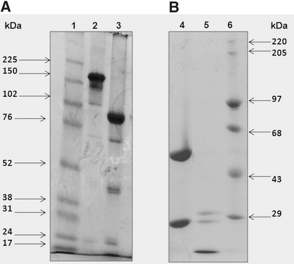
SDS-PAGE profile of trastuzumab and F(ab′)2-trastuzumab under
Conjugation and radiolabeling of trastuzumab and F(ab′)2-trastuzumab
The average number of CHX-A″-DTPA molecules conjugated to F(ab′)2-trastuzumab and intact trastuzumab was found to be 2 ± 0.8 and 4 ± 1.2, respectively, as determined by the spectroscopic assay using Y(III)-Arsenazo(III) complex. 15,30 Radiolabeling of the immunoconjugates with 177Lu was achieved within 15 min at ambient temperature. The RCP of 177Lu-CHX-A″-DTPA-trastuzumab as determined by SE-HPLC was 97% ± 1.1% with a retention time (Rt ) of 15.1 min (Fig. 2A-b), whereas the RCP of 177Lu-CHX-A″-DTPA-F(ab′)2-trastuzumab was 96% ± 1.0% with Rt of 16.1 min (Fig. 2B-d). It was observed that the radioactive peaks closely matched with the respective UV profile of trastuzumab (Fig. 2A-a) and F(ab′)2-trastuzumab (Fig. 2B-c). The RCP of 177Lu-CHX-A″-DTPA-trastuzumab as determined by PC was 96.3% ± 0.9%, whereas the RCP of 177Lu-CHX-A″-DTPA-F(ab′)2-trastuzumab was 95.2% ± 1.0%. The specific activity of 177Lu-CHX-A″-DTPA-trastuzumab and 177Lu-CHX-A″-DTPA-F(ab′)2-trastuzumab was determined to be 370 and 225 MBq/mg, respectively.
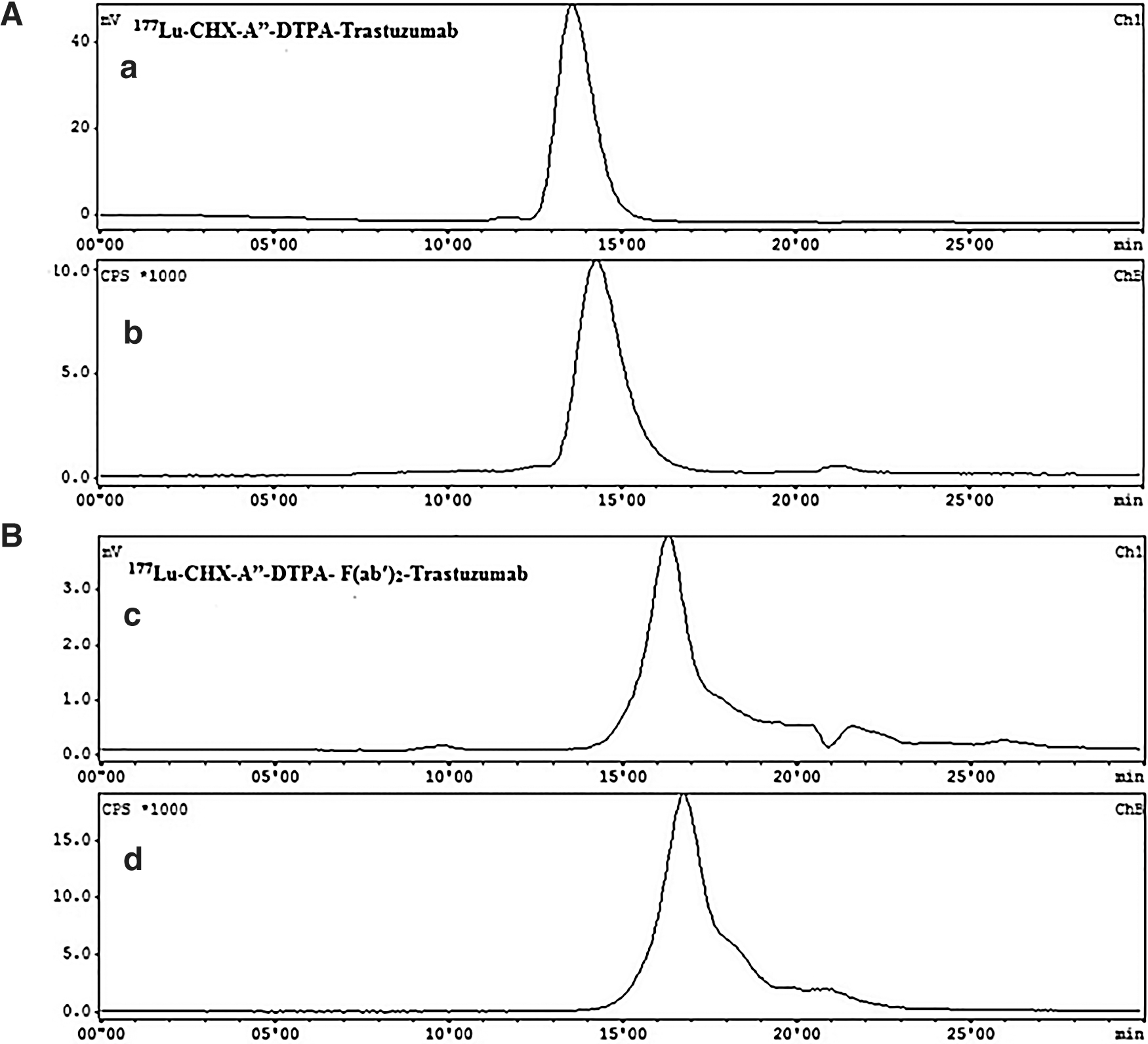
SE-HPLC pattern of
In vitro cell binding studies
Figure 3A, B represents the in vitro cell binding studies of 177Lu-CHX-A″-DTPA-trastuzumab and 177Lu-CHX-A″-DTPA-F(ab′)2-trastuzumab in HER2 expressing SK-OV-3, SK-BR-3, and MDA-MB-453 cells. SK-OV-3 cells exhibited a binding of 24.9% ± 1.8% and 21.7% ± 0.6% for 177Lu-CHX-A″-DTPA-trastuzumab (6.7 nM) and 177Lu-CHX-A″-DTPA-F(ab′)2-trastuzumab (6.7 nM), respectively, which reduced to 5.2% ± 2.0% (∼80% inhibition) and 2.4% ± 0.5% (∼88% inhibition), on co-incubation of cells with 70 nM of unlabeled trastuzumab and F(ab′)2-trastuzumab. The other breast cancer cells such as SK-BR-3 and MDA-MB-453 showed binding of 25.4% ± 0.4% and 17.0% ± 2.7% with 177Lu-CHX-A″-DTPA-trastuzumab and 23.0% ± 1.4% and 14.9% ± 1.1% with 177Lu-CHX-A″-DTPA-F(ab′)2-trastuzumab, respectively (Fig. 3A, B). Here also, an inhibition of binding was observed when the cells were co-incubated with the radioimmunoconjugates and excess of unlabeled trastuzumab and F(ab′)2-trastuzumab (70 nM) indicating the specificity of 177Lu-CHX-A″-DTPA-trastuzumab and 177Lu-CHX-A″-DTPA-F(ab′)2-trastuzumab to HER2 receptors. However, no significant binding or inhibition was observed with MDA-MB-231 (triple negative) cells (Fig. 3A, B).
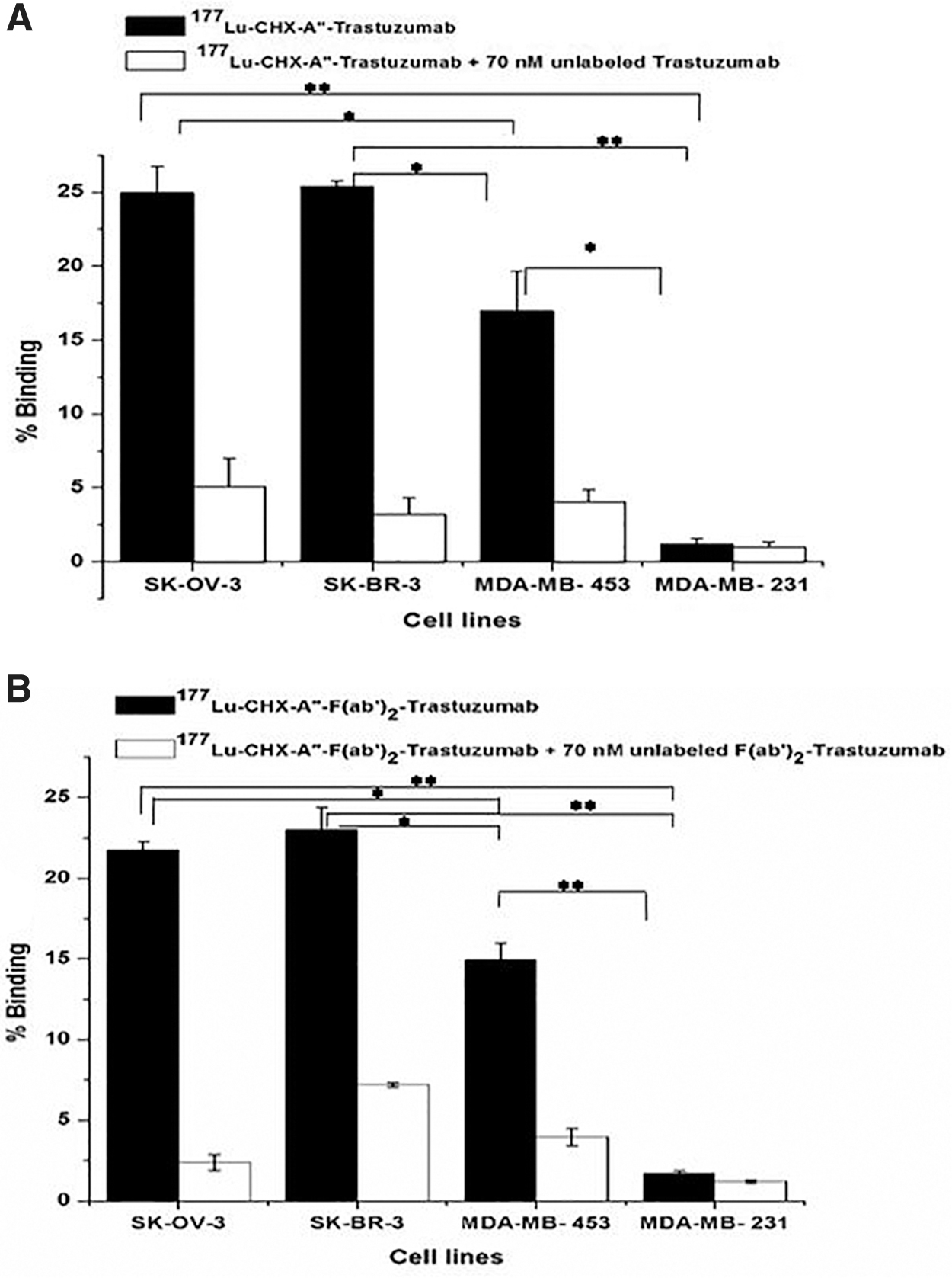
In vitro cell binding studies of
Cell membrane integrity studies
The toxicity of the radiolabeled complexes was estimated in terms of LDH release in the growth medium (Fig. 4A–C). The LDH release by SK-OV-3 cells treated with 37 MBq of 177Lu-CHX-A″-DTPA-trastuzumab was 19.6 ± 3.0 mU/mL, whereas the LDH release by SK-OV-3 cells treated with the same amount of activity of 177Lu-CHX-A″-DTPA-F(ab′)2-trastuzumab was 16.3 ± 0.4 mU/mL (Fig. 4A). In case of SK-BR-3 and MDA-MB-453, the LDH release was found to be comparatively higher at 33.6 ± 1.2 and 30.3 ± 7.2 mU/mL, respectively, when treated with the same amount of the activity of 177Lu-CHX-A″-DTPA-trastuzumab. Treatment of SK-BR-3 and MDA-MB-453 with 37 MBq of 177Lu-CHX-A″-DTPA-F(ab′)2-trastuzumab exhibited LDH release of 26.6 ± 0.7 and 35.3 ± 4.46 mU/mL, respectively (Fig. 4B, C). The % LDH release was comparatively less when the cells were exposed to less amounts of activity (3.7 MBq) of 177Lu-CHX-A″-DTPA-trastuzumab/177Lu-CHX-A″-DTPA-F(ab′)2-trastuzumab. The respective vehicle controls containing equivalent amount of unlabeled trastuzumab and F(ab′)2-trastuzumab also demonstrated low LDH release (Fig. 4A–C).
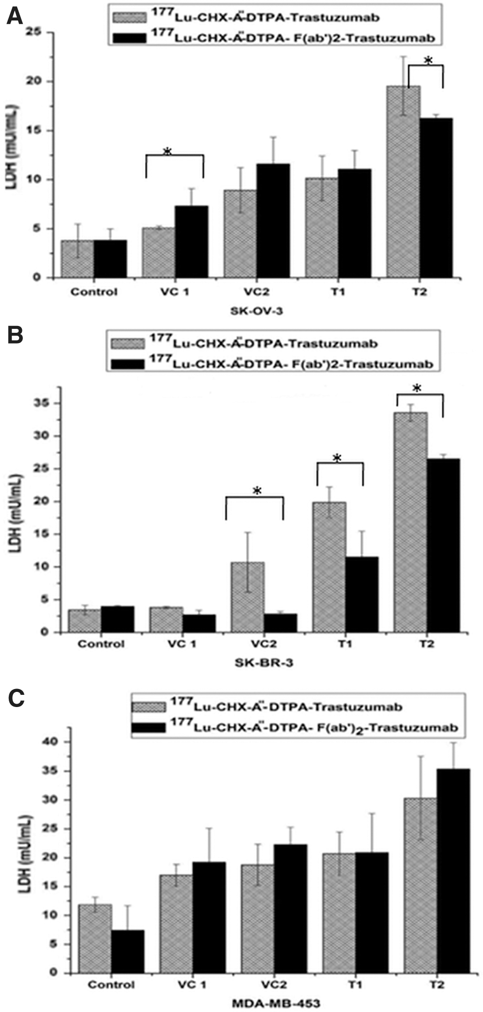
Cell toxicity determination by lactate dehydrogenase assay in
Cell viability study by flow cytometer
The cell viability was assessed by flow cytometer with two different amounts of activity of the radioimmunoconjugates in all the three cell lines. A typical flow cytometer dot plot viability data of MDA-MB-453 cells are shown in Figure 5. MDA-MB-453 cells treated with 37 MBq of 177Lu-CHX-A″-DTPA-trastuzumab and 177Lu-CHX-A″-DTPA-F(ab′)2-trastuzumab exhibited decrease in cell viability up to 17.1% ± 2.4% and 24.5% ± 4.9%, respectively (p < 0.05, n = 3), whereas SK-BR-3 cells treated with the same activity showed a decrease in cell viability up to 60.4% ± 2.6% and 70.4% ± 4.0%, respectively (p < 0.05, n = 3). Equivalent amount of cold trastuzumab and F(ab′)2-trastuzumab did not show significant change in cell viability as shown in Figure 6A, B. The cell viability in case of SK-OV-3 was higher compared with the other two cell lines—75.48% ± 6.30% and 85.8% ± 0.56% for 37 and 3.7 MBq of 177Lu-CHX-A″-DTPA-trastuzumab, respectively (p < 0.005, n = 3), whereas the cell viability when treated with 37 and 3.7 MBq of 177Lu-CHX-A″-DTPA-F(ab′)2-trastuzumab was found to be 76.09% ± 2.60% and 87.45% ± 0.08%, respectively (p < 0.005, n = 3) (Fig. 6A, B).
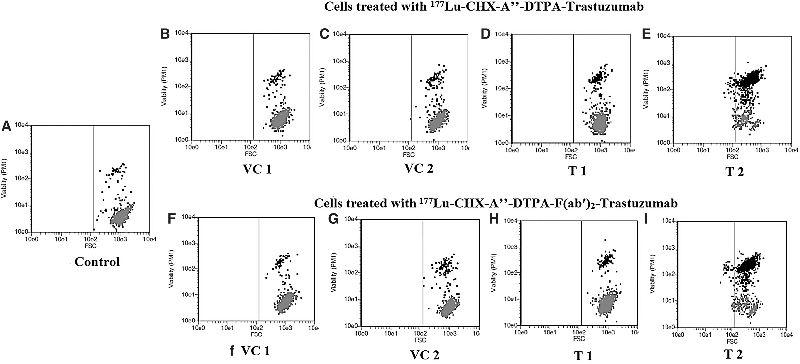
Cell viability estimation of MDA-MB-453 cells by flow cytometry wherein cells were treated with 3.7 and 37 MBq of 177Lu-CHX-A″-DTPA-trastuzumab/177Lu-CHX-A″-DTPA-F(ab′)2-trastuzumab along with their respective vehicle controls. The viability (PM2) versus FSC dot plot represents the viable cells at the bottom of the plot and dead cells at upper region of the plot. Here,
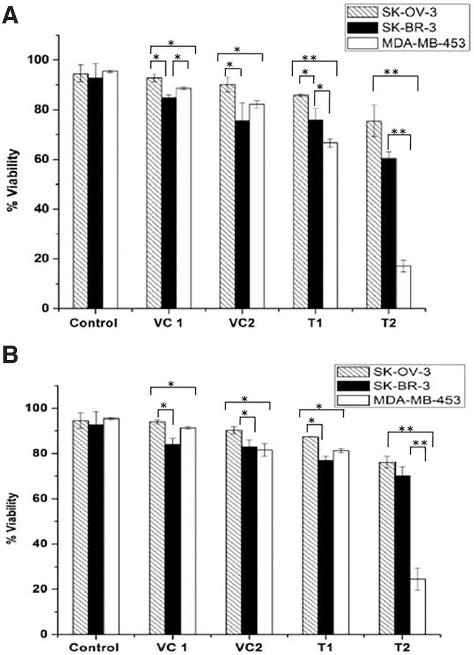
Percentage cell viability analyzed by flow cytometry in SK-OV-3, SK-BR-3, and MDA-MB-453 cell lines in response to treatment given with
Study of apoptotic cell death
In this study, data obtained by flow cytometric apoptosis analysis confirm that in all three cell lines, the mode of cell death after 48 h of treatment with different activities of 177Lu-CHX-A″-DTPA-trastuzumab and 177Lu-CHX-A″-DTPA-F(ab′)2-trastuzumab was largely by apoptosis, although a small extent of necrotic death was also observed. Apoptosis induction appeared to be more pronounced at higher doses, which is evident by an increase in the apoptotic cell population (Annexin V+/7-AAD−) in a dose-dependent manner (Fig. 7). Cytotoxicity was found to be higher in MDA-MB-453 cells compared with SK-BR-3 and SK-OV-3 cells. Treatment of MDA-MB-453 cells with 37 MBq of 177Lu-CHX-A″-DTPA-trastuzumab and 177Lu-CHX-A″-DTPA-F(ab′)2-trastuzumab induced apoptotic population of 13.7% ± 2.7% and 23.0% ± 5.4%, respectively (p < 0.05, n = 3), compared with their corresponding vehicle controls, which induced apoptosis up to only 4.9% ± 1.7% and 7.2% ± 1.6%, respectively (p < 0.05, n = 3) (Fig. 7). Unlike other assays, apoptosis induction in SK-BR-3 and SK-OV-3 cells was more pronounced than MDA-MB-453 cells (20.9% ± 3.3% and 19.65% ± 4.03%, respectively, compared with 13.7% ± 2.6%) on treatment with 37 MBq of 177Lu-CHX-A″-DTPA-trastuzumab [(Fig. 8A, p < 0.005 and p < 0.05, respectively, n = 3) dot plot data not shown]. However, the overall cytotoxicity, which includes both the apoptotic and necrotic populations, was observed to be higher in MDA-MB-453 cell lines compared with SK-BR-3 cell lines at all doses of treatment. Furthermore, as expected, SK-OV-3 cells exhibited comparatively low apoptosis in response to different doses (activity) of radioimmunoconjugates (Fig. 8A, B). In case of necrotic or dead (Annexin V+/7-AAD+) cell populations, the necrotic events increased with increase in the activity of 177Lu-CHX-A″-DTPA-trastuzumab and 177Lu-CHX-A″-DTPA-F(ab′)2-trastuzumab given to all the three cell lines, although a significant population of cells were found to be apoptotic. Figure 7 represents the dot plot representing necrotic or late apoptotic population in the upper right quadrant for MDA-MB-543 cells.
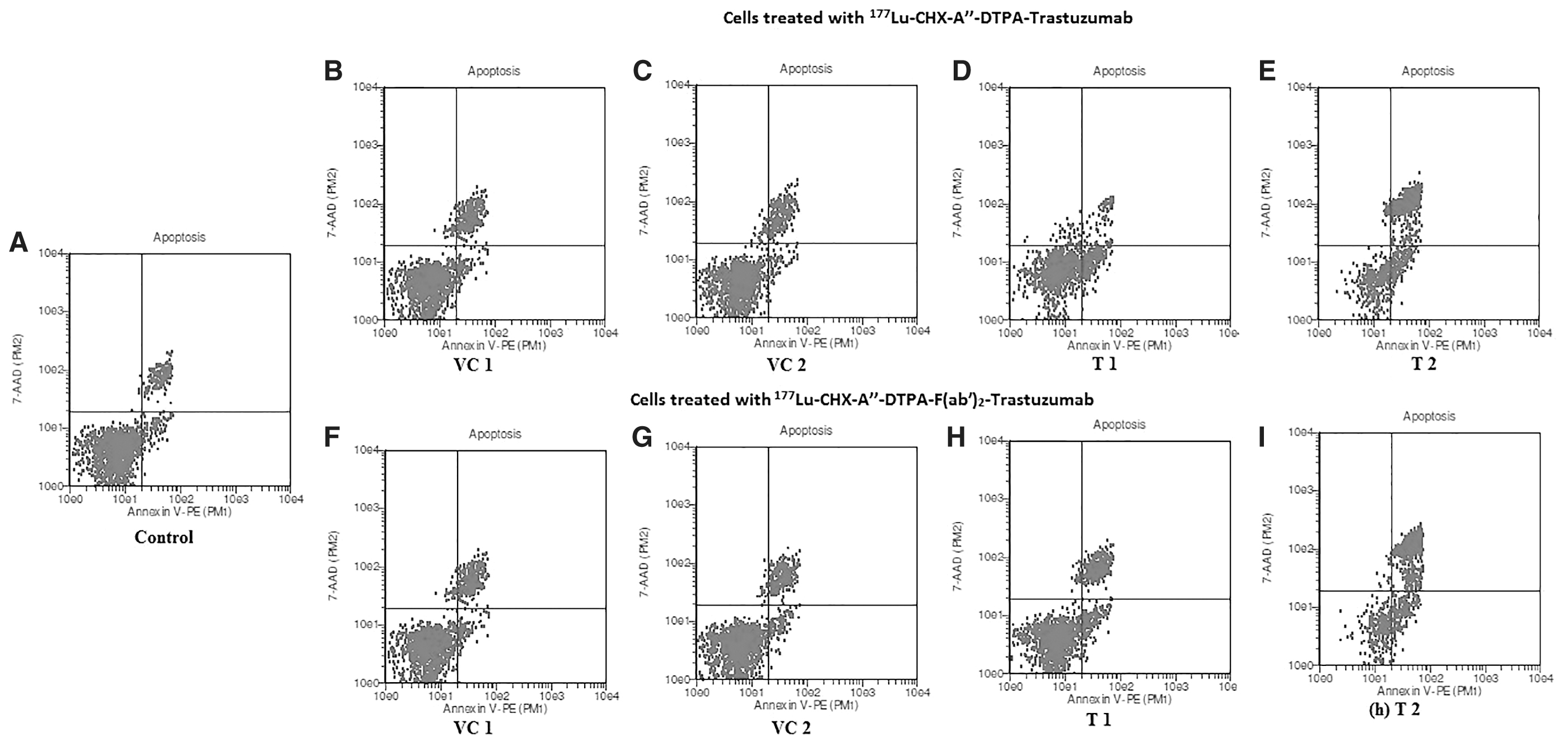
Mode of cell death analyzed by flow cytometry. MDA-MB-453 cells were treated with 3.7 and 37 MBq of 177Lu-CHX-A″-DTPA-trastuzumab/177Lu-CHX-A″-DTPA-F(ab′)2-trastuzumab along with their respective vehicle controls. The 7-AAD−−/Annexin V− dot plot represents viable cells in lower left quadrant, apoptotic cells 7-AAD−/AnnexinV+ in lower right quadrant and dead or necrotic cells (7-AAD+/Annexin V+) in upper right quadrant. Here,
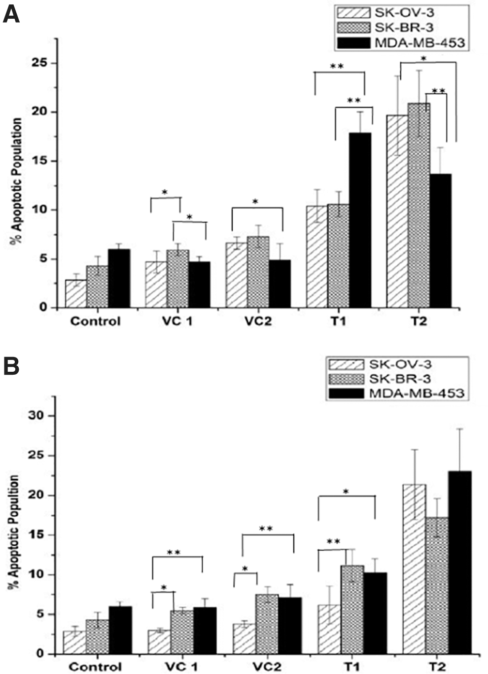
Apoptotic cell population estimation by flow cytometry in SK-OV-3, SK-BR-3, and MDA-MB-453 cells treated with
Discussion
Breast and ovarian cancers are major health problem worldwide among females. HER2 overexpression is generally associated with worse outcomes in different therapeutic modalities that are presently available. 36,37 Resistance to immunotherapeutic agents such as trastuzumab may present de novo, and clinical reports indicate primary or secondary resistance to trastuzumab in up to 70% of HER2-overexpressing breast cancer patients. 6 Although the exact mechanism of resistance against trastuzumab is still not known, identification of resistance mechanisms and the incorporation of new targeted drugs have resulted in improved outcomes. The possible mechanisms of resistance currently identified are (1) overgrowth of HER2/neu-negative cells under trastuzumab selective pressure, (2) HER2/neu extracellular domain shedding or alterations in antibody binding sites, (3) activation of downstream growth and survival pathways, and (4) overexpression of alternative HER2 ligands or receptors. 6 Treatment with radiolabeled trastuzumab/F(ab′)2-trastuzumab could be an effective therapeutic modality especially in the cases where breast cancer has acquired resistance due to activation of downstream growth and survival pathways and overexpression of alternative HER2 ligands or receptors. It could also be effective with other chemotherapeutic therapy agents in combinatorial therapeutic approaches. However, radioimmunotherapeutic modalities have limitations of slow pharmacokinetics and poor penetration in solid tumor vasculature. Several treatment modalities are being studied for improving the pharmacokinetics and antibody uptake to eventually enhance the efficacy of radioimmunotherapeutic agents in cases of solid tumors.
In present work, the authors have studied the preliminary therapeutic efficacy, in terms of cytotoxicity, of 177Lu-CHX-A″-DTPA-trastuzumab and 177Lu-CHX-A″-DTPA-F(ab′)2-trastuzumab under in vitro conditions in HER2-overexpressing cell lines. Trastuzumab-F(ab′)2-fragments, which were the precursors for the preparation of 177Lu-CHX-A″-DTPA-F(ab′)2-trastuzumab, were purified and characterized by SDS-PAGE under reducing and nonreducing conditions. Conjugation at a molar ratio of ligand to antibody 10:1 demonstrated that 4 ± 1.2 and 2 ± 0.8 ligand molecules were conjugated to trastuzumab and F(ab′)2-trastuzumab, respectively. Higher ligand to antibody ratio was not preferred, as increasing the ligand to antibody ratio could adversely affect the immunoreactivity of the radioimmunoconjugate. 15,38,39 Rasaneh et al. reported an immunoreactivity of 89% ± 1.4% in MCF-7 cell lines when 2.1–2.7 DOTA molecules were conjugated to trastuzumab. 40 Similarly, Guleria et al. reported a systematic study on effect of BFCA to antibody ratio on immunoreactivity with rituximab-DOTA and observed maximum immunoreactivity with ∼1.62 BFCAs conjugated per rituximab molecule. 39
Radiolabeling of trastuzumab/F(ab′)2-trastuzumab conjugate with 177Lu was achieved with high RCP of >98% and good specificity. The in vitro cell binding and inhibition studies in three HER-2-positive cell lines (SK-OV-3, SK-BR-3, and MDA-MB-453) and a triple negative cell lines (MDA-MA-231) confirmed the specificity of both 177Lu-CHX-A″-DTPA-trastuzumab and 177Lu-CHX-A″-DTPA-F(ab′)2-trastuzumab toward HER-2 receptors. The cell binding data revealed that there was no significant difference (p > 0.05) in terms of specificity of 177Lu-labeled trastuzumab/F(ab′)2-trastuzumab indicating their similar affinity toward the receptors. Ray et al. reported an uptake of 60.8% ± 6.8% of 177Lu-CHX-A″-DTPA-trastuzumab in SK-OV-3 cell lines with lower chelate to antibody ratio (∼1.7:1), 41 whereas an uptake of 28%–35% was reported with 131I-trastuzumab in MDA-MB-453 cells by Kameswaran et al. 31 A lower uptake with 177Lu-labeled formulations in this study may be due to the covalent conjugation of trastuzumab with BFCA. The binding of the radioimmunoconjugtes in SK-OV-3 and SK-BR-3 cells was higher as expected due to high expression of HER2 receptors (∼1 × 10⁶) in these cells compared with the moderate expression of the receptors in MDA-MB-453 cells. 31 –33
Ionizing radiation generates both direct and indirect damage to biological molecules eventually leading to cellular toxicity, and it is well documented that therapeutic radiopharmaceuticals result in oxidative damage to the cell membranes. 42 –44 Consequently, the amount of LDH released from cells is directly proportional to the extent of cell membrane damage, which indicates the extent of cell toxicity. The release of LDH in cell culture media due to membrane damage was found to be radiation specific, which increases with increase in the amount of activity used for treatment. The membrane damage propensity was not dependent on the size of the antibody per se under in vitro conditions since both formulations induced similar amount of LDH release.
The direct and indirect damages caused by β− radiations are not only limited to the cell membrane but also result in DNA damage, ultimately leading to cell death. 45 –47 The tendency of the β-particle to cause multiple damages along its track depends mainly upon its range. The β-particle range is usually in the order of several tens to hundreds of cell diameters whereby only a small fraction of the released β-energy is absorbed by the targeted cell itself while the rest of the energy is deposited to the surrounding cells, eventually killing both targeted and nontargeted cells. 48 The killing of nontargeted cancer cells by the energy from the β-emitting source is by the cross-fire cytotoxic effect, which would be beneficial under in vivo condition in case of radioimmunotherapy. The cross-fire effect plays an important and beneficial role particularly in case of large tumors where most of the β-particle energy gets deposited on targeted and nontargeted cancer cells. However, in case of metastasis and tumor cell clusters with sizes below the β-range, the ratio between the tumor dose and the dose to the surrounding tissues would decrease. 48,49 Since cross-fire effect is an important radiobiological aspect to decide the therapeutic efficacy under in vivo condition, the present comparative in vitro cytotoxicity study assumed that the cross-fire effect is present equally in all the samples of trastuzumab/F(ab′)2-trastuzumab. The assumption is based on the fact that equal number of cells are treated with identical amounts of activity of 177Lu-trastuzumab/F(ab′)2-trastuzumab under identical condition. The cytotoxicity was found to be radiation specific with no significant difference between 177Lu-CHX-A″-DTPA-trastuzumab and 177Lu-CHX-A″-DTPA-F(ab′)2-trastuzumab in terms of inducing cell death particularly when treated with high 177Lu activity. Furthermore, the cellular toxicity due to treatment with different amounts of activity of radioimmunoconjugates was inversely proportional to the HER2 expression by the cells. This observation was in agreement with reports from the external beam fractionated radiation dose by few groups wherein there is inverse relationship between HER2 expression and therapeutic efficacy of radiation. 50,51 The MDA-MB-453 cells were found be more radiosensitive compared with SK-BR-3 cells, and the ovarian cancer cell line SK-OV-3 was found to be highly radioresistance. These findings were reported by other groups indicating the differential radiosensitivity of breast and ovarian cancer cell lines. 52,53 However, additional and extensive molecular studies are essential to support these observations. Since cytotoxicity induced by both the radioimmunoconjugates is similar in this study, 177Lu-CHX-A″-DTPA-F(ab′)2-trastuzumab with its added advantages of improved in vivo pharmacokinetics and penetration capabilities in solid tumor vasculature would be more desirable over intact 177Lu-CHX-A″-DTPA-trastuzumab.
The primary objective while treating malignancies with radiation therapy is to deprive tumor cells of their proliferative potential. One approach to achieve this is by inducing tumor cell death by various modes. Literature survey reveals number of mechanisms by which cell death occurs such as apoptosis, necrosis, autophagy, and mitotic catastrophe. 54 DNA damaging agents such as anticancer drugs, β− radiation, and γ irradiation have shown to activate apoptosis pathways in leukemia and solid tumors. 55,56 In the present study, data obtained by flow cytometric apoptosis analysis confirm that in all three cell lines, the mode of cell death after 48 h of treatment with different amounts of 177Lu-CHX-A″-DTPA-trastuzumab and 177Lu-CHX-A″-DTPA-F(ab′)2-trastuzumab was largely by apoptosis, although necrotic cell death was also observed. Apoptosis induction appeared to be more pronounced at higher doses, which is evident by an increase in the apoptotic cell population (Annexin V+/7-AAD−) in a dose-dependent manner. Zhang et al. studied apoptosis induction with 131I in human thyrocytes and reported an increased apoptosis induction in human thyrocytes in a time-dependent manner when exposed to 7.4, 14.8, and 22.2 MBq of 131I indicating the therapeutic efficacy of iodine-131. 57 Similarly, Yong et al. reported 177Lu-trastuzumab induced apoptosis in human colon carcinoma-treated xenografts, which is in agreement to the in vitro findings. 45 Kumar et al. also demonstrated a dose-dependent apoptosis induction and G2/M arrest with 177Lu-DOTMP in human osteosarcoma (MG-63) cell lines indicating the apoptosis inducing tendency of 177Lu. 58 Analysis of the apoptotic data further confirmed that induction of overall cytotoxicity by radiation is inversely proportional to HER2 overexpression. The findings of this study are akin to the observations by other authors that overexpression of HER2 suppresses apoptosis owing to mechanisms that disrupt both the intrinsic and extrinsic apoptotic pathways. 59 –61 These findings indicate that the probable mechanism of cell death by therapeutic radiopharmaceuticals could be by both apoptosis and necrosis. Understanding the mechanism of cell death not only opens the path of multimodality therapeutic approach but also provides new targets for the development of therapeutic agents. 50 The findings of this study indicate that both 177Lu-CHX-A″-DTPA-F(ab′)2-trastuzumab and 177Lu-CHX-A″-DTPA-trastuzumab are equally effective under in vitro conditions and could be employed with other apoptosis-inducing chemotherapeutic drugs for combinational therapy. However, other radiobiological phenomena such as bystander effect, cross-fire effect, and abscopal effect responsible for killing of cancer cells under in vitro and in vivo conditions need to be explored extensively to render radioimmunotherapy with these formulations more effective.
Conclusions
Trastuzumab and F(ab′)2-trastuzumab could be radiolabeled with 177Lu with good RCP, and the radioimmunoconjugates were specific to HER2-positive cells. The cellular toxicity exhibited by both 177Lu-CHX-A″-DTPA-trastuzumab and 177Lu-CHX-A″-DTPA-F(ab′)2-trastuzumab was similar in triggering membrane damage, inducing apoptosis, and causing cell death particularly at high radiation doses of 177Lu-CHX-A″-DTPA-trastuzumab and its 177Lu-CHX-A″-DTPA-F(ab′)2-trastuzumab. These in vitro results indicate that 177Lu-CHX-A″-DTPA-F(ab′)2-trastuzumab could perhaps be a potential theranostic agent; however, its in vivo efficacy needs to be studied extensively.
Footnotes
Acknowledgments
The authors are grateful to Dr. P.K. Pujari, Associate Director, Radiochemistry and Isotope group, BARC for his support to the program. Acknowledgements are due to the staff of Radiochemicals Section, Radiopharmaceuticals Division for the regular supply of 177Lu.
Authors' Contributions
R.S.: performance of all the experiments—fragmentation of antibodies, characterization, radiolabeling, in vitro experiments, and article writing. M.K.: conceptualization of project, data analysis, and article editing. A.D.: inputs and editing of article. It is hereby confirmed that all the co-authors have reviewed and approved the article before submission.
Disclosure Statement
There are no existing financial conflicts.
Funding Information
Research carried out at Bhabha Atomic Research Centre is funded solely by the government of India.