
Abstract
Background:
MAGI2-AS3 has been reported to be a tumor suppressor in breast cancer and bladder cancer. This study analyzed the role of MAGI2-AS3 in non-small cell lung cancer (NSCLC).
Results:
The authors found that MAGI2-AS3 and suppressor of cytokine signaling 1 (SOCS-1) were both downregulated in NSCLC. MAGI2-AS3 and SOCS-1 were significantly and positively correlated in NSCLC tumor tissues. During follow-up, low levels of MAGI2-AS3 and SOCS-1 were found to be significantly correlated with patients' poor survival. In NSCLC cells, MAGI2-AS3 overexpression mediated the upregulated, while miR-155 expression mediated the downregulated SOCS-1 overexpression. RNA binding analysis showed that MAGI2-AS3 may be a sponge of miR-155. Cell proliferation revealed decreased cell proliferation rate of NSCLC cells after MAGI2-AS3 and SOCS-1 overexpression. MiR-155 played an opposite role and reduced the effects of MAGI2-AS3 overexpression.
Conclusion:
Therefore, MAGI2-AS3 upregulates cytokine signaling 1 by sponging miR-155 to inhibit NSCLC cell proliferation.
Introduction
As the most common type of malignancy, lung cancer causes about 1.6 million deaths worldwide every year, which is the sum of the deaths causes by breast, colon, and prostate cancers. 1,2 More than 85% cases of lung cancer are non-small cell lung cancers (NSCLCs). 1,2 Based on histopathological findings, NSCLCs can be further divided into large cell carcinoma, squamous cell carcinoma, and adenocarcinoma. 3 Therefore, clinical treatment of NSCLC should be based on both histopathological subtype and disease stage. 4,5 However, most NSCLC patients are diagnosed at advanced stages, which lack radical therapeutic approaches. 6 Therefore, novel therapeutic targets are needed.
Smoking is the major risk factor of NSCLC, while this disease also affects never-smokers, 7 indicating the involvement of genetic factors. 8 Suppressor of cytokine signaling 1, or SOCS-1, is a negative regulator of cytokine. 9 In human cancers, including NSCLC, SOCS-1 was downregulated and the expression of SOCS-1 regulates antitumor immunity to suppress tumor growth. 10,11 In effect, oncogenic miRNAs, such as miR-155, target SOCS-1 to aggregate the conditions of cancer patients. 12 Long (>200nt) noncoding RNA (lncRNA) MAGI2-AS3 has been characterized as a tumor suppressor in breast and bladder cacner. 13,14 The authors' bioinformatics analysis showed that miR-155 may bind MAGI2-AS3. This study was therefore carried out to investigate the interactions between miR-155 and MAGI2-AS3 and explore their roles in the regulation of SOCS-1 in NSCLC.
Materials and Methods
NSCLC patients
The Pharmacy School of Jiangsu University admitted a total number of 133 patients with NSCLC between January 2011 and March 2013. This study selected 62 NSCLC cases (gender: 38 males and 34 females; age: 34–68 years; and mean age: 52.1 ± 6.3 year). Inclusion criteria: (1) all patients should be newly diagnosed cases and (2) no therapies received. Exclusion criteria: (1) complications with other clinical disorders; (2) recurrent NSCLC; and (3) therapies were performed. Based on AJCC staging, the 62 patients included 18, 18, and 26 cases at clinical stage II, III, and IV, respectively. All the 62 NSCLC patients signed informed consent. This study passed the review board of Pharmacy School of Jiangsu University Ethics Committee.
Specimen collection
Before the initiation of therapies, lung biopsy was performed on all the 62 patients. During biopsy, nontumor (contain less than 1% cancer cells) and NSCLC (contain more than 95% cancer cells) tissue samples were collected from each patient.
NSCLC cells and lipofectamine 2000-mediated transfections
H23 (ATCC) human NSCLC cell line was used to perform all in vitro experiments. Cells were cultivated under conditions of 37°C, 5% CO2, and, 95% humidity in RPMI-1640 medium (10% FBS). To perform overexpression experiments, pcDNA3.1 vector expressing MAGI2-AS and SOCS-1 was constructed by GenePharma (Shanghai, China). MiRNA negative control and miR-155 mimic were also from GenePharma. H23 cells were harvested and counted, followed by transfection of 10 nM vector (empty vector as negative control, NC) and/or 35 nM miRNA (miRNA negative control as NC) into 5 × 10⁵ cells using lipofectamine 2000 (GenePharma). Subsequent experiments were performed at 24 h post-transfections. Control (C) cells were H23 cells without transfections.
RNA interaction prediction
The interaction between MAGI2-AS and miR-155 was predicted using IntaRNA (
RNA extractions and qPCR
All tissue samples were ground in liquid nitrogen to make a fine powder. Total RNAs in 0.02 g tissue and 5 × 10⁵ cells were extracted using Ribozol (Sigma-Aldrich). To harvest miRNAs, 80% ethanol was used to precipitate and wash RNA samples. Following digestion using DNase I, reverse transfections were performed using AMV Reverse Transcriptase XL (Clontech) and qPCR reaction mixtures were prepared using QuantiTect SYBR Green PCR Kits (Qiagen) with GAPDH as endogenous control to measure the expression levels of MAGI2-AS3 and SOCS-1 mRNA. To detect the expression levels of miR-155, reverse transcriptions and qPCR were performed using All-in-One™ miRNA qRT-PCR Detection Kit (Genecopoeia) with U6 as endogenous control. All PCR reactions were repeated thrice and 2−ΔΔCT method was used to normalize all data.
Western blot
Total proteins in 5 × 10⁵ cells were extracted using RIPA (GenePharma) and quantified using a BCA kit (GenePharma). Protein samples were first denatured in boiled water for 10 min, followed by electrophoresis (10% SDS-PAGE gel) to separate different proteins. Following gel transfer (PVDF membranes), blocking was performed in PBS containing 5% FBS for 1 h, followed by membrane blotting with GAPDH (1: 1000, ab37168; Abcam) and SOCS-1 (1: 1000, ab62584; Abcam) for 12 h at 4°C. After that, second blotting was performed using HRP goat (IgG) (1:1000; ab6721; Abcam) secondary antibody. All signal developments were performed using RapidStep™ ECL detection reagent (EMD Millipore) and Image J v1.46 software was used to normalize signals.
Cell proliferation assay
At 24 h post-transfections, H23 cells were harvested and RPMI-1640 medium (10% FBS) was mixed with the cells with a ratio of 1 mL medium per 3 × 10⁴ cells to prepare single-cell medium. A 96-well plate (100 μL per well) was used to cultivate H23 cells with the conditions of 37°C, 95% humidity, and 5% CO2. At 2 h before the end of cell culture, CCK-8 solution (Sigma-Aldrich) was added into cell culture medium with 10 μL per well. After the termination of cell culture, DMSO was added into the cell culture medium with 10 μL per well and OD values were measured at 450 nm.
Data analysis
Data presented in this article were all mean values of three biological replicates. Explorations of differences among multiple cell transfection groups were performed using ANOVA (one-way) and Tukey test. Explorations of differences between nontumor and NSCLC tissues were performed using paired t test. Correlations were analyzed using linear regression. Patients were divided into high and low MAGI2-AS3/SOCS-1 level groups according to the median value. Survival curves were plotted using K–M plotter and compared using log-rank test. p < 0.05 was statistically significant.
Results
MAGI2-AS3 and SOCS-1 were positively correlated in NSCLC
The authors first analyzed the expression data of MAGI2-AS3 and SOCS-1 in TCGA dataset. They found that MAGI2-AS3 was downregulated in both adenocarcinoma (LUAD, 4.55 vs. 23.83) and squamous cell carcinoma (LUSC, 2.96 vs. 24.19) compared to nontumor tissues (
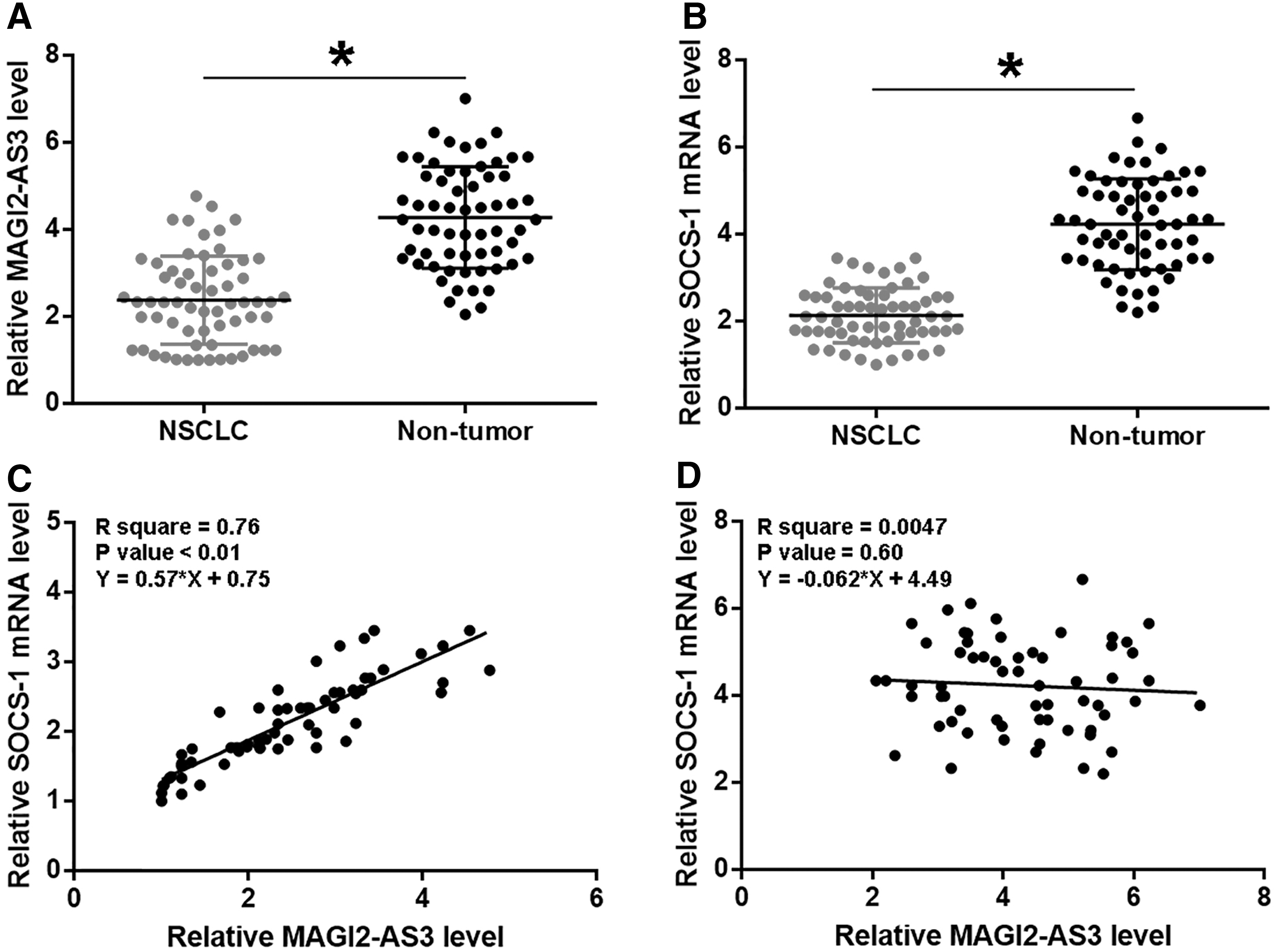
MAGI2-AS and SOCS-1 were positively correlated in NSCLC. Levels of MAGI2-AS3
Low levels of MAGI2-AS3 and SOCS-1 mRNA predicted poor survival
Survival curves of high and low MAGI2-AS3/SOCS-1 level groups were plotted and compared using the methods aforementioned. It was observed that the overall 5-year survival rate (disease specific) of patients in low MAGI2-AS3 level group was significantly lower compared with high MAGI2-AS3 level group (Fig. 2A). Similarly, patients in low SOCS-1 also showed significantly lower overall survival rate compared to patients in high SOCS-1 level group (Fig. 2B).
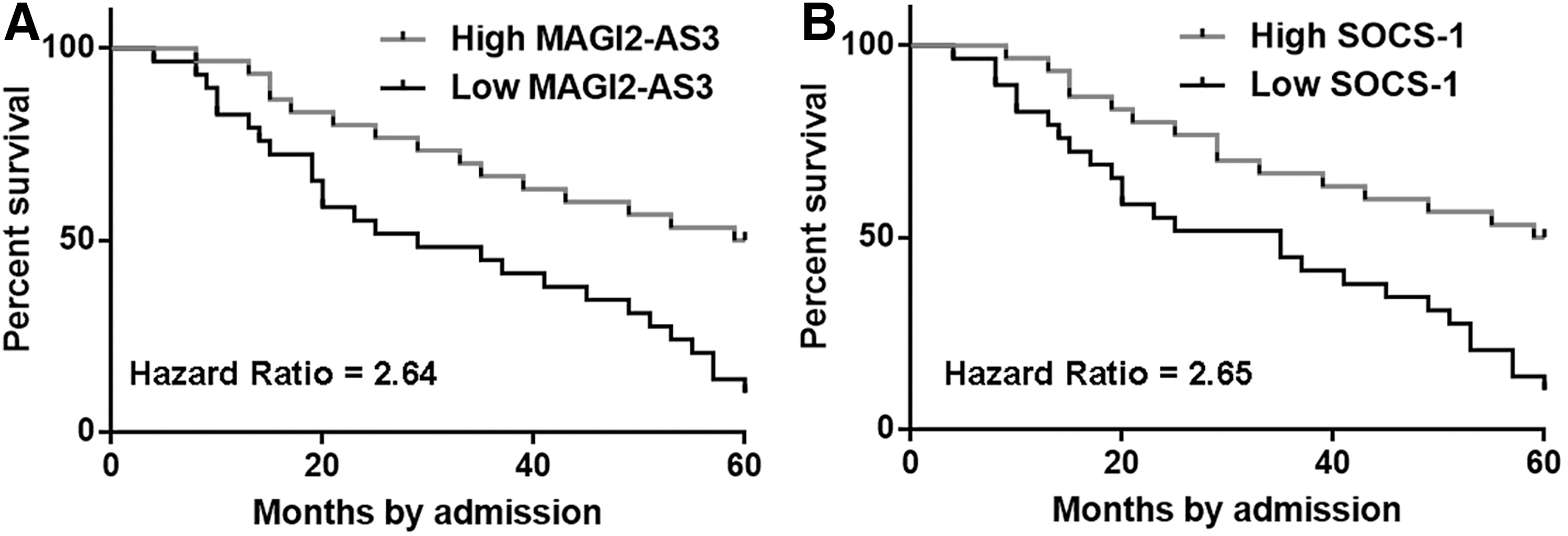
Low levels of MAGI2-AS3 and SOCS-1 mRNA predicted poor survival. Patients were divided into high and low MAGI2-AS
MAGI2-AS3 upregulated SOCS-1 by sponging miR-155 in H23 cells
H23 cells were transfected with MAGI2-AS3 and SOCS-1 expression vectors, as well as miR-155 mimic. Expression levels of MAGI2-AS3, miR-155, and SOCS-1 were measured by performing qPCR. Comparing to C and NC (empty pcDNA3.1 vector or miRNA NC) groups, expression levels of MAGI2-AS3, miR-155, and SOCS-1 mRNA were significantly increased at 24 h post-transfections (Fig. 3A, p < 0.05). MiR-155 can target SOCS-1. RNA interaction prediction by IntaRNA showed that miR-155 may bind MAGI2-AS3 (Fig. 3B). Compared to C and NC (empty pcDNA3.1 vector or miRNA NC) two controls, overexpression of miR-155 and MAGI2-AS3 failed to affect the expression levels of each other (Fig. 3C). Moreover, MAGI2-AS3 overexpression mediated the upregulated, while miR-155 expression mediated the downregulated SOCS-1 overexpression at both mRNA (left) and protein levels (middle and right), and miR-155 expression reduced the effects of MAGI2-AS3 overexpression (Fig. 3D).
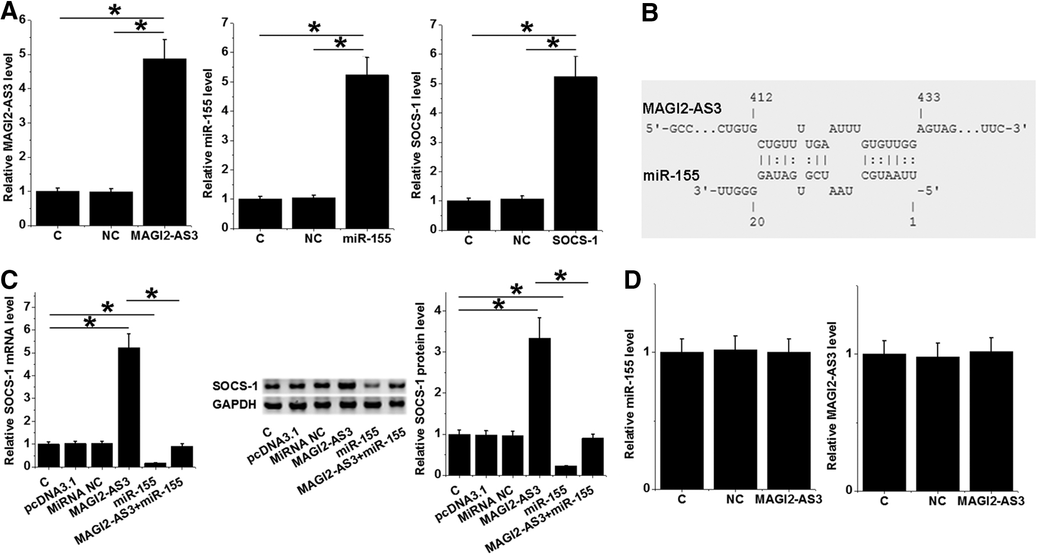
MAGI2-AS3 upregulated SOCS-1 by downregulating miR-155 in H23 cells. H23 cells were transfected with MAGI2-AS3 and SOCS-1 expression vectors, as well as miR-155 mimic. Overexpression of MAGI2-AS3, miR-155, and SOCS-1 was measured by performing qPCR
MAGI2-AS3 interacted with miR-155 and SOCS-1 to suppress the proliferation of H23 cells
The effects of MAGI2-AS3, miR-155, and SOCS-1 overexpression on cell proliferation were explored by performing cell proliferation assay. Compared to C group, significantly decreased cell proliferation rate of H23 cells was observed after MAGI2-AS3 and SOCS-1 overexpression. MiR-155 played an opposite role and reduced the effects of MAGI2-AS3 overexpression (Fig. 4, p < 0.05).

MAGI2-AS3 interacted with miR-155 and SOCS-1 to suppress the proliferation of H23 cells. The effects of MAGI2-AS3, miR-155, and SOCS-1 overexpression on cell proliferation were explored by performing cell proliferation assay. Cell proliferation rates were measured at 24, 48, 72 and 96 h after the innitiation of cell culture time points. Mean values of three biological replicates were presented, *p < 0.05.
Discussion
This study analyzed the functions of MAGI2-AS3 in NSCLC. The authors found that MAGI2-AS3 was downregulated in NSCLC and predicted the poor survival of NSCLC patients. Moreover, MAGI2-AS3 may sponge miR-155 to downregulate SOCS-1, thereby inhibiting cancer cell proliferation.
Two previous studies have characterized MAGI2-AS3 as a tumor-suppressive lncRNA in two types of cancers. 13,14 In breast cancer, MAGI2-AS3 is downregulated and MAGI2-AS3 expression targets Fas/FasL signaling to suppress the growth of breast cancer. 13 In bladder cancer, MAGI2-AS3 is also downregulated and regulates CCDC19 expression to suppress cancer progression. 14 Consistently, this study also observed the downregulation of MAGI2-AS3 in NSCLC tissues compared to nontumor lung tissues. In addition, cell proliferation was significantly enhanced after MAGI2-AS3 overexpression. Therefore, MAGI2-AS3 is also a tumor suppressor in NSCLC.
The authors' bioinformatics analysis showed that miR-155 can bind MAGI2-AS3. It is known that miRNAs can target other RNAs to induce the degradation or inhibit mRNA translation. 15 However, overexpression experiments showed that miR-155 and MAGI2-AS3 have no effect on the expression of each other. Therefore, miR-155 may not target MAGI2-AS3 in NSCLC. It has been well established that lncRNAs can be the endogenous sponge of miRNAs to attenuate their functions. 16,17 In this study, the authors observe that MAGI2-AS3 overexpression led to the upregulation of SOCS-1, which is the direct target of miR-155. 12 Therefore, MAGI2-AS3 may sponge miR-155 to upregulate SOCS-1, which can inhibit tumor growth. 10,11 However, it is known that miR-155 can target multiple genes in cancer biology, such as FOXO3a. 18 The authors' future studies will explore the involvement of other targets of miR-155 in MAGI2-AS3-mediated proliferation inhibition of NSCLC cells.
The authors' 5-year long-term follow-up study revealed that MAGI2-AS3 expression detection may be used to assist the prognosis of NSCLC patients. The accurate prognostic assignment of NSCLC patients may improve their survival by guiding the determination of therapeutic approaches. Their future studies will focus on the accuracy of the use of MAGI2-AS3 as a prognostic marker for NSCLC. Due to the experimental design, Charlson's comorbidity index was not included in this study. The authors' future studies will try to include this index to further confirm their conclusions.
In conclusion, MAGI2-AS3 was downregulated in NSCLC and may sponge miR-155 to upregulate SOCS-1, thereby inhibiting cancer cell proliferation.
Footnotes
Disclosure Statement
The authors declare that they have no competing interests.
Funding Information
The authors received financial support from the Jiangsu Provincial Traditional Chinese Medicine Administration (YB2017101) and Talent Start Fund of Affiliate Hospital of Jiangsu University (jdfyRC2017002).