
Abstract
Background:
It is known that LINC00974 is an oncogenic long noncoding RNA in liver cancer.
Results:
The authors observed in this study that LINC00974 was upregulated in gastric cancer (GC) and positively correlated with CDK6. Survival analysis showed that high levels of LINC00974 and CDK6 predicted poor survival. In GC tissues, LINC00974 and CDK6 were positively correlated. In GC cells, LINC00974 overexpression led to upregulated, whereas LINC00974 siRNA silencing led to downregulated CDK6. Analysis of cell cycle progression and proliferation showed that LINC00974 and CDK6 overexpression promoted and siRNA silencing inhibited G1-S transition and cell proliferation.
Conclusion:
Therefore, LINC00974 upregulates CDK6 to promote cell cycle progression in GC.
Introduction
Gastric cancer (GC) is a common malignancy and major cause of cancer deaths. In 2012, GC affected ∼951,600 new cases and caused ∼723,100 deaths worldwide. 1 A considerable portion of GC patients are diagnosed with inoperable disease and postoperative recurrence is common, leading to poor survival. 2 As the most common types of GC, gastric adenocarcinoma (GAC) is a major of public health burden. 3,4 Many risk factors such as Helicobacter pylori seropositivity and lifestyle have been identified for GC, 4,5 whereas pathogenesis of this disease remains hardly known. 6 The unknown pathogenesis is the major challenge in the development of novel therapies.
Genetic factors are the most critical players in the tumorigenesis and progression of GC. 7 Cyclin-dependent kinases, or CDKs, interact with their cyclin partners to regulate cell cycle progression. 8 Different CDKs participate in the regulation of different cell phases, for instance, CDK6 mainly acts on G1 phase, whereas CDK2 mainly plays its roles in S phase. 9 In cancers, CDKs are overexpressed and promote cancer cell cycle progression, which in turn accelerates cell division and tumor growth. 10 Therefore, inhibiting the expression of CDKs is a potential target for cancer therapies. It is known that the expression of certain CDKs, such as CDK6, can be regulated by certain (>200 nt) long noncoding RNAs (lncRNA), 11,12 which encode no proteins but regulate cancer-related gene expression. 13 LINC00974 is a recently identified regulator in liver cancer cell proliferation, indicating its possible interactions with CDKs. 14 This study aimed to analyze the interaction between LINC00974 and CDK6 in GC.
Materials and Methods
GC patients
In this study 66 GC patients (all were adenocarcinoma cases; gender: 36 men and 30 women; age: 32–67 years; mean age: 47.3 ± 5.0 years) were selected from the 122 GC patients admitted to Baotou Cancer Hospital between July 2010 and April 2014. This study passed the review board of Baotou Cancer Hospital before enrolling patients. Inclusion criteria: (1) new GC cases; (2) no therapies were initiated. Exclusion criteria: (1) recurrent GC; (2) history of malignancies; (3) other severe clinical disorders were observed. After admission, all the 66 GC patients were educated with the principle of this study and the potential publication of data from this study. All patients signed informed consent. According to the staging system established by AJCC, the 66 GC patients included 19, 20, and 27 cases at stages II–IV, respectively.
Follow-up
From the day of admission, survival conditions of all patients were monitored for 5 years. Follow-up was performed in a monthly manner through telephone. The patients died of other causes or lost before the end of follow-up were excluded from subsequent survival analysis.
GC tissues and cells
Before the initiation of therapies, nontumor gastric tissues and GC tissues were collected from each patient by performing fine needle gastric biopsy. The weight of each tissue sample ranged from 0.02 to 0.032 g. Based on histopathological examination results, GC tissues contained >95% cancer cells and nontumor tissues contained <5% cancer cells.
AGS human GC cell line (GAC, ATCC) was used. Cells were cultivated in a mixture of 90% F-12K Medium and 10% fetal bovine serum (FBS). Cell culture conditions were 37°C, 5% CO2, and 95% humidity.
Transient transfections
LINC00974 and CDK6 expression vectors were constructed using pcDNA3.1 vector (Sangon, Shanghai, China). Negative control (NC) siRNA and LINC00974 siRNA were also from Sangon. To perform transfections, AGS cells were harvested at confluence of 70%–80%, followed by transfection of 10 nM vector (empty vector as NC group) or 35 nM siRNA (NC siRNA as NC group) into 6 × 10⁵ cells using Lipofectamine 2000 (Thermo Fisher Scientific). Cells were collected at 24 h post-transfection to perform the following experiments. Untransfected cells were control (C) cells.
RNA extractions and qPCR
Total RNAs in tissues (ground in liquid nitrogen) and AGS cells were extracted using RNAzol (Sigma-Aldrich). After digestion with DNase I, total RNAs were subjected to reverse transcriptions using AMV Reverse Transcriptase (Sangon). The synthesized cDNA and SYBR® Green master mix (Takara, Japan) were used to prepare qPCR mixtures with GAPDH as endogenous control to measure the expression levels of LINC00974 and CDK6 mRNA.
Cell cycle assay
AGS cells were collected 24 h after transfection and trypsinization was performed. Pre-cold PBS was used to wash cells and ethanol (75%) was then used to incubate with the cells at 4°C for 4 h. After washing with pre-cold PBS again, cells were stained with BD Pharmingen™ PI/RNase for 30 min. Finally, cells were separated by flow cytometer and 105 events were included in each experiment.
Cell proliferation assay
At 24 h after transfection, AGS cells were collected and mixed with the mixture of 90% F-12K Medium and 10% FBS with a ratio of 4 × 10⁴ cells per 1 mL. Cells were mixed well with cell culture medium to prepare single cell suspensions. A 96 cell culture plate was used to cultivate cells with 0.1 mL per well at 37°C, followed by the addition of 10 μL CCK-8 solution (Sigma-Aldrich) at 4 h before the end of cell culture. After cell culture was terminated, OD values were measured at 450 nm.
Western blot
Total proteins in 5 × 10⁵ cells were extracted using RIPA solution (Thermo Fisher Scientific). Electrophoresis was performed using 10%-PAGE gel to separate different proteins after protein denaturing in boiling water for 5 min. Gel transfer was performed using PVDF membranes, and blocking was performed in PBS containing 5% FBS for 2 h at room temperature. Membranes were first blotted with GAPDH (1:900, ab9485; Abcam) or CDK6 (1:900, ab22349; Abcam) rabbit primary antibodies for 12 h at 4°C, followed by blotting with goat IgG-HRP (1:1500, MBS435036; MyBioSource) secondary antibody for 2 h at room temperature. Membranes were then incubated with ECL (Sigma-Aldrich) for 5 min at room temperature to develop signals. Data were processed using Image J v1.46 software.
Statistical analysis
All data analyses were performed using mean values of the data of three biological replicates. Differences between GC and nontumor tissues were explored by paired t-test. Differences among multiple cell groups were analyzed by ANOVA (one-way) and Tukey test. Correlations were analyzed by linear regression. The 66 GC patients were grouped into high and low LINC00974/CDK6 level groups according to the median expression level. Survival curves were plotted using Kaplan–Meier plotter and compared by log-rank test. p < 0.05 was statistically significant.
Results
LINC00974 and CDK6 mRNA were positively correlated in GC
Expression levels of LINC00974 and CDK6 mRNA in both nontumor and GC tissues were measured by qPCR and compared by paired t-test. Comparing with nontumor group, expression levels of LINC00974 (Fig. 1A) and CDK6 mRNA (Fig. 1B) were significantly higher in GC group (p < 0.05). Correlations between LINC00974 and CDK6 mRNA were analyzed by linear regression. It can be observed that expression level of LINC00974 was significantly and inversely correlated with that of CDK6 mRNA in GC tissues (Fig. 1C). However, the correlation was not significant in nontumor tissues (Fig. 1D).

LINC00974 and CDK6 mRNA were positively correlated in GC. Expression levels of LINC00974
High levels of LINC00974 and CDK6 mRNA predicted poor survival
Survival curves of both high and low LINC00974/CDK6 level groups were plotted using Kaplan–Meier plotter and compared by log-rank test. It can be observed that survival rate of patients in high LINC00974 level group was significantly lower than that of patients in low LINC00974 level group (Fig. 2A). In addition, survival rate of patients in high CDK6 mRNA level group was also significantly lower than that of patients in low LINC00974 level group (Fig. 2B).
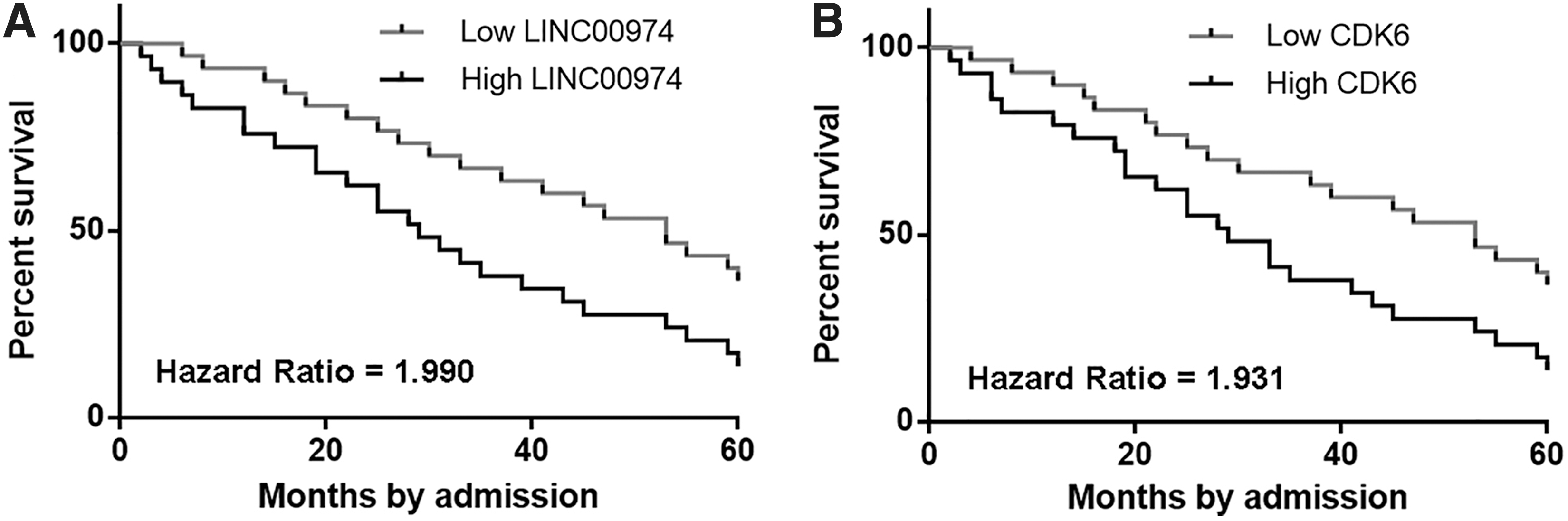
High levels of LINC00974 and CDK6 mRNA predicted poor survival. Survival curves of both high and low LINC00974
LINC00974 negatively regulated CDK6 in AGS cells
AGS cells were transfected with LINC00974 and CDK6 expression vectors as well as LINC00974 siRNA. Expression levels of LINC00974 and CDK6 mRNA were measured by qPCR. Comparing with two controls (C and NC), expression levels of LINC00974 and CDK6 mRNA were significantly altered at 24 h post-transfection (Fig. 3A, p < 0.05). Moreover, LINC00974 overexpression led to upregulated, whereas LINC00974 siRNA silencing led to downregulated CDK6 at both mRNA and protein levels (Fig. 3B, p < 0.05). However, CDK6 overexpression failed to affect LINC00974 expression.
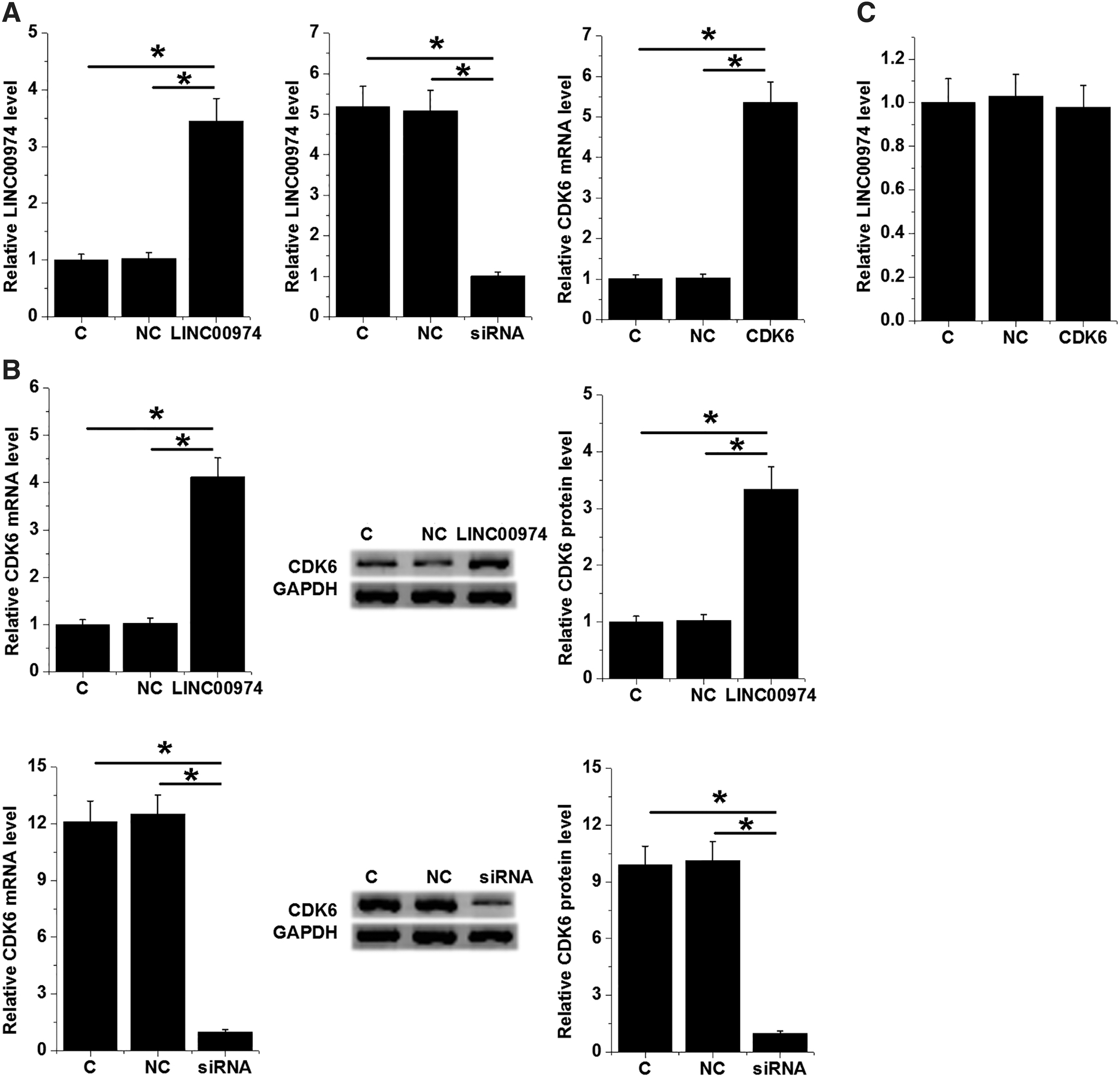
LINC00974 negatively regulated CDK6 in AGS cells. AGS cells were transfected with LINC00974 and CDK6 expression vectors as well as LINC00974 siRNA. Altered expression levels of LINC00974 and CDK6 mRNA were detected by qPCR at 24 h post-transfection
LINC00974 positively regulated AGS cell cycle progression and proliferation through CDK6
The effects of LINC00974 and CDK6 overexpression as well as LINC00974 siRNA silencing on AGS cell progression and proliferation were analyzed by cell cycle assay and cell proliferation assay, respectively. Comparing with NC and C groups, LINC00974 overexpression promoted and siRNA silencing inhibited G1-S transition (Fig. 4A, p < 0.05; reflected by the percentage of cells at G1 and G2 phases) and cell proliferation (Fig. 4B, p < 0.05). In addition, CDK6 overexpression attenuated the effects of LINC00974 overexpression (p < 0.05).
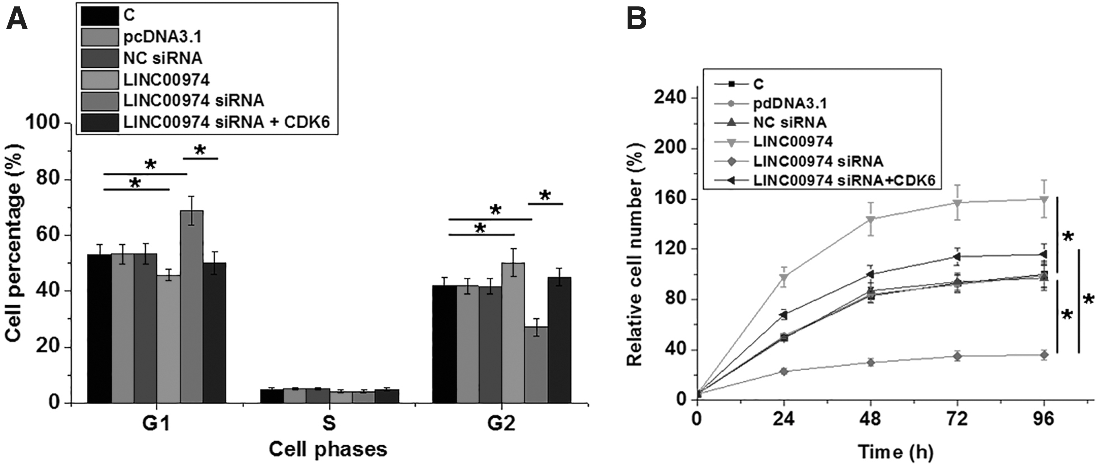
LINC00974 positively regulated AGS cell cycle progression and proliferation through CDK6. The effects of LINC00974 and CDK6 overexpression as well as LINC00974 siRNA silencing on AGS cell progression and proliferation were analyzed by cell cycle assay
Discussion
This study mainly investigated the roles of LINC00974 in GC. The authors found that LINC00974 was upregulated in GC and predicted poor survival. In addition, LINC00974 may positively regulate CDK6 to promote the cell cycle progression and cell proliferation.
The functions of LINC00974 have only been analyzed in oral fibrogenesis and hepatocellular carcinoma (HCC). In oral fibrogenesis, LINC00974 is upregulated and promotes disease progression. 15,16 In HCC, LINC00974 is also upregulated and activates Notch and transforming growth factor-beta (TGF-β) pathways to promote cancer cell proliferation and metastasis. 14 Based on the authors' knowledge, the involvement of LINC00974 in GC is still unknown. This study is the first to report the upregulation of LINC00974 in GC. In addition, the authors also observed the accelerated G1-to-S phase transition and GC cell proliferation after LINC00974 overexpression. Therefore, LINC00974 is an oncogenic lncRNA in GC.
In this study the authors showed that LINC00974 can upregulate the expression of CDK6. However, the mechanism of the interaction between them is unclear. Previous studies have shown that CDK6 has crosstalk with TGF-β and NORTH signaling, 17 while LINC00974 regulates TGF-β and NORTH pathways. 14 Therefore, TGF-β and NORTH may mediate the interaction between LINC00974 and CDK6. LncRNAs are known to sponge miRNAs to upregulate the expression of their downstream targets. 18 Therefore, LINC00974 may sponge certain miRNAs to upregulate CDK6. The authors' future studies will explore this possibility.
Clinical treatment of GC is still challenged by the low early diagnostic rate. 1 Early diagnosis is challengeable owing to the lack of sensitive and specific markers, and this situation is unlikely to be changed in near future. 2 Accurate prognostic assignment may be another approach to improve the survival of GC patients by guiding the selection of treatment approaches and the development of care program. In this study the authors showed that high levels of LINC00974 and CDK6 can be used as the markers of poor survival of GC patients. However, the accuracy remains to be further analyzed.
This study is limited by a small sample size. Future studies with a larger sample size are needed to further confirm the conclusions. In addition, animal models are needed to verify the authors' observations under in vivo conditions.
In conclusion, LINC00974 was upregulated in GC and may positively regulate CDK6 to promote GC cell proliferation and cell cycle progression.
Footnotes
Disclosure Statement
No competing financial interests exist.
Funding Information
No funding was received for this article.