
Abstract
Background:
This study aims at investigating the effect of the Weifufang, an effective prescription for the treatment of gastric cancer developed by the Traditional Chinese Medicine (TCM)/Combination of TCM and Western Medicine Department of the Hunan Cancer Hospital, on gastric cancer xenografts in nude mice and its effect on the PTEN gene; it also aims at exploring the possible tumor suppression mechanism.
Methods:
Nude mice with xenografts were treated with different concentrations of the Weifufang for 2 weeks, and changes in tumor volume were observed. The histopathology of the tumor was detected by hematoxylin and eosin staining; PTEN gene expression in tumor tissues was detected by immunohistochemistry (IHC) and western blot.
Results:
After 2 weeks of treatment, tumor inhibition rates in the 5-flourouracil (5-FU) group, and in the Weifufang low-, middle-, and high-dose groups were 30.67%, 19%, 49.52%, and 29.36%, respectively. The IOD of the PTEN gene was detected by IHC. The values in the water group, the 5-FU group, and the Weifufang low-, middle-, and high-dose groups were 0.013 ± 0.004, 0.085 ± 0.062, 0.041 ± 0.024, 0.128 ± 0.032, and 0.061 ± 0.052, respectively. Except for the 5-FU group, the differences between the gastric compound middle dose-group and the other groups were statistically significant (p < 0.05). Results of PTEN expression detection by western blot: The expression levels in the water group, 5-FU group, and the Weifufang low-, middle-, and high-dose groups were 0.2240 ± 0.0172, 0.4200 ± 0.0228, 0.2760 ± 0.0163, 0.3840 ± 0.0133, and 0.3040 ± 0.0211, respectively. Except for the 5-FU group, differences between the Weifufang middle-dose group and the other groups were statistically significant (p < 0.05).
Conclusion:
The Weifufang may inhibit the growth of gastric cancer xenografts by upregulating PTEN gene expression. The middle-dose group had the best effect.
Introduction
Gastric cancer is recognized as one of the most common malignant tumors that severely threatens the health of humans worldwide. According to the 2014 World Cancer Report released by the World Health Organization, 1 gastric cancer is the fifth most common cancer in the world, and its mortality ranks third in the world. It was estimated that new cases of gastric cancer reached 952,000 in 2012, and that the number of deaths was ∼723,000; among these, more than 70% occurred in developing countries, especially in China. 2 Gastric cancer has become the second most common cancer in China, and its mortality ranks third. 3 In recent years, neoadjuvant chemotherapy and molecular targeted drug therapy have prolonged the survival of patients with advanced gastric cancer to a certain extent, and improved their quality of life. However, the 5-year survival rate remained lower than 10%. 4 The occurrence and development of gastric cancer is related to the activation of oncogenes and the inactivation of tumor suppressor genes.
Phosphatase and tensin homolog deleted on chromosome 10 (PTEN) was the first tumor suppressor gene found with phosphatase activity following the P53 gene, and the function area of tumor suppression is located in the N-terminal phosphatase domain. 5 Studies have revealed that PTEN in involved in the regulation of tumors through the following pathways: PI3K/AKT pathway, Shc/MAPK pathway, and FAK/p130cas pathway. 6,7 Further, studies have revealed that PTEN expression presented a decrease in digestive tract tumors, hematological tumors, urinary tract tumors, respiratory tumors, nervous system tumors, motor system tumors, and reproductive system tumors. 8 –16 PTEN gene mutation, loss of heterozygosity and over methylation, microRNA, and microsatellite instability constitute the basic molecular biology of the downregulation of PTEN gene expression. 15 PTEN is involved in the occurrence and development of gastric cancer. Further, PTEN expression level is correlated with the differentiated degree of stomach cancer tissue and lymph node metastasis; whereas PTEN expression level is low in poorly differentiated gastric cancer tissue and lymph node metastasis.
The Weifufang is an effective prescription for the treatment of gastric cancer, and it was developed by the Traditional Chinese Medicine (TCM)/Combination of TCM and Western Medicine Department of Hunan Cancer Hospital through long-term clinical practice. 16 –20 This compound exhibits a relatively good antitumor effect in clinics, effectively improves the quality of life of patients, and prolongs survival. However, the antitumor effect of compound Chinese herbal medicine is an extremely complex biological process. To explore its possible mechanism, further studies are needed. In this study, we observed the changes in size of the transplanted tumor in nude mice with gastric cancer treated with different doses of the Weifufang, observed the histopathology of tumors by hematoxylin and eosin (H&E) staining, and detected the expression of the PTEN gene in tumor tissues by immunohistochemistry (IHC) and western blot. Therefore, the effect and possible mechanism of the Weifufang on the growth of the transplanted tumors in nude mice were preliminary studied.
Materials and Methods
Materials
Animals and cell lines: BALB/c-nu nude mice (n = 52, SPF class) were provided by the Hunan Provincial Tumor Hospital Experimental Animal Center (Experimental animal license number: SYXK [Hunan] 2014-0016; Experimental animal production license number: SCXK [Hunan] 20110003). All nude mice were male and weighted within 18–20 g. Each nude mouse was fed in a single cage with standard forage at a temperature of 22°C–24°C with a humidity of 50 ± 10%, and 12 h of light was provided per day. All animals successfully passed through a week of quarantine. The experimental process was in line with the relevant provisions of the Guiding Opinions on the Management of Laboratory Animals established by the Ministry of Science and Technology of the People's Republic of China in 2006. The experiment was reviewed and approved by the Animal Ethics Committee of Hunan Provincial Tumor Hospital. The human gastric cancer BGC-823 cell line was provided by Boster Co. Ltd. (Wuhan, China).
The Weifufang (provided by the TCM dispensary of Hunan Provincial Tumor Hospital, each medicinal herb had a clear origin and batch, and was identified by the Department of Medicine, Medicine School, Hunan University of Chinese Medicine): Astragalus, 20 g; Codonopsis pilosula, 15 g; Atractylodes macrocephala, 10 g; Poria cocos, 10 g; Nutgrass Galingale Rhizome, 10 g; Radix Curcumae, 10 g; Sappan Wood, 10 g; Rhizoma Curcumae, 10 g; Zaoxiu Paris Root Rhizoma Paridis, 10 g; Barbed Skullcap Herb, 15 g; Ligustrum lucidum, 10 g; South Dodder Seed, 10 g; Oldenlandia, 15 g; Liquorice Root, 5 g; Chicken's Gizzard-membrane, 10 g; fry malt, 15 g; and fry Rice-grain Sprout, 15 g. According to the Standard Decoction Theory, 21 the percentage concentration of the standard decoction = the weight of the medicinal material/the amount of juice. The amount of water used for decoction was 5 mL of water per gram of TCM, in which 70% of this water was added in the first boil and 30% of this water was added in the second boil. Herbs were soaked for ∼30 min and boiled for 20–30 min; the decoction obtained in the two boils was mixed, filtrated, and concentrated to the desired concentration. The decoction was transferred into cans, sterilized, and placed in a refrigerator at 4°C.
Main reagents and related materials
RPMI-1640, fetal bovine serum (FBS), phosphate-buffered saline (PBS), myllicin, and trypsin were purchased from Hyclone; Tris, ammonium persulfate, sodium dodecyl sulfate, tetramethylethylenediamine, Tween-20, acrylamide, glycine and bisacrylamide, Ponceau, paraffin, and neutral gum were purchased from Sigma; hematoxylin, PBS, and citrate buffer were purchased from Wellbio; the two-step immunohistochemistry kit and diaminobenzidine kit were purchased from ZSGB-BIO (Beijing, China); horseradish peroxidase (HRP)-labeled goat anti-mouse immunoglobulin G (IgG) and HRP-labeled goat anti-rabbit IgG were purchased from Proteintech; Super ECL Plus Detection Reagent was purchased from Thermo Pierce; RIPA lysate was purchased from Applygen Technologies, Inc. (Beijing, China); protease inhibitors were purchased from Merck (German); protein phosphatase inhibitor was purchased from Roche (Swiss); developer and fixer were purchased from Wellbiology (China); and 5-flourouracil (5-fu) was purchased from Shanghai Xudong Haipu Pharmaceutical Co. Ltd., batch number: H31020593, diluted with 0.9% normal saline at a ratio of 1:9, and stored in a refrigerator at 4°C.
Experimental methods
Cell culture
Cells were cultured in RPMI-1640 containing 10% FBS at 37°C with 5% CO2. Cells that reached the logarithmic growth phase (2 × 10⁷/L cell suspension) were selected for the experiment.
Modeling
The prepared single-cell suspension of human gastric cancer (concentration: 2 × 10⁷ cells/mL) was inoculated subcutaneously in the dorsal side of the armpit of two nude mice (tumor-supplying mice) at 0.2 mL for each mouse (equivalent to 4 × 10⁶ cells/mL). When a subcutaneous tumor with a long diameter of ∼1.5 cm was observed in each nude mouse, tumor-supplying mice were sacrificed by cervical dislocation. The tumor mass was stripped, cut into small pieces at ∼1 × 1 mm, and passaged in mice to the third generation (two mice for one generation). Nude mice were fasted for 6–8 h before the operation, and anesthesia was induced by an intraperitoneal injection of 0.1 mL/10 g of 5% chloral hydrate. The third generation of the tumor mass was inoculated in subcutaneous tissue with a trocar 22 to establish a subcutaneously transplanted gastric tumor model of nude mice. The whole process was strictly performed under aseptic conditions, and the inoculation was completed within 30 min. The situation of nude mice and the growth of subcutaneous tumors were observed daily until the size of hard tumor nodules reached ∼3 × 3 mm.
Conversion of dose and concentration between humans and mice
According to the ratio of the body surface areas between humans and mice, the equivalent dose of crude drug for mice was 0.52 g (this is one fold of the dose; a double dose was 1.04 g, and a 1/2-fold dose was 0.26 g). The calculated concentrations of the decoction were as follows: the concentration of each ml of crude drug = 2.6 g/(mL · d) in the high-dose group, 1.3 g/(mL · d) in the middle-dose group, and 0.65 g/(mL · d) in the low-dose group.
Groupings and observation
The 47 successfully modeled nude mice were divided into five groups according to the random number table: negative control group (n = 7), mice treated with distilled water; Weifufang low-dose group (n = 10), mice that received a 1/2-fold dose; Weifufang middle-dose group (n = 10), mice that received a one-fold dose; Weifufang high-dose group (n = 10), mice that received a double dose; and Weifufang 5-FU positive control group (n = 10). The general condition of animals (routine indicators: activity, reactivity, spirit, skin, stool, and urination) was observed and recorded. The long and short diameters of the tumor in nude mice were measured by using a Vernier caliper. Then, the tumor volume was calculated by using the formula V = ab 2/2 23 (a and b are the maximum and shortest diameters of the tumor, respectively). Observation was continued for 14 d, and the body weight of nude mice was measured and recorded every 3 d with an electronic balance.
Drug treatment and calculation of tumor inhibition rate
The amount of gavage for mice was 0.2–0.3 mL/10 g per session. Therefore, a 20-g mouse was infused with a volume of 0.4 mL, once a day at 9
H&E staining
Tumor tissues were removed, fixed with 4% paraformaldehyde for 24–72 h, dehydrated, treated with xylene until they become transparent, embedded by paraffin, and sliced in 8-μm-thick sections by using a microtome. The sections were pastered on gummed paper, dewaxed, and stained. The prepared sections were placed under the microscope to observe for pathological changes in tumor tissue. The tissue sections were observed by pathology experts of Hunan Human Provincial Tumor Hospital and Hunan University of Chinese Medicine.
Detection of PTEN gene expression in tumor tissues by SP IHC
Specific steps were performed according to kit instructions. A negative control group was set to exclude false positive and false negative. When the nucleus/cytoplasm was stained to yellow or brownish yellow (when the color was deep, it was brown), cells were considered to be positive for the PTEN gene. When only the cytoplasm was stained and the nuclei were not stained, or the nucleus and cytoplasm were not stained, these were considered negative cells. Image acquisition was conducted by using an ordinary computer. The image analysis software used was Image-Pro-Plus. Average IOD was the ratio of the accumulated optical density and the area of the sample under the visual field (for visual field of a 400 × light microscope). The tissue sections were observed by pathology experts of Hunan Provincial Tumor Hospital and the Hunan University of Chinese Medicine.
Detection of PTEN gene expression in tumor tissues by Western blot
The quantitative analysis of tumor tissues and protein was performed according to kit instructions, and the experiment was performed in triplicate. The target bands were analyzed by using the Quantity-one image analysis system. The target protein expression levels were corrected by using β-actin.
Statistical methods
Univariate analysis of variance was performed by using statistical software SPSS 17.0. The multiple comparisons of means between two samples were performed by using least significant difference. Measurement data were expressed as mean ± standard deviation (x ± SD). p < 0.05 was considered statistically significant.
Results
Establishment of the subcutaneously transplanted tumor model of nude mice
Among these 50 nude mice, 47 mice were successfully modeled, whereas 3 mice died (bite marks were observed in some mice fed in the same cage, and these mice revealed obvious emaciation; no obvious pathological changes in organs was found by anatomy, hence mortality by mutual biting was considered).
Observation of the general situations of nude mice in each group during the 2-week drug treatment
The general situations of nude mice in the gastric compound low- and middle-dose groups were normal. Nude mice in the 5-FU group and Weifufang high-dose group were treated for 1 h each time. After treatment, all mice were rarely activated, stood packed together tightly, and subsequently returned to the normal state. The nude mice treated with distilled water had slightly poor mental activity, presented with folds in the skin, had poor response to external stimulation, and were rarely activated. This revealed significant emaciation in the later stage of the treatment.
Measurement of changes in body weight and tumor volume, tumor weight, and the calculation of the tumor growth IR during drug treatment
As shown in Figure 1, the weight of nude mice in each group initially exhibited an increasing trend, decreased, and further decreased in the late stage. This was most obvious in the negative control group, followed by the Weifufang low-dose group. Further, this exhibited a steady trend in the late stage in the Weifufang high-dose group, whereas this initially exhibited a decreasing trend, followed by slight increase in the late stage, in the 5-FU positive control group and Weifufang middle-dose group. The results just cited revealed that nude mice in the negative control group were not treated by the drugs, and the reason may be that the load of the tumors and physical consumption may be high, which decreased the weight in the late stage. The weight of nude mice in the Weifufang high-dose group revealed a steady trend in the late stage, and the reason may be that the antitumor effect of the TCM and the effect of tumors on physical consumption form a state of confrontation. The weight of nude mice in the 5-FU positive control group and Weifufang middle-dose group decreased, and it increased slightly in the late stage. The reason may be that the effect of the antitumor of the drug resisted the effect of tumors on physical consumption, and this may improve the quality of life of nude mice. This study revealed that the Weifufang can improve the survival and quality of life of nude mice with gastric cancer xenografts, and it may have a certain antitumor effect.
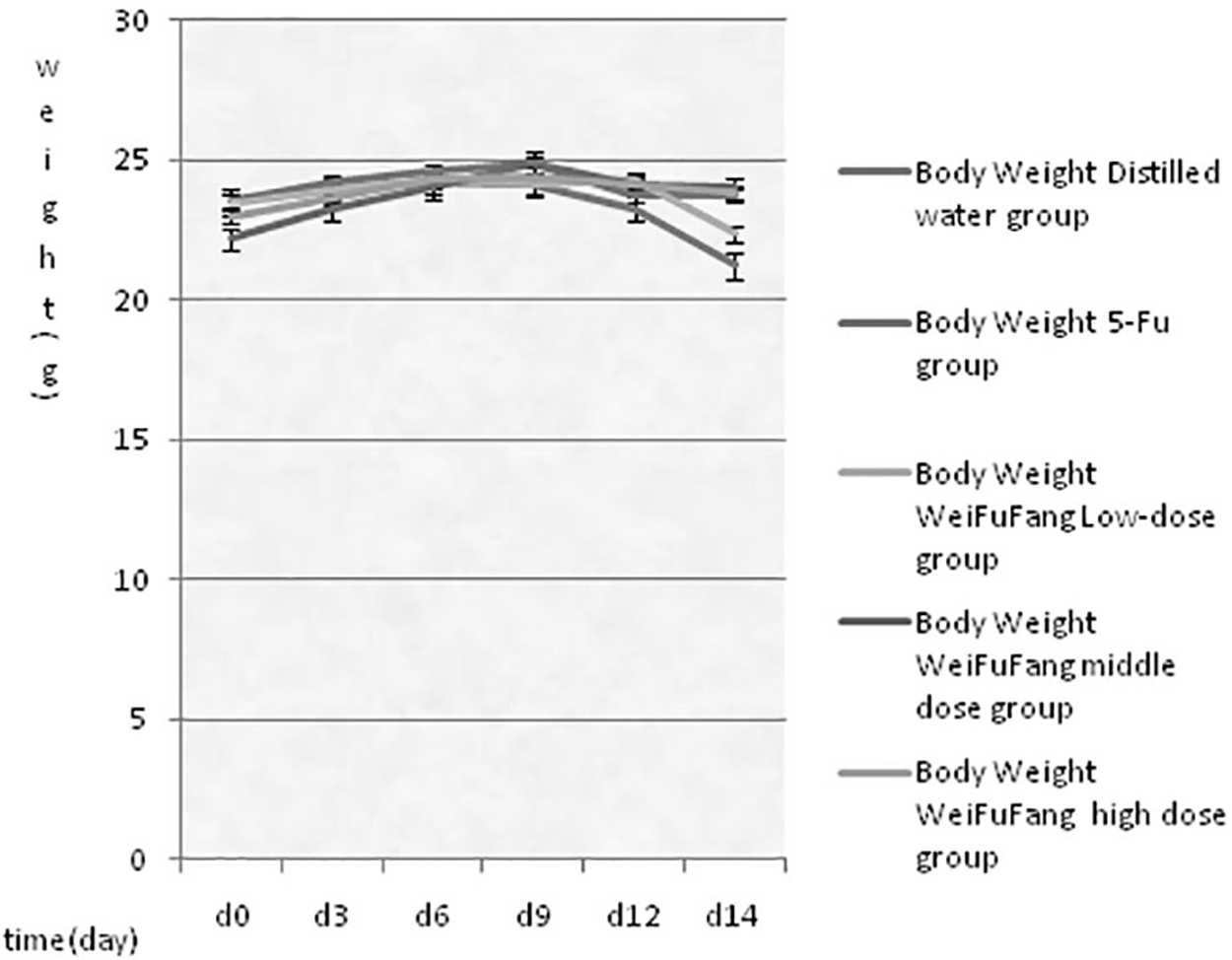
The tendency of the average body weight to change with time in each group.
As shown in Figure 2, the growth rate of nude mice in the negative control group was faster, and the average volume was 172 mm3. The growth rates of nude mice in the other groups increased at the early stage of drug treatment, and they decreased in the late stage of drug treatment. This decrease was obvious in the 5-FU positive control group and Weifufang middle-dose group at the late stage of the drug treatment. The average volumes in the negative control group, 5-FU positive control group, and in the Weifufang low-, middle-, and high-dose groups were 172, 80, 108, 52, and 110 mm3, respectively. These results revealed that the inhibitory effect on the growth of subcutaneous tumors in nude mice was significantly better in the drug treatment groups than that in the negative control group, and the best effect was observed in the Weifufang middle-dose group.
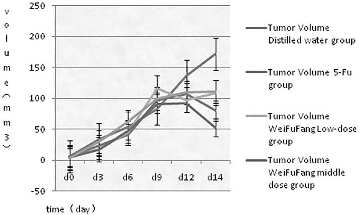
The tendency of the average tumor volume to change with time in each group.
As shown in Figure 3, the order of tumor weights was as follows: negative control group > Weifufang low-dose group > Weifufang high-dose group > 5-FU positive control group > Weifufang middle-dose group. Thus, the inhibitory effect on the growth of subcutaneous tumors in nude mice was significantly better in the drug treatment groups than in the negative control group, and the antitumor effect in the Weifufang middle-dose group was the best.
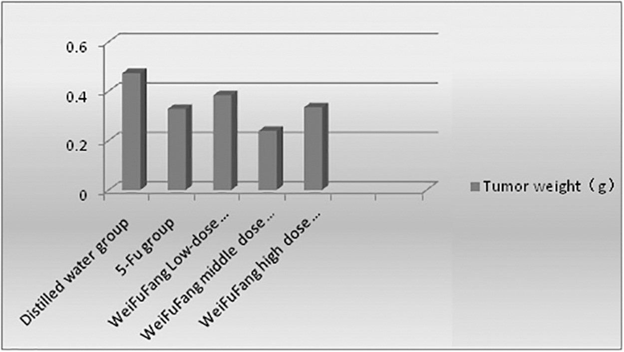
Average tumor weight histogram of animals in each group.
As shown in Table 1, before treatment, statistical analysis results revealed that differences in the weight and volume of xenografts in nude mice among all groups were not statistically significant (p > 0.05). This suggests that the baseline level was the same (Table 1).
Changes of Indexes Before and After Intervention (
Compared with negative control group p < 0.05.
Compared with Weifufang dose group p < 0.05.
Compared with 5-FU group p < 0.05.
5-FU, 5-flourouracil.
After treatment, statistical analysis results revealed that tumor growth was inhibited in the drug treatment groups, differences were statistically significant compared with the negative control group (p < 0.05), and differences between the Weifufang middle-dose group and other drug treatment groups were statistically significant (p < 0.05). This study revealed that the growth of solid tumors can be inhibited by drug treatment, and the antitumor effect in the Weifufang middle-dose group was the best (Table 1).
H&E staining of xenograft tissues
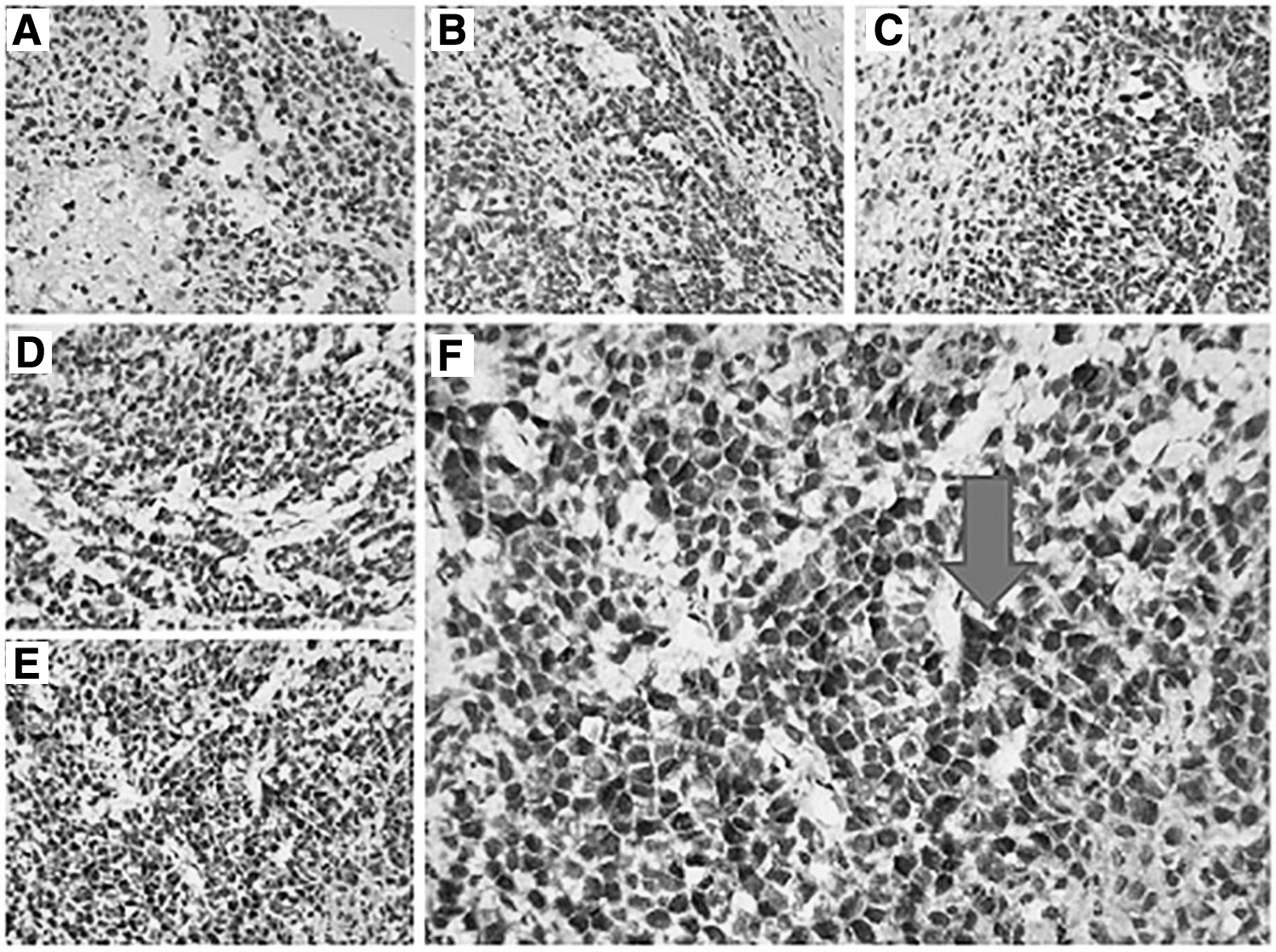
As shown in Figure 4, under a 400 × light microscope, tumors in nude mice in the negative control group had typical morphological characteristics of malignant tumor cells: large nuclei, deep staining, multiple nucleoli, visible tumor giant cells, obvious nuclear abnormalities, common pathological mitosis (the average was 6–8/field of a 400 × microscope), irregularly arranged tumor cells, and crumby or strip-shaped cancer nests. The following morphological characteristics of tumor cells were found in the Weifufang groups and 5-FU positive control group: Tumor cells were deep stained, had large nuclei and multiple nucleoli, and had obvious nuclear abnormalities; the number of tumor giant cells, pathological mitosis, and tumor cells decreased, compared with those in the negative control group. In particular, in the Weifufang middle-dose group, almost no tumor giant cells were observed; the tumor cells were round or elliptic, and they had superficial staining and nuclear divisions. Further, pathological nuclear fissions were rare (the average was 3–4/field of a 400 × microscope), the number of tumor cells relatively decreased, the formation of an obvious glandular cavity could be observed, and cells had an organ-like differentiation.

Hematoxylin and eosin staining of tumor tissue in each group. Pathological nuclear fissions  . 5-FU, 5-flourouracil.
PTEN gene expression in tumor tissues after drug treatment by IHC
As shown in Figure 5, brownish yellow or deep brownish yellow stains of the PTEN gene were observed in the nucleus and cytoplasm. The expression of the PTEN gene was weak in the negative control group, but it was enhanced in the drug treatment groups. In particular, the expression of the PTEN gene was relatively stronger in the gastric compound middle-dose group (in the control group, the PTEN gene was not expressed).
Immunohistochemical staining of tumor tissue in each group PTEN.
As shown in Table 2, the order of IOD values in each group is as follows: Weifufang middle-dose group > 5-FU positive control group > Weifufang high-dose group > Weifufang low-dose group > negative control group. The difference in the Weifufang middle-dose group was not statistically significant when compared with the 5-FU positive control group (p > 0.05), but it was statistically significant when compared with the other groups (p < 0.05). Further, the difference in the negative control group was statistically significant when compared with the 5-FU positive control group and the Weifufang middle-dose group (p < 0.05), but it was not statistically significant when compared with the Weifufang low- and high-dose groups (p > 0.05). In addition, the difference in the 5-FU positive control group was statistically significant when compared with the gastric compound high-dose group (p < 0.05). The expression of the PTEN gene was upregulated in the drug treatment groups, and the Weifufang middle-dose group and the 5-FU positive group exhibited the best effect.
Average IOD Value of PTEN in Each Group was Measured by Immunohistochemistry (
Compared with Weifufang middle-dose group p < 0.05.
Compared with 5-FU group p < 0.05.
Compared with Weifufang high-dose group p < 0.05.
Compared with Weifufang middle-dose group p < 0.05.
SD, standard deviation.
PTEN gene expression in tumor tissues after drug treatment by Western blot
As shown in Figure 6, compared with the negative control group, the expression of the PTEN gene increased in the drug treatment groups. Further, the expression of the PTEN gene was relatively stronger in the 5-FU positive control group and the gastric compound middle-dose group, whereas this expression was relatively weak in the Weifufang high- and low-dose groups.
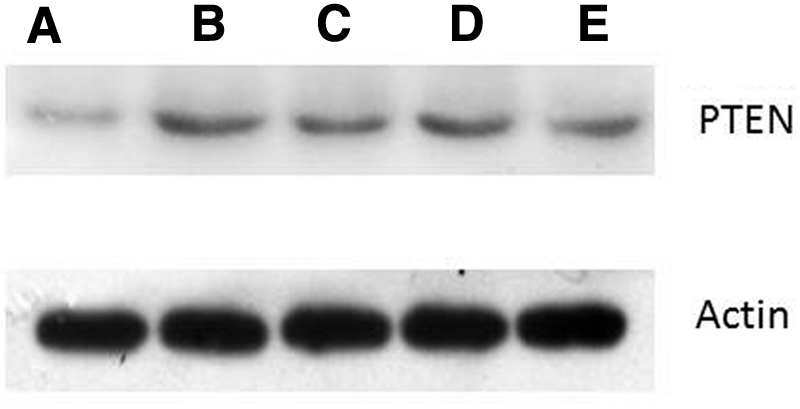
Compared with the negative control group, the expression of the PTEN gene increased in the drug treatment groups.
As shown in Table 3, the difference in the negative control group was not statistically significant when compared with the Weifufang low-dose group (p > 0.05), but it was statistically significant when compared with the other groups (p < 0.05). Further, the difference in the 5-FU positive control group was not statistically significant when compared with the Weifufang middle-dose group (p > 0.05), but it was statistically significant when compared with the Weifufang low- and high-dose groups (p < 0.05). The difference in the Weifufang middle-dose group was statistically significant when compared with the gastric compound low- and high-dose groups (p < 0.05). The difference between the Weifufang low- and high-dose groups was not statistically significant (p > 0.05). The results confirm that the expression of the PTEN gene was upregulated in the drug treatment groups, and the best effect was observed in the 5-FU positive control group and the Weifufang middle-dose group.
Average Value of PPAR-γ in Each Group was Measured by Western Blot (
Compared with negative control group p < 0.05.
Compared with 5-FU group p < 0.05.
Compared with Weifufang middle-dose group p < 0.05.
Compared to distilled water p < 0.01.
Discussion
The effect of compound Chinese herbal medicine on tumors requires multi-level, multi-target, and multi-link comprehensive regulation. Studies have revealed that compound Chinese herbal medicine can inhibit the proliferation of tumor cells, induce cell apoptosis, inhibit tumor angiogenesis, inhibit telomerase activity, and regulate immune function. At present, the multi-target effect of compound Chinese herbal medicine in the field of tumor treatment has been widely recognized at home and abroad. However, its specific anticancer mechanism is complex, and the mechanism of compound Chinese herbal medicine needs to be studied.
As a classical tumor suppressor gene, PTEN has been confirmed to be related to the occurrence and development of gastric cancer. Recently, there have been reports on the effect of Chinese herbal medicine on gastric cancer through the regulation of the PTEN gene. For example, studies on ursolic acid, 6 resveratrol, 24 and malol 6 have confirmed that compound Chinese herbal medicine controlled gastric cancer by regulating PTEN. This suggests that, at present, the management of gastric cancer by TCM is related to the PTEN gene, which has become a hot topic.
To further explore the mechanism of the Weifufang, in this experiment, PTEN gene expression was detected by IHC and western blot to confirm whether the tumor suppression effect and mechanism of the compound are correlated to the expression of the PTEN gene.
The results of this experiment revealed that after nude mice with xenografts were treated with the Weifufang, the tumor suppression effect was dose dependent. Further, by comparing the weight, tumor volume, and tumor weight of nude mice, these results confirmed that the Weifufang had a certain antitumor effect, when compared with the negative control group. This effect was superior in the Weifufang middle-dose group compared with that in other groups, and the differences were statistically significant (p < 0.05). PTEN gene expression was detected by IHC and western blot, and results revealed that the Weifufang may inhibit the growth of gastric cancer xenografts by upregulating the expression of the PTEN gene. In this study, through an in vivo experiment, it was preliminarily confirmed that the Weifufang had an inhibitory effect on gastric cancer xenografts in nude mice, and it had a certain effect on the PTEN gene. This result was consistent with the results of previous studies on ursolic acid, resveratrol, and malol.
The Weifufang has effects of detoxifying, dissipation of blood stasis, tonifying the qi, and strengthening body resistance. Among these herbs, Astragalus, Atractylodes macrocephala, Codonopsis pilosula, and Poria cocos have effects of tonifying the qi and tonifying the spleen; whereas Rhizoma Cyperi, radix curcumae, rhizoma curcumae, and Sappan wood have effects of regulating the flow of vital energy, invigorating the circulation of blood, and dissipating blood stasis. Further, Sculellaria barbata, Bistortae, Rhizoma, and Oldenlandia have effects of clearing heat and detoxifying; and Ligustrum lucidum and Barbed Skullcap Herb have effects of tonifying the kidney and liver. After more than 30 years of clinical practice and a number of clinical observations conducted by the investigators, it was confirmed that the Weifufang has a good clinical effect on patients with gastric cancer. We found through this in vivo experiment that the mechanism of the Weifufang is correlated to the expression level of the PTEN gene. In view of the complex composition of compound Chinese herbal medicine, we speculate that this is only one of the mechanisms of the Weifufang. The multi-component and theory of the compatibility of Monarch, Minister, Assistant, and Guide of the compound Chinese herbal medicine reflect the overall concepts of TCM. This is inosculated with multiple genes and multiple factors involved in the pathological process of the occurrence and development of gastric cancer. Therefore, it is worthy to further study the other mechanisms of the Weifufang. In the future, we will further study the effect of the Weifufang on the PTEN gene in gastric cancer, further conduct in-depth studies carried out along the PPAR γ/PTEN gene pathway, to provide new ideas for the study on the mechanism of compound Chinese herbal medicine, and provide a theoretical basis for TCM in the prevention and treatment of gastric cancer.
Authors' Contributions
J.-Q.H., S.-R.Z., D.-F.L., J.-Y.T., and P.-L.X. acquired data. J.-Q.H., S.-R.Z., D.-F.L., J.-Y.T., and M.Z. drafted the article. All authors contributed substantially to its revision. All authors take responsibility for the article as a whole. All authors read and approved the final article.
Footnotes
Acknowledgment
The authors are particularly grateful to all the people who had rendered help on this article.
Ethical Approval
All applicable international, national, and/or institutional guidelines for the care and use of animals were followed.
Disclosure Statement
There are no existing financial conflicts.
Funding Information
Li Yueheng old Chinese medicine studio (Hunan provincial finance department Fund Project No. 119 [2014]); Key fund project of Hunan administration of traditional Chinese Medicine (grant no. 201625).