
Abstract
Background:
Long noncoding RNA WT1-AS has been demonstrated as a potential tumor suppressor in gastric cancer. However, the functions of WT1-AS in other types of cancer remain unclear. Our study was therefore performed to explore the role of WT1-AS in triple-negative breast cancer (TNBC).
Materials and Methods:
Tissue specimens were obtained from 62 TNBC patients included in this study. A TNBC cell line BT-549 was used as the cell model of TNBC. Gene expression was detected by qPCR and Western blot. Overexpression experiments were used to analyze gene interactions. Transwell assays were used to explore the effects of transfections on cell invasion and migration.
Results:
We found that WT1-AS was downregulated in TNBC tissues than in nontumor tissues and decreased with increase in clinical stages. Transforming growth factor β1 (TGF-β1) was upregulated in TNBC tissues and inversely correlated with WT1-AS. TGF-β1 overexpression did not significantly affect WT1-AS in BT-549 cells, but WT1-AS negatively regulated the expression of TGF-β1. WT1-AS overexpression caused inhibited migration and invasion of TNBC cells. TGF-β1 overexpression showed opposite functions and reduced the effects of WT1-AS overexpression.
Conclusion:
WT1-AS may downregulate TGF-β to inhibit the migration and invasion of TNBC cells.
Introduction
In 2018, breast cancer caused 2,088,849 new cases, which accounts for ∼11.6% of all cancer cases, and caused 626,679 deaths, which accounts for ∼6.6% of all cancer deaths. 1 Breast cancer now is and will be the second most common type of malignancy and the second leading cause of cancer mortalities in the expected future. 2,3 Breast cancer can be divided into several subtypes, such as hormone receptor-positive, HER2-positive and triple-negative types. 4 Triple-negative breast cancer (TNBC) as a subtype of breast cancer is characterized by the lack of the expression of epidermal growth factor receptor 2 and hormone receptors. 4 Compared with other types of breast cancer, TNBC is more aggressive and the prognosis is generally poor. 5
Genome-wide genetic studies through next-generation sequencing approach have revealed that genetic alterations are critical players in TNBC. 6,7 Transforming growth factor β (TGF-β) signaling is altered during cancer biology, and the altered signaling transduction of TGF-β may inhibit (early-stage) and promote (late-stage) cancer progression at different cancer stages. 8,9 Therefore, regulation of TGF-β signaling is a potential approach for cancer treatment. It is known that the functions of TGF-β signaling can be achieved through the interactions with long noncoding RNAs (lncRNAs), which are defined as the noncoding RNA transcripts longer than 200 nt, 10,11 which have critical roles in cancer progression by regulating gene expression. 12 LncRNA WT1-AS in a recent study has been proven to be a tumor suppressor in gastric cancer. 13 Our preliminary transcriptome analysis showed that WT1-AS was downregulated in TNBC and negatively correlated with TGF-β1 mRNA (data not shown). This study aimed to investigate the role of WT1-AS in TNBC and explore its interaction with TGF-β signaling.
Materials and Methods
TNBC patients
This study passed the review board of Shanxi Provincial People's Hospital. Research subjects of this study were 62 TNBC patients (33–67 years; mean = 49.7 ± 4.9 years), who were selected from the 114 TNBC cases diagnosed and treated in the aforementioned hospital. Inclusion criteria were as follows: (1) no therapies were initiated within 3 months; and (2) newly diagnosed TNBC cases. Exclusion criteria were as follows: (1) patients complicated with other clinical disorders; (2) recurrent TNBC; (3) therapies were performed; and (4) history of malignancies. AJCC staging system was used to stage the 62 patients. Based on clinical findings, the 63 patients included 12, 20, 18, and 12 cases at stages I–IV, respectively. The patients were informed of the details of experimental design. They provided informed consent for the potential publication of the experimental data derived from this study.
TNBC tissue samples and cells
Breast biopsy under the guidance of magnetic resonance imaging was performed on all the 62 TNBC patients before the initiation of therapies. During biopsy, TNBC and nontumor tissue samples were collected (0.018–0.026 g). Histopathological examinations were performed to test all tissue samples. All nontumor tissue samples contained <1% cancer cells, and all TNBC tissue samples contained >95% cancer cells.
BT-549 human TNBC cell line (ATCC) was used. To prepare cell culture medium, RPMI-1640 medium was mixed with fetal bovine serum (FBS) in a ratio of 9:1. Cell culture conditions were 37°C, 5% CO2, and 95% humidity.
Transient cell transfection
BT-549 cells were harvested at 70%–80% confluence to perform transfections. pcDNA3.1 vector was used to construct WT1-AS and TGF-β1 expression vectors, and the vector construction service was provided by Sangon (Shanghai, China). Negative control (NC) siRNA and WT1-AS siRNA were from Sangon. All transient transfections were mediated by Lipofectamine 2000 (Invitrogen) to transfect 10 nM vectors (empty vector as NC group) or 40 nM siRNAs (NC siRNA as NC group) into 3 × 10⁶ cells. Control (C) group for all transfections was untransfected cells. Transfections were performed in three biological replicates. At 24 h post-transfection, cells were harvested and used for subsequent experiments (significant silencing started from 12 h post-transfection and lasted until 100 h post-transfection).
RNA extractions and qPCR
Total RNAs in 3 × 10⁵ BT-549 cells (collected at 24 h post-transfection) or 0.015 g tissue samples (ground in liquid nitrogen) were extracted using RNAzol reagent (Sigma-Aldrich). Tetro Reverse Transcriptase (Bioline) was used to perform all reverse transcriptions. To measure the expression levels of WT1-AS and TGF-β1 mRNA, KAPA SYBR FAST qPCR Master Mix (Kapa Biosystems) was used to prepare all qPCR mixtures with GAPDH as endogenous control. PCRs were performed in three replicates and 2−ΔΔCT method was used to normalize Ct values.
Western blot
Total proteins in 3 × 10⁵ BT-549 cells (collected at 24 h post-transfection) were extracted by RIPA solution (Thermo Fisher Scientific) and quantified by bicinchoninic acid assay (Thermo Fisher Scientific). Before electrophoresis (10% sodium dodecyl sulfate – polyacrylamide gel electrophoresis), all protein samples were denatured by incubating with boiling water for 5 min. After electrophoresis, proteins were transferred to polyvinylidene difluoride membranes, followed by blocking with 5% nonfat milk at 24°C for 90 min. After that, anti-TGF-β1 (1:1500, ab50716; Abcam) and anti-GAPDH (1:1500, ab37168; Abcam) rabbit primary antibodies were used to incubate with membranes for 18 h at 4°C. Then, immunoglobulin G–horseradish peroxidase (1:1000, MBS435036; MyBioSource) goat secondary antibody was used to incubate with the membranes for another 2 h at 24°C. Finally, ECL Western Blotting Substrate (Thermo Fisher Scientific) was used to incubate with membranes for 10 min at 24°C to develop signals. ImageJ v1.48 software was used to normalize data.
Transwell assays
Single-cell suspensions were prepared by mixing 1 mL mixture of 90% RPMI-1640 medium and 10% FBS with 3 × 10⁴ cells. Effects of transfections on the invasion and migration of BT-549 cells were analyzed using Transwell assay. To mimic in vitro cell invasion, Matrigel (200 mg/mL, 356234; Millipore) was used to coat up chamber membranes. The upper chamber was filed with cell suspension, and the lower chamber was filled with a mixture of 80% RPMI-1640 medium and 20% FBS. Cells were cultivated under aforementioned conditions for 12 h. After staining with 0.5% crystal violet (Sigma-Aldrich) for 2 h at 24°C, cells were counted under a light microscope. The numbers of invading and migrating cells were normalized to cell proliferation rates (OD values measured at 450 nM after the addition of CCK-8 solution) at 12 h after the initiation of experiments.
Statistical analysis
All experiments were performed in three biological replicates. Mean values were calculated and were used for all cell analyses. Correlations were analyzed by linear regression. Differences were explored between two types of tissues or among multiple patient and cell groups by performing paired t test and analysis of variance (ANOVA) (one-way) in combination with Tukey test, respectively. p < 0.05 was statistically significant.
Results
WT1-AS was downregulated in TNBC and was affected by clinical stages
qPCR and unpaired t test were performed to measure and compare the expression levels of WT1-AS in nontumor and TNBC tissues. Compared with nontumor tissues, significantly lower expression levels of WT1-AS were observed in TNBC tissues (Fig. 1A, p < 0.05). ANOVA (one-way) in combination with Tukey test was used to compare expression levels of WT1-AS in TNBC tissues among different clinical stages. It can be observed that expression levels of WT1-AS decreased significantly with the increase in clinical stages (p < 0.05; Fig. 1B).
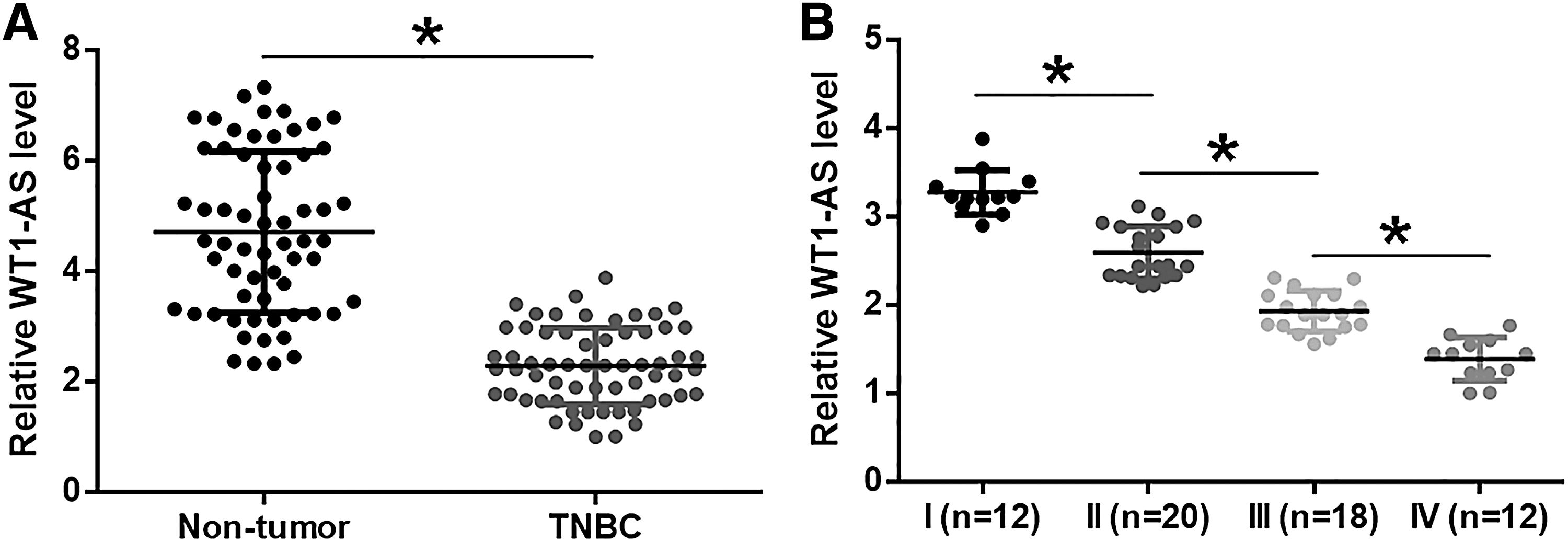
WT1-AS was downregulated in TNBC and was affected by clinical stages. qPCR and unpaired t test were performed to measure and compare the expression levels of WT1-AS in nontumor and TNBC tissues
TGF-β1 mRNA and WT1-AS were inversely correlated in TNBC tissues
qPCR and unpaired t test were performed to measure and compare the expression levels of TGF-β1 mRNA in nontumor and TNBC tissues. Compared with nontumor tissues, significantly higher expression levels of TGF-β1 mRNA were observed in TNBC tissues (p < 0.05; Fig. 2A). The correlation between TGF-β1 mRNA and WT1-AS was analyzed by linear regression. It can be observed that expression levels of TGF-β1 mRNA were significantly and inversely correlated with the expression levels of WT1-AS in TNBC tissues (Fig. 2B). However, the correlation between them was not significant in nontumor tissues (Fig. 2B).
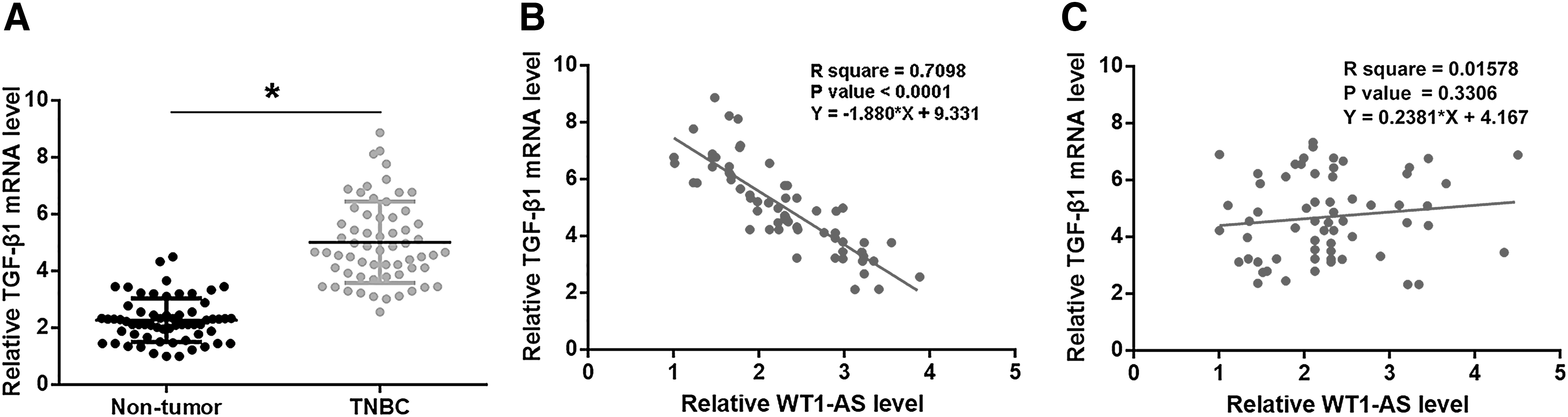
TGF-β1 mRNA and WT1-AS were inversely correlated in TNBC tissues. qPCR and unpaired t test were performed to measure and compare the expression levels of TGF-β1 mRNA in nontumor and TNBC tissues
WT1-AS negatively regulated the expression of TGF-β1 in BT-549 cells
WT1-AS and TGF-β1 vectors and WT1-AS siRNA were transfected into BT-549 cells. Expression levels of WT1-AS and TGF-β1 were measured at 24 h post-transfection by qPCR. Compared with two controls C and NC, expression levels of WT1-AS and TGF-β1 mRNA were significantly altered (Fig. 3A, p < 0.05). Moreover, compared with two controls, WT1-AS expression was not significantly altered after TGF-β1 overexpression (Fig. 3B). However, WT1-AS overexpression led to downregulated expression, whereas WT1-AS siRNA silencing led to upregulated expression of TGF-β1 at both mRNA and protein levels (p < 0.05; Fig. 3C).
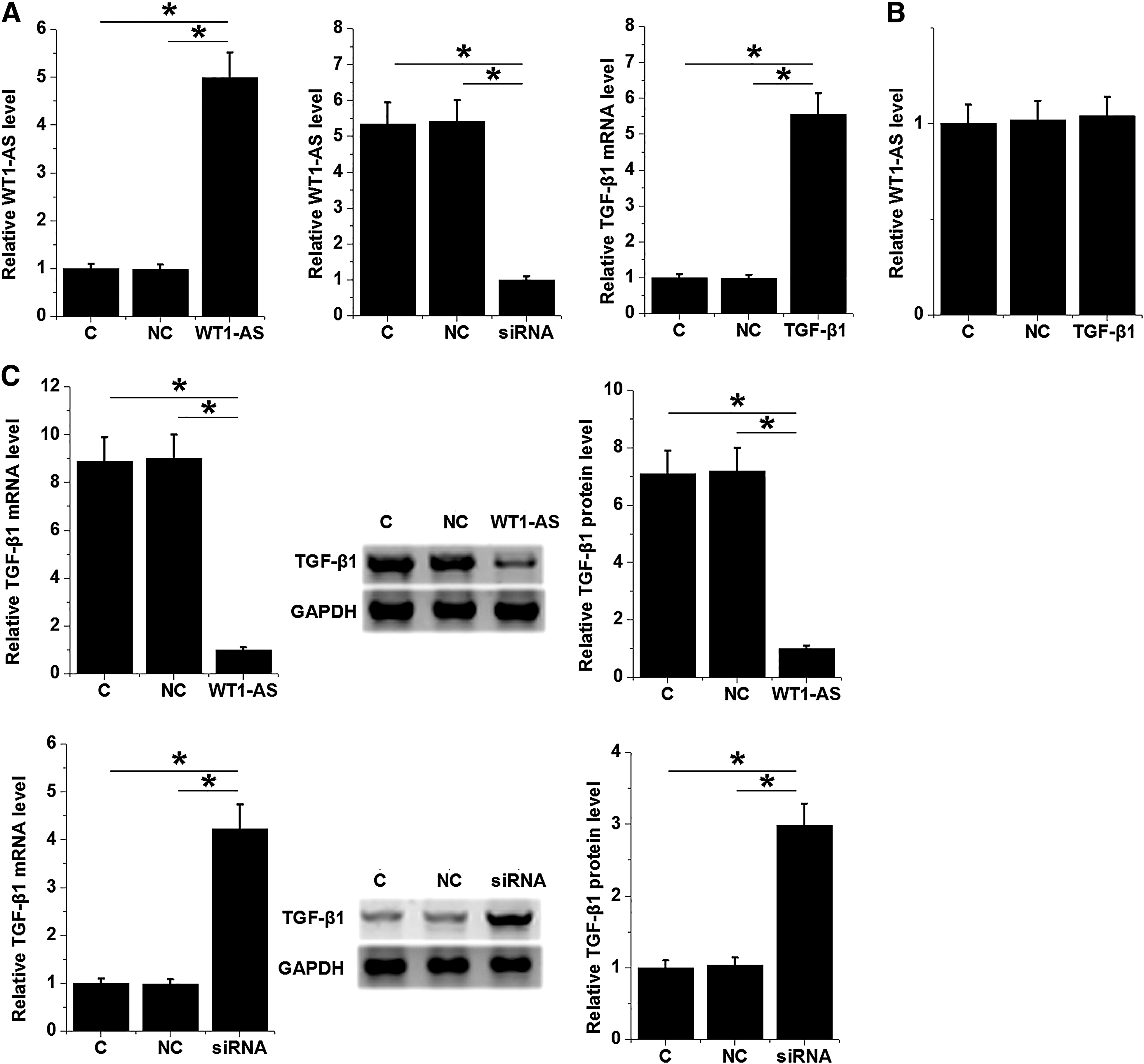
WT1-AS negatively regulated the expression of TGF-β1 in BT-549 cells. WT1-AS and TGF-β1 vectors and WT1-AS siRNA were transfected into BT-549 cells. Transfections were confirmed by qPCR at 24 h post-transfection
WT1-AS suppressed BT-549 cell invasion and migration through TGF-β1
Transwell assays were performed to analyze the effects of transfections on the invasion (Fig. 4A) and migration (Fig. 4B) of BT-549 cells. Compared with two controls C and NC, WT1-AS overexpression caused inhibited invasion and migration of TNBC cells. TGF-β1 overexpression and WT1-AS siRNA silencing showed opposite functions and TGF-β1 overexpression reduced the effects of WT1-AS overexpression on cell behaviors (p < 0.05).

WT1-AS suppressed BT-549 cell invasion and migration through TGF-β1. Transwell assays were performed to analyze the effects of transfection of WT1-AS and TGF-β1 expression vectors and WT1-AS siRNA on the invasion
Discussion
In this study we investigated the roles of WT1-AS in TNBC. We found that WT1-AS was downregulated in TNBC and negatively regulated the invasion and migration of cancer cells by negatively regulating the expression of TGF-β1.
The expression pattern and functions of WT1-AS have only been investigated in gastric cancer and cervical cancer. 13 –15 In gastric cancer, WT1-AS was downregulated and is at least partially responsible for the accelerated invasion and proliferation of cancer cells. 13 WT1-AS was also downregulated in cervical cancer, and the downregulated WT1-AS not only promoted the aggressiveness of cancer and predicted the poor survival of cancer patients. 14,15 Our study is the first to report the downregulation of WT1-AS in TNBC. We also observed that expression levels of WT1-AS decreased with the increase in cancer stages and WT1-AS negatively regulated the invasion and migration of cancer cells. Therefore, our data suggested that WT1-AS was a tumor suppressor in TNBC.
Although TGF-β plays different roles at different cancer development stages, 16 it is generally believed that the activation of TGF-β can induce epithelial–mesenchymal transition and promote cancer metastasis at advanced cancer stages. 17 Consistently, our study also observed the upregulation of TGF-β1 in TNBC, and the overexpression of TGF-β1 led to the promoted cancer cell invasion and migration. Our data further confirmed the oncogenic roles of TGF-β1 in TNBC. Our study showed that WT1-AS could negatively regulate TGF-β1 to participate in the regulation of the behaviors of TNBC cells. It is known that WT1-AS can upregulate p53 to inhibit cancer progression, 14 and the interactions between p53 and TGF-β1 have been well studied. 18,19 In addition, p53 signaling is altered during the development of TNBC. Specifically, p53 gene in BT-549 cells has R249S mutation that contributes to cancer progression. 20 Therefore, the altered p53 signaling in TNBC may mediate the interaction between WT1-AS and TGF-β1. This is consistent with the observation that WT1-AS and TGF-β1 mRNA were inversely correlated in TNBC tissues but not in nontumor tissues.
In conclusion, WT1-AS plays tumor suppressive roles in TNBC, and it can negatively regulate TGF-β1 to suppress cancer cell invasion and migration.
Footnotes
Acknowledgments
The authors thank the financial support from Scientific research funding for returned overseas in Shanxi Province (2014-082).
Disclosure Statement
No competing financial interests exist.
Funding Information
No funding was received for this article.