
Abstract
Renal cell carcinoma (RCC) is a common cancer, and extensive research suggests that microRNA may play an important role in the progression of RCC. The emphasis of this article was to reveal the function and mechanism of microRNA-1293(miR-1293) in the development of RCC tumors. First, the authors carried out bioinformatics analysis. The differential expression of miR-1293 in RCC tumor and normal cells was analyzed using the data from The Cancer Genome Atlas database, and Kaplan-Meier survival analysis was carried out to test the survival rate. Subsequently, the miR-1293 expression in RCC cell lines was examined by quantitative real-time PCR. Then Cell counting kit-8 and Transwell assays were executed to detect the function of miR-1293 in RCC. Bioinformatics prediction, western blotting, and dual-luciferase reporter assay were set to check the target gene of miR-1293. Finally, they conducted rescue experiments to verify whether the regulation of miR-1293 on the biological function of RCC cells was achieved by regulating hydrocyanic oxidase 2 (HAO2). Bioinformatics results showed that miR-1293 was highly expressed in RCC, and the miR-1293 high-expression group showed a lower survival rate than the miR-1293 low-expression group, which suggested that the high expression of miR-1293 was related to unfavorable prognosis in RCC. Subsequent assays evidenced that upregulation of miR-1293 expression significantly increased the cell viability and promoted cell migration and invasion in RCC. Silencing miR-1293 expression showed opposite results. Furthermore, HAO2 was confirmed to be a direct target gene of miR-1293 by dual-luciferase reporter assay, and miR-1293 negatively regulated the expression of HAO2. Moreover, rescue experiments evidenced that miR-1293 reduced the cell viability, invasion, and migration of RCC by regulating HAO2. In sum, miR-1293 can regulate the viability, invasion, and migration of RCC tumor cells by targeting HAO2, suggesting that miR-1293 can be used as a new biomarker for clinical treatment of RCC.
Introduction
Renal cell carcinoma (RCC) is considered to be the most common primary renal malignancy, accounting for 90%–95% of all renal cancer cases, with a mortality rate of 25%. 1,2 In recent years, surgical resection is the most recommended treatment for RCC. 3 However, due to its high metastasis rate and recurrence rate after surgery, the prognosis of some patients is still not optimistic. 4 Since RCC is insensitive to chemotherapy and radiotherapy, no effective postoperative adjuvant therapy has been developed. 5 Hence, it is urgent to develop new and effective tumor biomarkers and therapeutic targets to improve the treatment status of RCC.
MicroRNAs (miRNAs) are short-chain noncoding RNAs, which can transform the expression of gene by connecting with specific sites on the 3′UTR of target mRNA. 6,7 This mechanism enables miRNAs to participate in the regulation of biological behaviors such as proliferation, apoptosis, invasion, and migration of cancer cells, thus playing a carcinogenic or anticancer role. 8,9 Extensive research has confirmed that miRNAs can be involved in the regulation of the process of RCC. MiR-193a-3p was overexpressed in RCC and played a role in promoting tumor development by targeting PTEN. 10 miR-106b-5p activated Wnt/beta-catenin signaling pathway in RCC, thus promoting cancer progression. 11 miR-122 accelerated the metastasis of cancer cells in CCRCC. 12 On the contrast, miR-411 has a tumor suppressive effect in RCC. 13 It was revealed by bioinformatics analysis that hsa-mir-1293 was highly expressed in PRCC, and could be used as a prognostic indicator of PRCC. 14 However, the detailed role of miR-1293 in PRCC remains largely unknown, the specific biological function of it in RCC needs further study.
On the basis of existing literature, the authors carried out studies in an effort to quest whether miR-1293 plays a role in regulating cell viability, migration, and invasion in RCC, and to further explore its mechanism, hoping to supply a potential biomarker for the therapy of RCC.
Materials and Methods
Data collection
The expression data of RCC (PRCC and ccRCC) were downloaded from The Cancer Genome Atlas (TCGA) database (
Cell lines, culture, and transfection
Human renal cell carcinoma ACHN, OS-RC-2, 786-O, and Caki-2 cell lines and normal control cell HK-2 (American Tissue Culture Collection) were conventionally cultured in RPMI-1640 medium with 10% fetal bovine serum, penicillin (100 U/mL), and streptomycin (0.1 mg/mL) at 37°C with 5% CO2.
Si-HAO2#1 (sequence: CCATATTTCCCACATTTCTA), Si-HAO2#2 (sequence: CCCTTGGAGCTAAGTGCATTT), si-con (sequence: AATTCTCCGAACGTGTCACGT), and pcDNA3.1-HAO2 were synthesized by Shanghai GenePharma Co., Ltd. (Shanghai, China). miR-1293 mimic/inhibitor and corresponding NC were provided by Shanghai GenePharma Co., Ltd. Cells at 80% confluence were inoculated into the six-well plate 24 h before transfection to give 50%–90% confluence on the transfection day. Transfection was conducted according to the instructions of Lipofectamine 2000 transfection kit.
RNA extraction and quantitative real-time PCR
TRIzolRNA extraction kit (Life Technologies, CA) was used to extract total RNA from cells, and reverse transcription was carried out using Prime Script RT Kit (RR036A; Takara Biotechnology Co., Ltd., Dalian, China). The expression level of mir-1293/hao2 was detected by quantitative real-time PCR (qRT-PCR) using SYBR Green RT-qPCR kit (Kapa Biosystems, Wilmington, MA), and the procedure was as follows: predenaturation at 95°C for 5 min, followed by 40 cycles of 95°C for 30 s and then at 65°C for 45 s. The expression of miRNA and mRNA was calculated by 2−△△Ct method. Repeat three times independently. The primer sequence was as follows:
MiR-1293: F: 5′-GGTGGTCTGGAGATTTG-3′,
R: 5′-GAACATGTCTGCGTATCTC-3′;
U6: F: 5′-CTCGCTTCGGCAGCACA-3′,
R: 5′-AACGCTTCACGAATTTGCGT-3′
HAO2: F-5′-TGAAGGTGGAGCAGATGACAGC-3′
R: 5′-TGAAGGTGGAGCAGATGACAGC-3′
β-actin: F: 5′-GCAGGAGTATGACGAGTCCG-3′
R: 5′-CGGACTCGTCATACTCCTGC-3′
Western blot
Cells were lysed by RIPA lysate with protease inhibitor to extract protein, and then using BCA kit to determine the protein concentration. Subsequently, about 20 μg of protein was added to each well in a vertical electrophoresis tank, and protein samples were isolated by sodium dodecyl sulfate–polyacrylamide gel electrophoresis. After that, the separated protein was transferred to PVDF membrane and sealed for 1 h with 5% skimmed milk powder. Afterward, the blotting membranes were incubated with primary antibody overnight at 4°C and then washed with TBST for 3 × 5 min. Hereafter, membranes were incubated at room temperature with second antibody for 1 h. The signals were developed using an ECL Plus Western Blotting Detection System (Amersham Biosciences). QUANTITY ONE software was used to scan the gray value after imaging, and β-actin was used as an internal consultation. The relative expression of each protein was calculated by the objective protein/internal reference.
CCK8 assay
After digesting the cells, the cells were counted and the concentration of each group was adjusted to 1000/100 μL. Then, 100 μL of cell suspension was added to each well in a 96-well plate. Conventional culture was carried out in a CO2 incubator at 37°C. Cell viability was measured at 0, 24, 48, and 72 h. For cell viability detection, 10 μL of Cell counting kit-8 (CCK-8) solution (Dojindo, Japan) was added to each well and routinely cultured for 1.5 h. The OD value was detected with a 450 nm excitation light by a microplate reader, and a viability curve was drawn. Each experiment was conducted three times.
Bioinformatics prediction
The miRNA target gene prediction site miRTarBase (
Dual-Luciferase reporter assay
Dual-luciferase report was used to further confirm the predicted results of bioinformatics software. In the first place, 3′UTR of HAO2 containing the wide-type putative target sites (WT) and 3′UTR of HAO2 containing the mutant target sites for miR-1293 (MUT) were subcloned into the pmirGLO luciferase vector to establish the HAO2-WT and HAO2-MUT luciferase reporter vectors. Then, miR-1293mimic, miR-1293 mimic NC, and HAO2-WT and HAO2-MUT were co-transfected into HEK-293T, ACHN, and 786-O cells, respectively. The cells were separately planted in a 96-well plate at a density of 1 × 10⁴/well after transfection, and culture was continued for 48 h, followed by the addition of 100 μL of lysate per well. Dual-LuciferaseTM system was used to test the luciferase activity according to the manufacturer's instructions.
Transwell assay
Matrigel matrix glue was evenly applied to the upper chamber of Transwell chamber and placed overnight to form a film. One hundred microliters of cell suspension (1 × 10⁵ cells) was added to the upper chamber and 500 μL of the complete culture solution was added to the lower chamber, and then, the Transwell chamber was incubated in 5% CO2 incubator at 37°C. After overnight incubation, the chamber was taken out and the remaining cells in the upper room were wiped off with a cotton swab. After cleaning with phosphate buffered saline buffer, the invaded cells were fixed with 4% polyformaldehyde for 30 min. Then, the cells were stained using 0.1% crystal violet to stain for 20 min, followed by being dried with air, and the cell number was calculated under a microscope and randomly taken from five high-power fields. The tentative was duplicated thrice.
During the migration experiment, the upper chamber was not given a matrigel coating, and the remaining operation was the same as the invasion experiment.
Statistical analysis
In this study, statistical analysis software SPSS22.0 was used for data analysis, and student's t-test (two groups) or one-way analysis of variance with post-test of Dunnett (three groups and above) was used for comparison between groups. Kaplan-Meier survival analysis was used to plot the survival curves and log-rank test for comparison. The high-expression group and the low-expression group were grouped based on the median expression level of miR-1293. Pearson correlation coefficient analyses were used to detect the correlation between miR-1293 and HAO2 in RCC. p < 0.05 indicated that the difference has statistical significance.
Result
Upregulated expression of miR-1293 was related to unfavorable prognosis in RCC
The authors first analyzed the level of miR-1293 expression in PRCC and ccRCC on the basis of public data. The gene expression profiles and clinical data of PRCC and ccRCC were downloaded from TCGA database. The results from Figure 1A and C showed that miR-1293 was highly expressed both in PRCC (p < 0.001) and ccRCC (p < 0.05) than in the corresponding control group. To check the influence of miR-1293 expression on the survival rate of RCC, Kaplan-Meier survival analysis was carried out. From Figure 1B and D, the miR-1293 high-expression group showed significantly lower survival rate than the miR-1293 low-expression group, suggesting that miR-1293 high expression in PRCC (p < 0.005) and ccRCC (p < 0.05) was associated with poor prognosis. Taken together, these results indicated that miR-1293 was upregulated in RCC and higher expression was interrelated to poor outcomes.
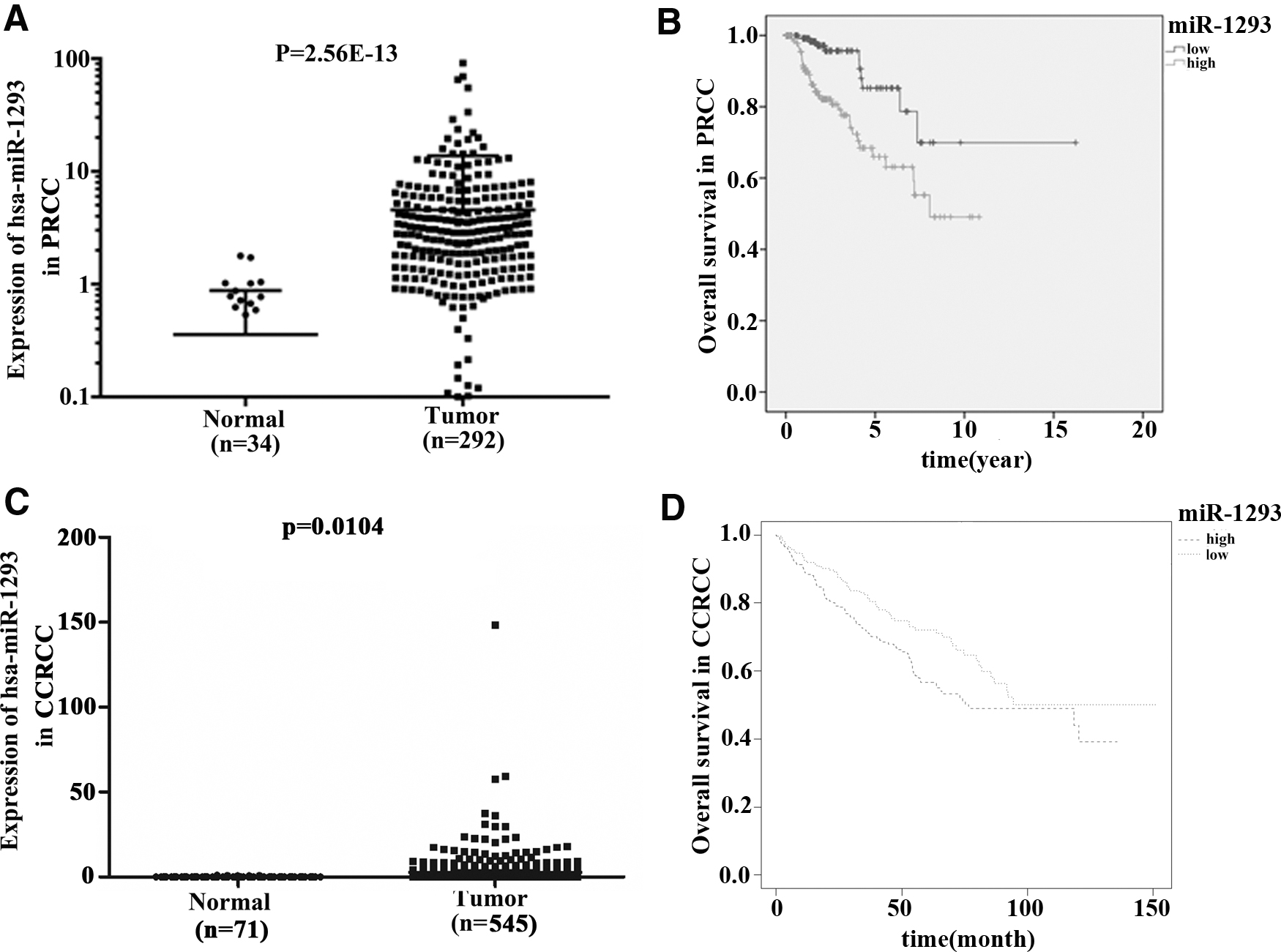
Overexpression of miR-1293 was associated with unfavorable prognosis in RCC.
The expression of miR-1293 was upregulated in RCC cell lines
To further examine the miR-1293 expression in RCC cells, the authors carried out a qRT-PCR experiment. As demonstrated in Figure 2A, miR-1293 was highly expressed in RCC cell lines ACHN, OS-RC-2, 786-O, and Caki-2 compared with the normal healthy cell HK-2 (p < 0.01). Interestingly, miR-1293 expressed differently in different RCC cell lines, of which 786-O cell has the highest expression and ACHN has the lowest expression.
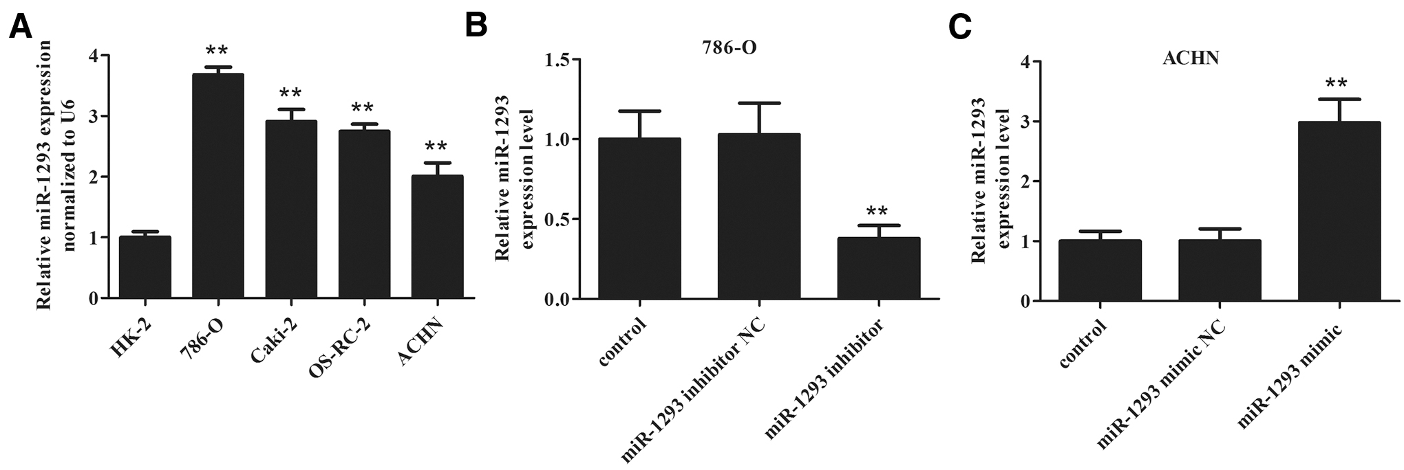
Detection of miR-1293 mimic and miR-1293 inhibitor transfection efficiency in RCC cell lines 786-O and ACHN.
To better verify the function of miR-1293 in RCC, they chose 786-O cells to perform knockdown experiments, and selected ACHN cells to carry out overexpression assays. ACNH cells were transfected with miR-1293 mimic NC and miR-1293 mimic, respectively, and 786-O cells were transfected with miR-1293 inhibitor NC and miR-1293 inhibitor. As described in Figure 2B and C, miR-1293 was successfully depleted in 786-O cells and upregulated in ACNH cells (p < 0.01). This provided a basis for the authors' subsequent experiments to detect the biological function of miR-1293 in RCC.
miR-1293 contributes to cell viability, invasion, and migration in RCC
To detect the biological function of miR-1293 in RCC, CCK8 and Transwell experiments were set up. The results of CCK8 experiment showed that inhibiting the expression of miR-1293 significantly (p < 0.01) decreased the viability of 786-O cells, while upregulating the expression of miR-1293 significantly (p < 0.01) increased the viability of ACNH cells (Fig. 3A, B). Moreover, it can be seen from Figure 3C and D that, after being transfected with miR-1293 inhibitor, the number of migrated and invaded 786-O cells decreased significantly compared with the control group (p < 0.01). Simultaneously, the number of ACHN cells that migrated and invaded to the lower chamber in miR-1293 mimic group was notably larger compared with the control group (Fig. 3E, F, p < 0.01). Transwell assay results suggested that downregulating the expression of miR-1293 can inhibit the invasion and migration of 786-O cells, and upregulation of miR-1293 has an opposite effect. From the above results, it can be concluded that the regulation of miR-1293 expression may alter the biological behavior of RCC tumors.
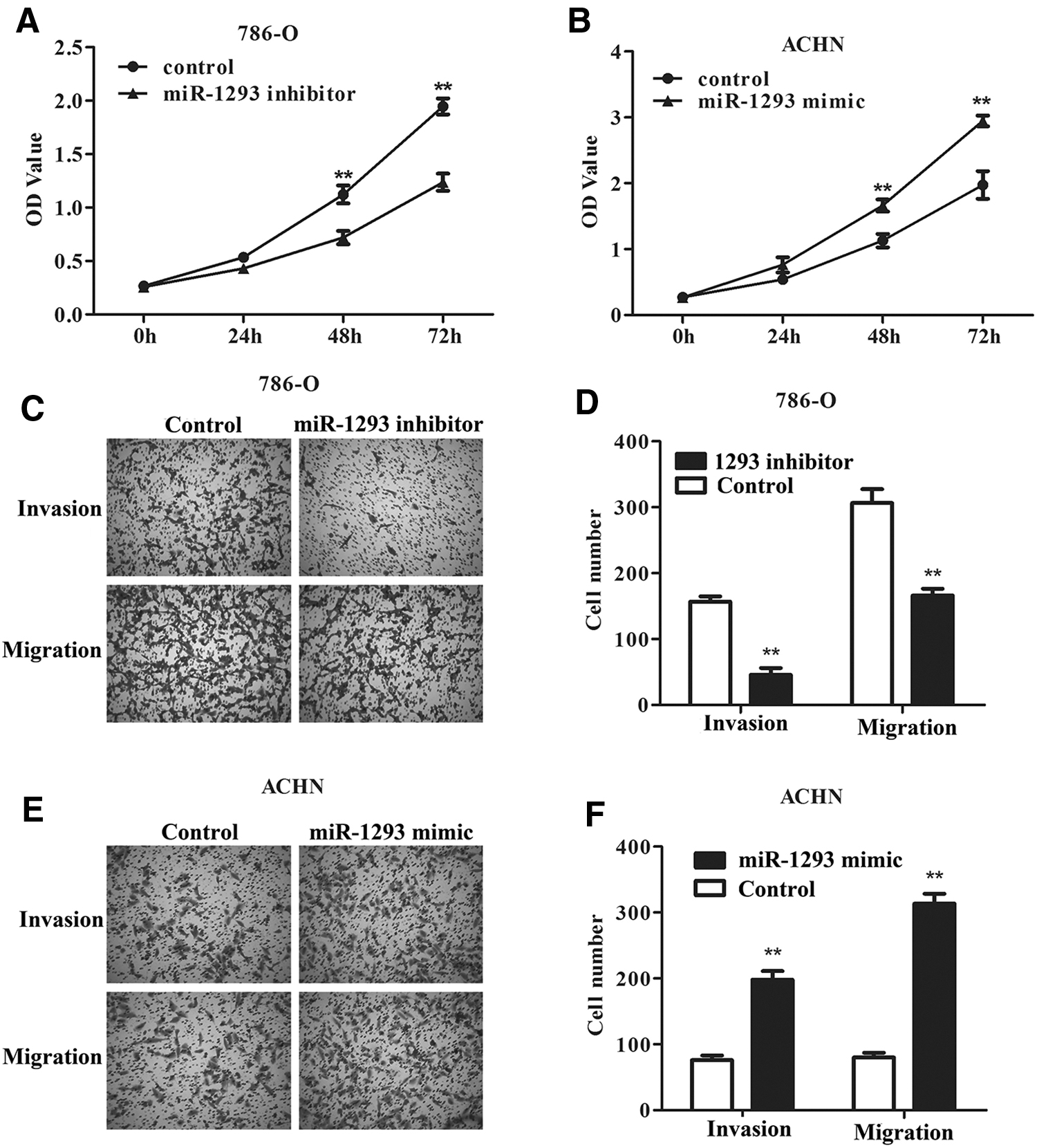
Effect of altering the expression of miR-1293 on the cell biological behaviors in 786-O and ACHN.
Targeted regulation of HAO2 by miR-1293
First, the authors used miRTarBase (
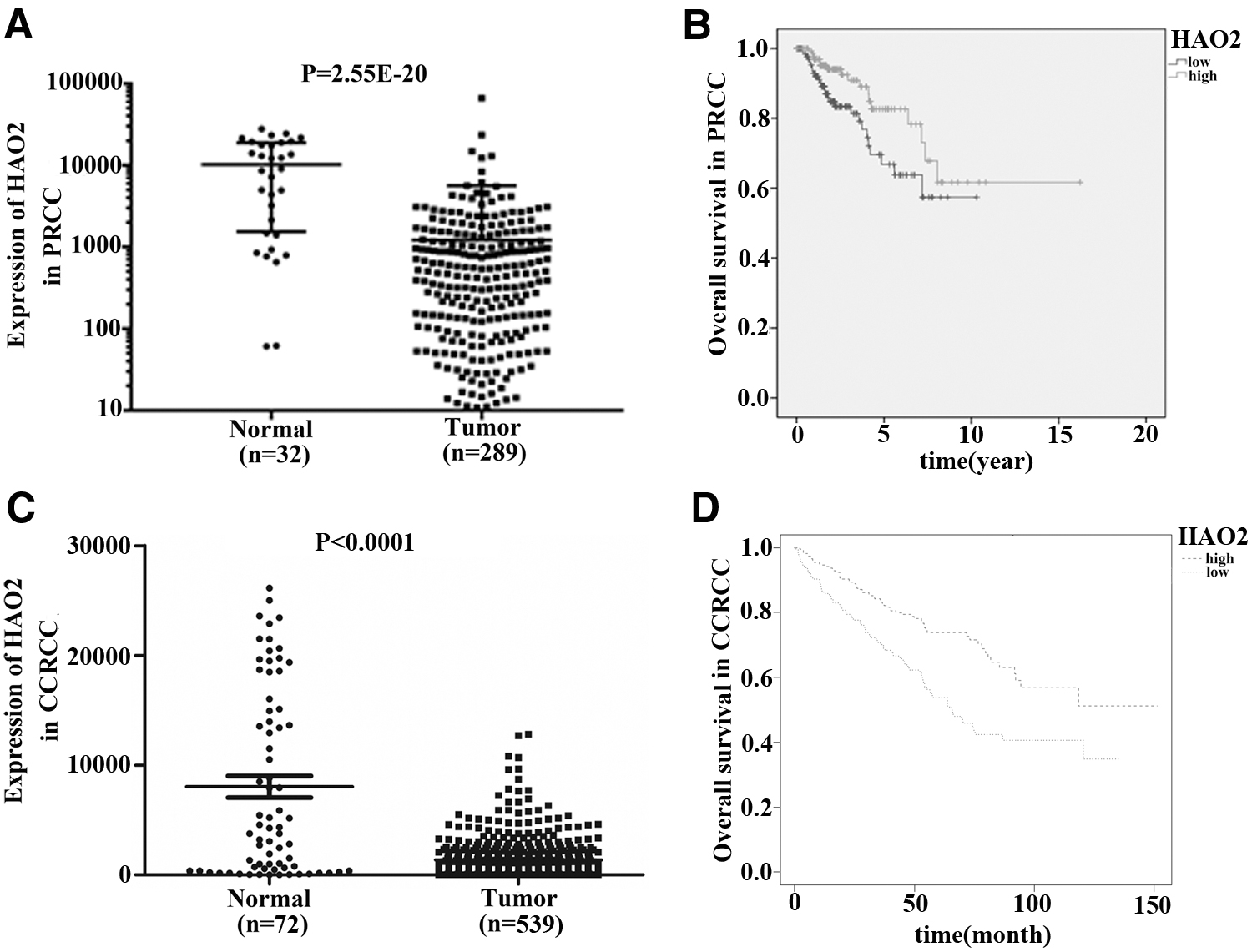
HAO2 was expressed lowly in RCC.
Then dual-luciferase reporter assay was carried out to verify whether HAO2 is a direct target gene of miR-1293. HEK 293T cell, ACHN cells, and 786-O cells were, respectively, transfected with HAO2-WT or HAO2-MUT and miR-1293 mimic or miR-1293 mimic NC to measure fluorescence intensity. The results showed that the fluorescence intensity of HAO2-WT and miR-1293 mimic co-transfected cells was significantly reduced (p < 0.01), while the fluorescence intensity of HAO2-MUT and miR-1293 mimic co-transfected cells was almost unchanged (Fig. 5A–D). These results indicated that miR-1293 directly targets HAO2.

HAO2 was a direct target gene of miR-1293.
To further verify the correlation between miR-1293 and HAO 2, Pearson correlation coefficient analysis was carried out. As shown in Figure 5D and E, miR-1293 showed a significant negative correlation with HAO2 levels both in PRCC (R = −0.2354, p = 0.0001) and ccRCC (R = −0.2956, p < 0.0001).
Subsequently, 786-O cells were separately transfected with si-HAO2#1 and si-HAO2#2, and then qRT-PCR assay and Western blot assay were used to detect the expression of HAO2 in the transfected cells. The result showed that the mRNA and protein expression level of HAO2 in 786-O cells transfected with si-HAO2#1 or si-HAO2#2 were both significantly declined than that in the si-con group (Fig. 6A–C, p < 0.01), and it can be clearly seen that the knockdown efficiency of si-HAO2#1 was higher compared with si-HAO2#2, so the authors selected si-HAO2#1 for the knockdown experiments.

The efficiency of knockdown of HAO2 in 786-O cells and overexpression of HAO2 in ACHN cells.
Next, a Western blot assay was used to test whether miR-1293 can target and regulate the level of HAO2 protein. As shown in Figure 6D and E, downregulation of miR-1293 obviously elevated the level of HAO2 protein in 786-O cells compared with the control, while knockdown of HAO2 showed an opposite result. In addition, in 786-O cells co-transfected with si-HAO2 and miR-1293 inhibitor, the level of HAO2 protein decreased significantly compared with 786-O cells transfected with miR-1293 inhibitor (p < 0.01). Simultaneously, upregulation of miR-1293 in ACHN cell can reduce the expression of HAO2, whereas overexpression of HAO2 can induce the expression of HAO2. Moreover, in ACHN cell co-transfected with pcDNA3.1-HAO2 and miR-1293 mimic, the level of HAO2 protein was markedly higher than that in ACHN cells transfected with miR-1293 mimic (Fig. 6F, G, p < 0.01). These findings illustrated that HAO2 is a direct target gene of miR-1293.
Effect of miR-1293/HAO2 on the function of RCC cells
Furthermore, the authors examined the effectiveness of miR-1293/HAO2 on RCC cell function. First, ACHN cells were transfected with miR-1293 mimic or miR-1293 mimic plus pcDNA3.1-HAO2, and 786-O cells were transfected with miR-1293 inhibitor or miR-1293 inhibitor plus si-HAO2. Cells treated with transfection reagent were regarded as control. Then, the viability, migration, and invasion of ACHN cells and 786-O cells were tested by CCK8 and Transwell experiments, respectively. The results indicated that in the miR-1293 inhibitor plus si-HAO2 group, the viability of 786-O cells and the number of 786-O cells migrating and invading into the lower chamber increased significantly compared with the miR-1293 inhibitor group (p < 0.01, Fig. 7A, C, and D), whereas ACHN cells co-transfected with miR-1293 mimic and pcDNA3.1-HAO2 significantly reduced cell viability, migration, and invasion compared with ACHN cells transfected with miR-1293 mimic (p < 0.01, Fig. 7B, E, and F). The above results indicated that miR-1293 regulates the function of RCC cells by regulating HAO2.
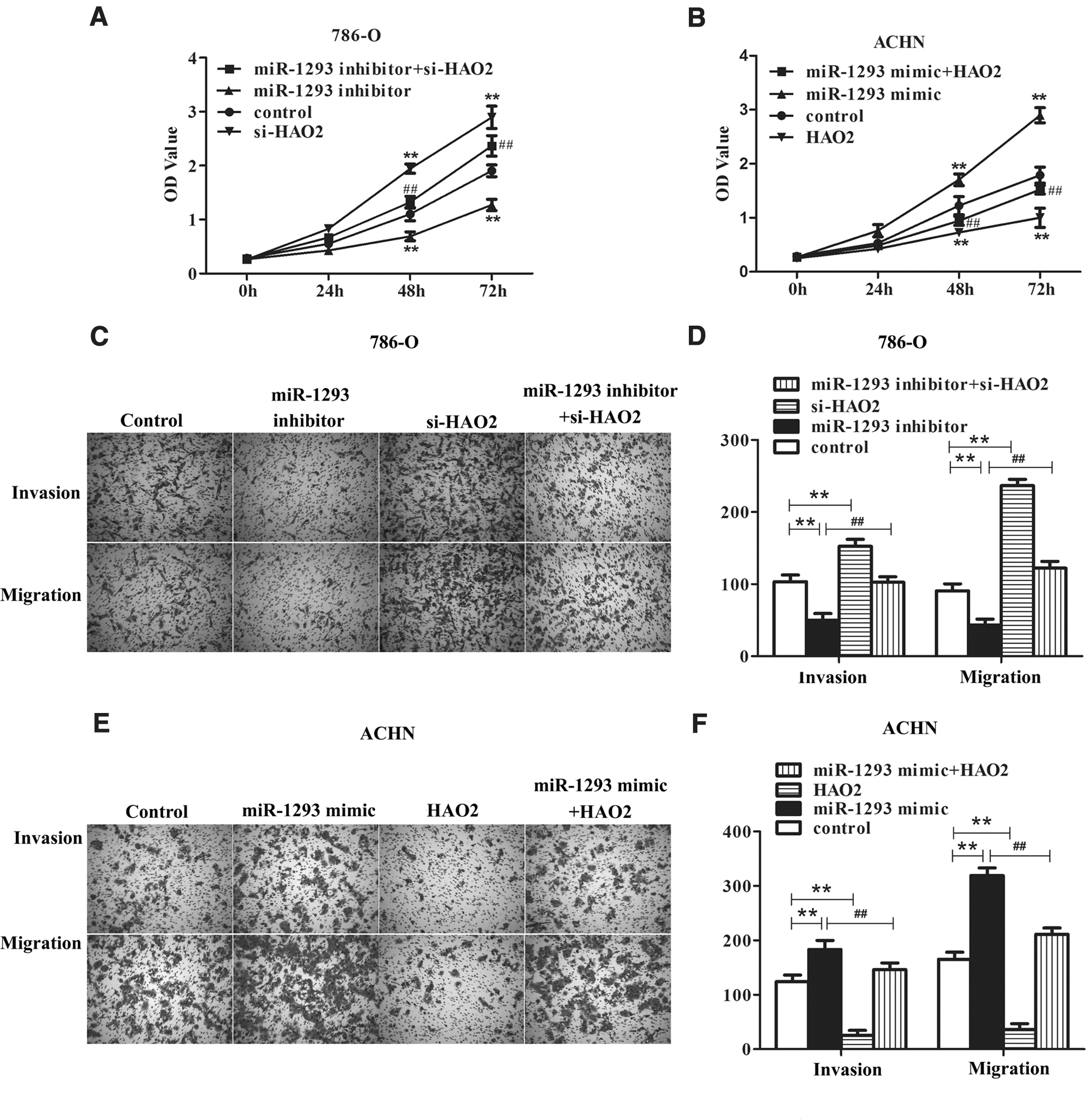
The effect of miR-1293/HAO2 on phenotype of RCC cells.
Effect of miR-1293/HAO2 on the EMT of RCC cells
To explore in depth the underlying mechanism of miR-1293/HAO2 axis functioning in RCC, the authors detected the changes in EMT-related proteins using Western blot analysis. As can be seen from Figure 8A and B, downregulation of miR-1293 can obviously increase the expression of E-cad in 786-O cells, while it decreased the expression of N-cad, Snail, and MMP9. In the meantime, knockdown of HAO2 showed an opposite effect. Furthermore, knockdown of HAO2 can reverse the influence of miR-1293 inhibition on the procession of EMT in 786-O cells, p < 0.01. Conversely, the expression of E-cad was markedly declined in ACHN cells transfected with miR-1293 mimic, whereas the expression of N-cad, Snail, and MMP9 was markedly induced. Moreover, ACHN cells co-transfected with the miR-1293 mimic and pcDNA3.1-HAO2 showed the opposite effect compared with the miR-1293 overexpression group (Fig. 8C, D,p < 0.01). Based on the above results, the authors discovered that miR-1293/HAO2 axis mediates the process of EMT in RCC.

The influence of miR-1293/HAO2 axis on EMT pathway of RCC cells.
Discussion
In this research, the authors found that miR-1293 was highly expressed in RCC, and high expression was associated with unfavorable prognosis. Upregulation of miR-1293 promoted the proliferation, migration, and invasion of RCC cells. Subsequently, they confirmed that HAO2 is a direct target gene of miR-1293 and miR-1293 was negatively correlated with the HAO2 in RCC. Finally, they confirmed that miR-1293 can regulate the phenotypic function of RCC by regulating HAO2.
As a member of miRNAs, there are few published data on the association between miR-1293 and cancer. De Sarkar et al. found that miR-1293 was overexpressed in gingivobuccal Cancer. 15 Some reports have confirmed miR-1293 as a prognostic marker in pancreatic adenocarcinoma, 16 head and neck squamous cell carcinoma, 17 PRCC, 14 and lung adenocarcinoma. 18 However, its biological functions in cancer cells have not been reported. Consistent with the literature, this study found that miR-1293 was overexpressed in RCC and was associated with adverse prognosis. Furthermore, the authors confirmed that upregulation of miR-1293 significantly enhanced the viability of RCC cells and promoted cell migration and invasion, whereas knockdown of miR-1293 had the opposite effect. These results further support the idea that miR-1293 may be a new biomarker of RCC.
HAO2 is a member of 2-hydroxy acid oxidase, which is mainly expressed in the liver and kidney. 19 Recently, there are two reports on the expression of HAO2 in tumors. Mattu et al. reported the anticancer effect of HAO2 for the first time in 2016, and found that the early imbalance of HAO2 in HCC may make it an effective diagnostic marker for hepatocellular carcinoma. 20 Xiao et al. confirmed that the expression of HAO2 in ccRCC was downregulated, and that overexpressed HAO2 could inhibit the proliferation, migration, and invasion of ccRCC by targeting KRAS. 21 There are also a few reports suggesting the role of HAO2 in other diseases, such as pharmacological effects in the treatment of blood pressure. 22 The authors have studied the downstream media function of HAO2 for the first time. In this study, they confirmed that HAO2 was the direct target of miR-1293. Upregulation of HAO2 can inhibit the promotion on the proliferation, migration, and invasion of RCC caused by overexpression of miR-1293, while knockdown of HAO2 has the opposite effect. This suggested that miR-1293 exerted its role in RCC progression by modulating HAO2. However, in vivo assays are urgently needed to prove these data.
In summary, the authors concluded that miR-1293 was upregulated in RCC and overexpression of miR-1293 can accelerate the viability, invasion, and migration of RCC tumor cells, which was possibly achieved by targeting HAO2. It was hoped to supply new biomarkers and ideas for the treatment of RCC, but the deeper mechanism remains to be solved. Further studies are still needed to explore the effects of miR-1293-HAO2 axis on RCC; thus, the authors will establish animal models and conduct in vivo experiments in the future.
Footnotes
Disclosure Statement
There are no existing financial conflicts.
Funding Information
No funding was received for this article.