
Abstract
Cancer Biotherapy and Radiopharmaceuticals
officially retracts the paper entitled, “miR-205 Promotes Apoptosis of Cervical Cancer Cells and Enhances Drug Sensitivity of Cisplatin by Inhibiting YAP1,” by Xingmei Li, Yuewen Li, Yuning Han, Bing Dong, Dan Liu, Liqun Che, Yu Liu, and Yuchun Wang (Cancer Biother Radiopharm. 2020;35(5):338–344; doi: 10.1089/cbr.2019.2983) due to the discovery that the paper was submitted from a paper mill. This is a violation of the journal's standard protocols and is considered an infraction against the rigorous standards of scientific publishing.
The Editor and Publisher of Cancer Biotherapy and Radiopharmaceuticals are committed to preserving the scientific literature and the community it serves, and does not tolerate any misconduct.
Introduction
Cervical carcinoma is a common clinical female malignant tumor in the reproductive system, which is second only to breast cancer and ranks second in female malignant tumors. 1 In recent years, the incidence of cervical cancer has become higher and higher with younger populations being predominant, posing a serious threat to the lives and health of patients. 2,3 Chemotherapy is considered the standard treatment for patients with advanced or recurrent cervical cancer, and cisplatin (CDDP) appears to treat the disease effectively. However, resistance to cisplatin may develop, thus substantially compromising the efficacy of cisplatin to treat advanced or recurrent cervical cancer. 4
Yes-associated protein 1 (YAP1) is the major effector and target protein of the classical Hippo-YAP signaling pathway, and regulates the expression of target genes by entering the nucleus in a transcriptional coactivated form and has a role in promoting cancer development in a variety of tumors, such as prostate cancer, 5 nonsmall cell lung cancer, 6 and colorectal cancer. 7 A number of studies have shown that the abnormal expression and functional dysfunction of YAP1 is associated with the occurrence, progression, and metastasis of cervical cancer, suggesting that YAP1 plays a role in the development and progression of cervical cancer. 8 –10
MicroRNA is an endogenous noncoding small-molecule single-stranded RNA of eukaryotes with a length of ∼22–25 nucleotides, which binds to the 3′-UTR of the target gene mRNA by complementary pairing to degrade or inhibit translation, thus regulating the expression of target gene mRNA. microRNA abnormalities play an important role in the occurrence, progression, metastasis, and drug resistance of various tumors such as colorectal cancer, bladder cancer, and lung cancer. 11 –13 A number of studies have shown that miR-205 expression and dysfunction play a crucial role in the regulation, progression, and metastasis of cervical cancer. 14 –16 Bioinformatics analysis showed that there was a targeted complementary binding relationship between miR-205 and YAP1 mRNA. In this study, we established CDDP-resistant cervical cancer cells, analyzed the expression of miR-205 and YAP1 in drug-resistant cells and parental sensitive cervical cancer cells, and explored whether miR-205 regulates YAP1 expression and plays a role in cervical cancer cell proliferation, apoptosis, and CDDP resistance.
Materials and Methods
Main reagents and materials
HEK293T cells were purchased from the Chinese Academy of Sciences cell bank; human normal cervical epithelial cells HCerEpiC were purchased from Beijing Beina Biological; cervical cancer Hela cells were purchased from Shanghai Jining Cell Culture Center; Dulbecco's modified Eagle's medium (DMEM) and serum-free Opti-MEM medium were purchased from Gibco; fetal bovine serum (FBS) was purchased from Shanghai Ikesaisheng; total RNA extraction reagent TRNzol Universal was purchased from Tiangen Biochemical; Lipo 2000 transfection reagent was purchased from Invitrogen; QuantiTect SYBR Green RT-PCR Kit was purchased from Qiagen, Germany; miR-205 Mimic, miR-205 inhibitor, and miR-NC were purchased from Guangzhou Ruibo Bio; rabbit anti-human YAP1 polyclonal antibody was purchased from American Abcam; rabbit anti-human β-actin antibody and CCK-8 reagent were purchased from Beijing Suo Labao Biological; horseradish peroxidase (HRP)-coupled goat anti-rabbit secondary antibody was purchased from Wuhan Boster Bio; Annexin V-fluorescein isothiocyanate (FITC)/ propidium iodide (PI) apoptosis detection kit and BeyoECL Plus chemiluminescence reagent were purchased from Jiangsu Biyuntian; EdU Flow Cytometry Kit was purchased from American Thermo; CDDP was purchased from MedChemExpress; luciferase activity assay kit Dual-Glo Luciferase Assay System was purchased from Promega; and pGL3 vector was purchased from Beijing Biovector.
Cell culture
HCerEpiC, Hela, and HEK293T cells were preserved in liquid nitrogen and thawed in a 37°C water bath. Then, cells were seeded in six-well plate in DMEM medium containing 10% FBS and cultured at 37°C with 5% CO2. After the cells were confluent, 0.25% trypsin was used to collect the cells that were then passaged according to 1:4–1:5 ratio. Cells in log-phase cell growth state were selected for analysis.
Establishment of CDDP drug-resistant cell model
CDDP drug-resistant cell model was established as follows: When Hela cells were in logarithmic growth phase, CDDP was added to the medium at a final concentration of 0.05 μg/mL. After the cell growth was stable for 2 weeks, the CDDP concentration was increased to 0.1 μg/mL and further gradually increased to 0.2, 0.4, and 0.8 μg/mL after 2 weeks until Hela cells could maintain stable growth in 0.8 μg/mL CDDP and this kind of cells was called Hela/CDDP against CDDP.
Cell proliferation analysis
Hela and Hela/CDDP cells were seeded in 96-well plates at a density of 10,000 cells per well, and treated with CDDP at 0, 0.1, 1, 10, and 100 μg/mL for 24 h after adherence. Ten microliters of CCK-8 solution was added to each well after 48 h of culture. After 4 h, the absorbance at 450 nm of each well was detected (A450). Inhibition rate = (1 − drug group A450 value)/control group A450 value × 100%. The SPSS software was used to calculate the drug concentration (IC50) required for 50% cell growth inhibition, and the resistance index (RI) = IC50 of the resistant cells/IC50 of the parental cells.
Dual luciferase activity assay
The polymerase chain reaction (PCR) product of the full-length 3′-UTR fragment of the YAP1 gene or the fragment containing the mutant was double-digested and ligated into the pGL3 vector and then transformed into the bacteria. The correct plasmids were sequenced and designated as pGL3-YAP1-WT and pGL3-YAP1-MUT, respectively. pGL3-YAP1-WT (or pGL3-YAP1-MUT) was transfected into HEK293T cells with MiR-205 mimic (or miR-205 inhibitor, miR-NC) using Lipofectamine 2000 and the cells were cultured at 37°C with 5% CO2 for 48 h followed by measuring the dual luciferase activity using the Dual-Glo Luciferase Assay System kit according to the kit instructions.
Cell transfection and grouping
Hela/CDDP cells were divided into three groups: miR-NC group, miR-205 mimic group, and miR-205 inhibitor group. The general procedure for transfection was as follows: 10 μL of Lip2000, 50 nmoL miR-NC, 50 nmoL miR-205 mimic, and 50 nmoL miR-205 inhibitor were diluted with 100 μL of serum-free Opti-MEM, respectively, and incubated for 5 min at room temperature followed by mixing of Lip2000 with miR-NC, miR-375 mimic, or miR-205 inhibitor and incubation at room temperature for 20 min. The transfectants were then separately added to the cell culture medium and cultured for 72 h followed by collection of cells for relevant analysis.
qRT-PCR detection of gene expression
Intracellular RNA was extracted using Trizol reagent and reversely transcripted into cDNA that was subjected to one-step quantitative real-time polymerase chain reaction (qRT-PCR) to detect gene expression using QuantiTect SYBR Green RT-PCR Kit in a 20 μL qRT-PCR reaction system including 10.0 μL 2 × QuantiTect SYBR Green RT-PCR Master Mix, 1.0 μL 0.5 μm/L of preprimer and postprimer, 2 μg RNA, 0.5 μL QuantiTect RT Mix and ddH2O complement Volume. The qRT-PCR reaction conditions were as follows: 45°C 5 min; 94°C for 30 s and 40 cycles of 95°C for 5 s; 60°C for 30 s. Gene expression was detected on a Bio-Rad CFX96 real-time PCR instrument.
Western blot
Cells in each treatment group were collected by trypsinization and 100 μL of RIPA lysate was added to extract protein. After quantitative determination by BCA assay, 40 μg protein was separated by SDS-PAGE (sodium dodecyl sulfate–polyacrylamide gel electrophoresis) gel (12% separation gel, 5% concentrated gel) (45 V, 150 min), transferred to PVDF membrane (250 mA, 100 min), blocked with 5% skim milk powder at room temperature, and incubated with primary antibody at 4°C overnight (YAP1, β-actin primary antibody dilution ratio is 1:1500, 1:8000). After washing the membrane three times with phosphate-buffered saline with Tween® (PBST), HRP-conjugated secondary antibody (diluted by 1:10,000) was added and incubated for 60 min at room temperature, followed by washing, addition of the BeyoECL Plus working solution for 2–3 min, and subsequent exposure and development.
Flow cytometry detection of cell proliferation
The above three transfected cells from miR-NC group, miR-205 mimic group, and miR-205 inhibitor group were collected by trypsinization and resuspended in DMEM complete medium containing 10% FBS. After incubation with 10 μM of EdU at 37°C for 2 h, cells were seeded in the culture plate and 0.8 μg/mL of CDDP was added and cultured for 48 h. The cells were collected by trypsinization, washed once with phosphate-buffered saline (PBS), fixed with paraformaldehyde, washed once with PBS, permeabilized followed by addition of 500 μL of test solution and incubation at room temperature for 30 min in the dark. After that, cells were centrifuged three times using 3 mL of permeabilized solution and resuspended in 500 μL of wash buffer followed by measuring cell proliferation by Beckman Coulter FC500MCL flow cytometry.
Detection of cell apoptosis
The above three transfected cells from miR-NC group, miR-205 mimic group, and miR-205 inhibitor group were collected by trypsinization and resuspended in DMEM complete medium containing 10% FBS. The cells were seeded in the culture plate and 0.8 μg/mL CDDP was added and treated for further 48 h. The cells were collected by enzymatic digestion, washed once with PBS, resuspended in 100 μL Annexin V Binding Buffer, and then stained with 10 μL of Annexin V-FITC and 5 μL of PI. After incubation for 15 min at room temperature, 400 μL of Annexin V Binding Buffer was added to detect cell apoptosis.
Statistical analysis
Statistical analysis was performed using SPSS 18.0. The measurement data were expressed as mean ± standard deviation. The Student's t-test was used to compare the measurement data between the two groups. The comparison between the measurement data of multiple groups was first analyzed by one-way analysis of variance, and then compared between the two groups by Bonferroni method, p < 0.05 was considered statistically significant.
Results
A targeted regulation relationship between miR-205 and YAP1 mRNA
Bioinformatics analysis revealed a complementary binding site between miR-205 and the 3′-UTR of YAP1 mRNA (Fig. 1A). The results of dual luciferase gene reporter assay showed that transfection of miR-205 mimic significantly reduced the relative luciferase activity in pGL3-YAP1-WT-transfected HEK293T cells, and transfection of miR-205 inhibitor significantly increased the relative luciferase activity in pGL3-YAP1-WT-transfected HEK293T cells, whereas miR-205 mimic or miR-205 inhibitor had no significant effect on the relative luciferase activity in pGL3-YAP1-MUT-transfected HEK293T cells (Fig. 1B), indicating that miR-205 can target and inhibit the expression of YAP1 mRNA.
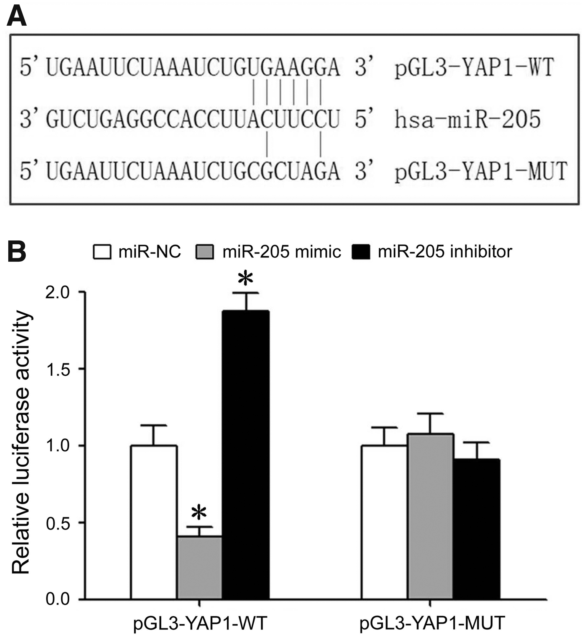
There is a targeted regulation relationship between miR-205 and YAP1 mRNA.
High resistance of Hela/CDDP cells
The IC50 of Hela cells was 1.89 ± 0.17 μg/mL, the IC50 of drug-resistant Hela/CDDP cells was 23.66 ± 2.29 μg/mL, and the resistance index of Hela/CDDP cells was 12.52 (Fig. 2 and Table 1).
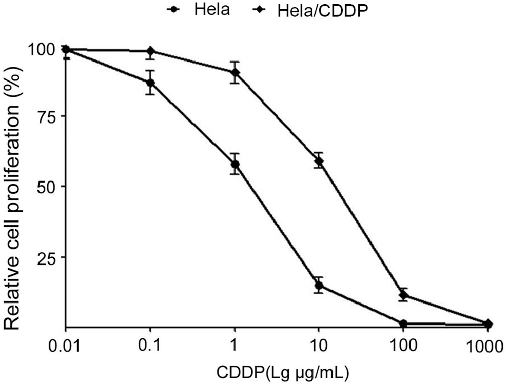
CCK-8 detects Hela, Hela/CDDP cell proliferation activity. CDDP, cisplatin.
IC50 of Hela, Hela/CDDP Cells
CDDP, cisplatin.
Abnormal expression of miR-205 and YAP1 in cervical cancer-resistant cells
The results of qRT-PCR showed that the expression of miR-205 in human parental cancer Hela cells was significantly lower than that in human cervical epithelial HCerEpiC cells and was significantly higher than that in drug-resistant Hela/CDDP cells (Fig. 3A). The results of qRT-PCR showed that compared with HCerEpiC cells, the expression of YAP1 mRNA in cervical cancer Hela cells was significantly increased and significantly higher in drug-resistant Hela/CDDP cells than that in Hela cells (Fig. 3B). Western blot analysis showed that compared with HCerEpiC cells, the expression of YAP1 protein in cervical cancer Hela cells was significantly increased and significantly higher in drug-resistant Hela/CDDP cells than that in Hela cells (Fig. 3C).

Abnormal expression of miR-205 and YAP1 in cervical cancer-resistant cells.
Overexpression of miR-205 inhibits YAP1 expression and reduces CDDP resistance in cervical cancer cells
The results of qRT-PCR showed that compared with the miR-NC group, YAP1 mRNA expression in HeLa/CDDP cells was significantly decreased in miR-205 mimic transfection group and significantly increased in HeLa/CDDP cells transfected with miR-205 inhibitor (Fig. 4A). Western blot analysis showed that compared with miR-NC group, the expression of YAP1 protein in Hela/CDDP cells was significantly decreased in miR-205 mimic transfection group, and significantly increased in Hela/CDDP cells transfected into miR-205 inhibitor group (Fig. 4B). The results of EdU staining showed that compared with the miR-NC group (27.2% ± 2.61%), the proliferation of Hela/CDDP cells in the miR-205 mimic transfection group (16.1% ± 1.01%) was significantly reduced, and significantly enhanced in the miR-205 inhibitor transfection group (36.3% ± 3.12%) (Fig. 4C). Flow cytometry results showed that compared with the miR-NC group (3.81% ± 0.31%), the apoptosis of Hela/CDDP cells in the miR-205 mimic transfection group (17.5% ± 1.26%) was significantly increased, and was significantly reduced in the miR-205 inhibitor transfection group (1.21% ± 0.12%) (Fig. 4D).
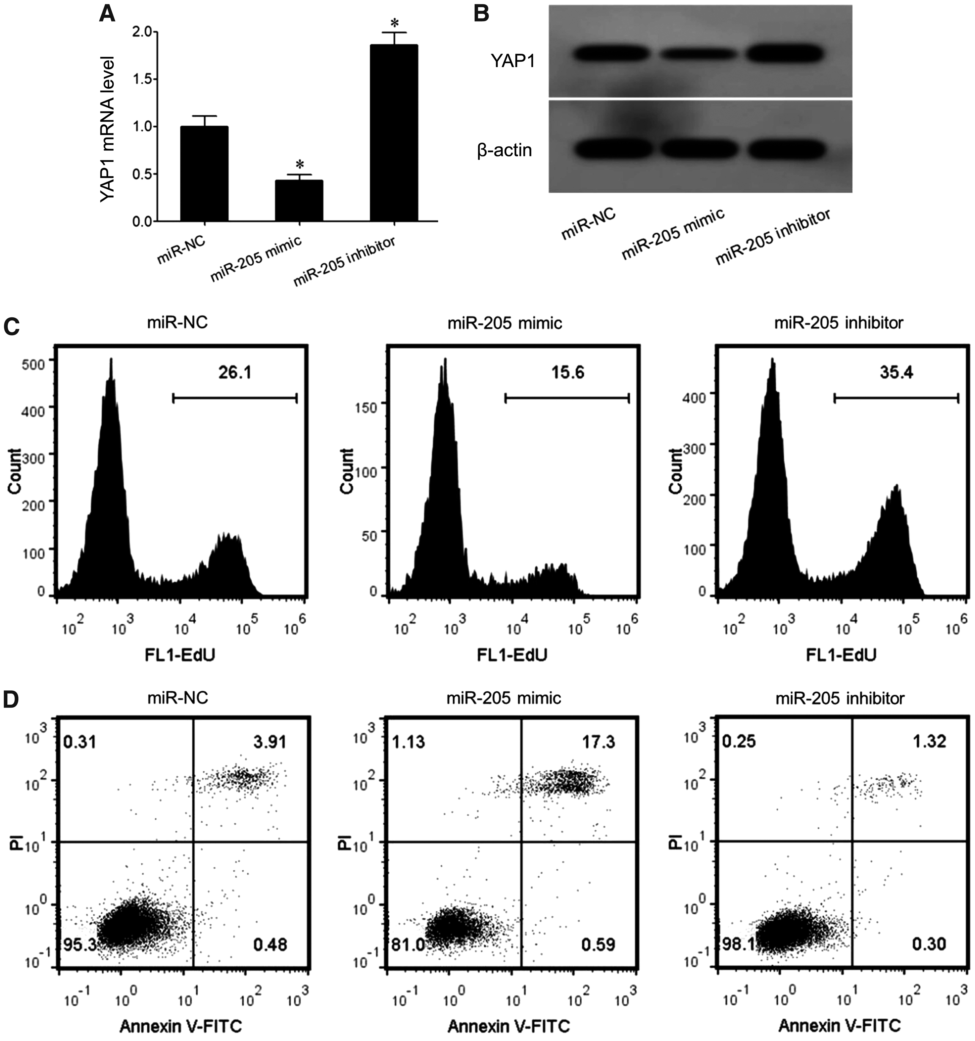
Overexpression of miR-205 inhibits YAP1 expression and reduces CDDP resistance in cervical cancer cells.
Discussion
Chemotherapy is an important approach in the treatment of cervical cancer, but a considerable number of patients in clinical practice are less sensitive to chemotherapy. Therefore, to find the signal molecules of abnormal changes in the process of cervical cancer resistance and explore the mechanism of cervical cancer drug resistance is beneficial to improve the treatment effect, prognosis, and the survival rate.
YAP1 is a multifunction intracellular connexin and transcriptional coactivator and is the main effector downstream of the Hippo-YAP signal transduction pathway. Its expression and functional activity are regulated by phosphorylation of the upstream kinase chain. 17 YAP1 specifically recognizes and binds to the transcription factor PPXY motifs in the nucleus, thereby regulating the transcription and expression of various genes, promoting cell proliferation, and inhibiting apoptosis. 18,19 Studies have shown that the expression and function enhancement of YAP1 is associated with the occurrence and progression of various types of tumors such as esophageal cancer, 20 gastric cancer, 21 and cervical cancer. 9 In cervical cancer, a number of studies have shown that the abnormal expression and functional dysfunction of YAP1 is associated with the occurrence, progression, and metastasis of cervical cancer, suggesting that YAP1 plays a role in the development and progression of cervical cancer. 8 –10
The abnormality of miR-205 is related to the occurrence, progression, and drug resistance of various tumors such as endometrial cancer, 22 pancreatic cancer, 23 and gastric cancer. 24 This study investigated whether miR-205 plays a role in regulating YAP1 expression and cervical cancer cell proliferation, apoptosis, and CDDP resistance.
In this study, the dual luciferase gene reporter assay showed that transfection of miR-205 mimic significantly reduced the relative luciferase activity in pGL3-YAP1-WT-transfected HEK293T cells, and transfection of miR-205 inhibitor significantly increased the relative luciferase activity in the pGL3-YAK1-WT-transfected HEK293T cells, confirming the targeted regulatory relationship between miR-205 and YAP1. According to the results of CCK-8 assay, the proliferation activity of Hela cells was significantly lower than that of Hela/CDDP cells under the same concentration of CDDP treatment, indicating that CDDP-resistant cervical cancer cells were successfully established. The results of gene and protein expression assay showed that the expression of miR-205 in cervical cancer Hela cells was significantly reduced in human normal cervical epithelial HCerEpiC cells, and was further reduced in drug-resistant Hela/CDDP cells. The expression of YAP1 mRNA and protein in cells was significantly increased in drug-resistant Hela/CDDP cells. The results showed that the decrease of miR-205 expression was associated with abnormal expression of YAP1, and the abnormal expression of miR-205 and YAP1 was not only related to the pathogenesis of cervical cancer, but also related to CDDP resistance. In the study of the relationship between miR-205 and cervical cancer, Pang and Yue 14 showed that compared with paracancerous tissues and normal cervical epithelial cells, cervical cancer patients and cervical cancer SiHa, Hela cell lines showed significantly decreased miR-205 level and decreased miR-205 expression was in association with histopathological grade, distant metastasis, and FIGO clinical stage. Consistent with this, this study revealed that the expression of miR-205 is associated with cervical cancer.
In this study, we further showed that transfection of miR-205 mimic significantly reduced the expression of YAP1 in Hela/CDDP-resistant cells, allowing Hela/CDDP-resistant cells to maintain stable growth in CDDP. The death rate was increased significantly, whereas the cell proliferation ability was significantly reduced and the drug resistance was decreased. Transfection of miR-205 inhibitor could increase the expression of YAP1 and enhance the drug resistance of Hela/CDDP cells. In the study of miR-205 regulating the biological effects of cervical cancer cells, Pang and Yue 14 showed that overexpression of miR-205 in cervical cancer SiHa and Hela cells can significantly inhibit cell proliferation, colony formation, and migration of cervical cancer cells. In addition, miR-205 overexpression can induce cell cycle arrest and promote cell apoptosis, and the anticancer effect of miR-205 is achieved by inhibition of the IFGR1 gene and its downstream signaling pathway. Yue et al. 15 study showed that treatment of cervical cancer SiHa, Hela cells with olmesartan can significantly increase the expression of miR-205 and inhibit the expression of its target gene VEGF-A, thereby inhibiting proliferation of SiHa and Hela cells and decreasing the clonal formation and invasion ability of cervical cancer cells. The results of Zhao et al. 25 showed that overexpression of DeltaNp63alpha protein can significantly inhibit the invasion and metastasis of cervical cancer and the anticancer effect of DeltaNp63alpha protein is through upregulating the expression of miR-205, leading to inhibition of the expression of miR-205 target gene ZEB1 and attenuating the EMT process of cervical cancer SiHa cells. In the study of the relationship between YAP1 and cervical cancer, Wang et al. 26 showed that the increased expression of YAP1 is related to the malignant biological characteristics of cervical cancer cells. Overexpression of YAP1 in cervical cancer cells can significantly promote cell proliferation and enhance the migration ability of cells. The results of Zhang et al. 10 showed that circulating RNA has-circ-0023404 can upregulate the expression of miR-136 and TFCP2 that activates YAP and promotes the proliferation, migration, and invasion of cervical cancer cells. The results of this study show that the increased expression of YAP1 is associated with cervical cancer, and is supported by the results of Wang et al. 26 and Zhang et al. 10 In this study, we found that miR-205 inhibits YAP1 expression and plays a role in reducing CDDP resistance in cervical cancer cells. However, whether miR-205 affects drug resistance in patients through regulating YAP1 expression remains unclear and it is required to verify this through collection of clinical tissue specimens of drug-resistant and drug-sensitive patients in the future.
Conclusions
The expression of miR-205 is related to the CDDP resistance of cervical cancer cells. Increasing the expression of miR-205 can downregulate the expression of YAP1, inhibit the proliferation and promote apoptosis of cervical cancer cells, and enhance the sensitivity to CDDP.
Footnotes
Disclosure Statement
There are no existing financial conflicts.
Funding Information
This study was supported by the Natural Science Foundation of Heilongjiang Province (No. H2016099) and the Special Research Fund for Doctorate of Qiqihar Medical University (No. QY2016B-03).